Key Points
EVs cause accumulation of activated maternal platelets within the placenta, resulting in a thromboinflammatory response and PE.
Activated maternal platelets cause NLRP3-inflammasome activation in trophoblast cells via ATP release and purinergic signaling.
Abstract
Preeclampsia (PE) is a placenta-induced inflammatory disease associated with maternal and fetal morbidity and mortality. The mechanisms underlying PE remain enigmatic and delivery of the placenta is the only known remedy. PE is associated with coagulation and platelet activation and increased extracellular vesicle (EV) formation. However, thrombotic occlusion of the placental vascular bed is rarely observed and the mechanistic relevance of EV and platelet activation remains unknown. Here we show that EVs induce a thromboinflammatory response specifically in the placenta. Following EV injection, activated platelets accumulate particularly within the placental vascular bed. EVs cause adenosine triphosphate (ATP) release from platelets and inflammasome activation within trophoblast cells through purinergic signaling. Inflammasome activation in trophoblast cells triggers a PE-like phenotype, characterized by pregnancy failure, elevated blood pressure, increased plasma soluble fms-like tyrosine kinase 1, and renal dysfunction. Intriguingly, genetic inhibition of inflammasome activation specifically in the placenta, pharmacological inhibition of inflammasome or purinergic signaling, or genetic inhibition of maternal platelet activation abolishes the PE-like phenotype. Inflammasome activation in trophoblast cells of women with preeclampsia corroborates the translational relevance of these findings. These results strongly suggest that EVs cause placental sterile inflammation and PE through activation of maternal platelets and purinergic inflammasome activation in trophoblast cells, uncovering a novel thromboinflammatory mechanism at the maternal-embryonic interface.
Introduction
Preeclampsia (PE) is a placenta-induced inflammatory disease and a leading cause of maternal and fetal morbidity and mortality.1,2 However, the mechanisms causing PE remain obscure and specific therapies are lacking.3 PE is associated with maternal proteinuria, hypertension, inflammation (eg, elevated interleukin-1β [IL-1β] levels), increased extracellular vesicles (EVs), platelet activation, and hypercoagulability.4-8 Platelet activation occurs before the onset of PE,9 yet attempts to link placental blood clots with the onset or outcome of PE overall failed.10 A proinflammatory function of platelets has been increasingly recognized in recent years,11 but the role of platelet-mediated proinflammatory processes, such as release of adenosine 5′-diphosphate (ADP), IL-1β, or EVs, for pregnancy complications such as PE remains unknown.12-14 Intriguingly, elevation of both activated and nonactivated platelet EVs in association with PE has been repeatedly reported.15,16
EVs isolated from pregnant women with PE differ phenotypically and functionally from those isolated from healthy pregnant controls.17-19 For example, ex vivo–generated EVs have an increased fms-like tyrosine kinase-1 (Flt-1)–to–soluble Flt-1 (sFlt-1) protein ratio if generated from PE placentae.19 In vitro, syncytiotrophoblast-derived EVs modulate the immune response, impair endothelial function, and convey procoagulant function (eg, platelet activation).19,20 These observations suggest that EVs have a pathogenetic function in PE. However, how EVs and platelet activation may promote a placental-specific disease process, resulting in systemic inflammation, is not understood. Here we show that EVs trigger accumulation of activated platelets specifically in the placenta, causing inflammasome activation within trophoblast cells via purinergic signaling, which is required and sufficient for a PE-like phenotype in pregnant mice.
Methods
Mice
Wild-type (WT) C57BL/6 mice and constitutive NACHT, LRR and PYD domains-containing protein 3 (NLRP3) and caspase-1 (Casp-1) knockout mice were obtained from The Jackson Laboratory. p45-NF-E2 and Gαq knockout mice have been previously published.21,22 All animal experiments were conducted following standards and procedures approved by the local animal care and use committee (Landesverwaltungsamt Halle, Germany).
Human tissues
Human placenta samples from pregnancies complicated with preeclampsia and normotensive control pregnancies were collected and provided by Universitätsklinikum Erlangen Frauenklinik (Erlangen, Germany) in accordance with the guidelines and with the approval of the local ethics committee and after obtaining informed consent of patients. PE was characterized by the development of hypertension, proteinuria, and an increased sFlt-1-to-placental growth factor ratio (>38).23,24
Generation and purification of procoagulant EVs
Mouse-derived SVEC cells (mouse endothelial cells; ATCC) or human-derived human umbilical vein endothelial cells (human endothelial cells; PromoCell) were serum-starved for 72 hours to generate EVs. Cell culture supernatant was collected, centrifuged at 200g for 10 minutes, followed by high-speed centrifugation at 20 000g for 45 minutes to pellet endothelial cell–derived EVs. For isolation of human or mouse platelet-derived EVs, citrated whole blood was collected, and platelet-rich plasma (PRP) was obtained after centrifugation at 160g for 20 minutes. The PRP was incubated with thrombin (1 nM final concentration) for 30 minutes followed by addition of excess hirudin to inactivate thrombin. Remaining platelets were pelleted by centrifugation at 2500g for 20 minutes and the supernatant was further centrifuged at 20 000g for 45 minutes to pellet platelet-derived EVs. After centrifugation, the EV pellet (for both EV types) was washed twice with phosphate-buffered saline (PBS) followed by centrifugation each time. The EV pellet was finally resuspended in PBS, aliquoted, and stored at −80°C until further use. Supernatant from the last wash was used as control for all experiments. Procoagulant activity (thrombin generation potential in nanomoles) of EVs (thawed once) was assessed using Zymuphen MP-Activity enzyme-linked immunosorbent assay (ELISA). EVs used for experiments were likewise only thawed once. EV concentration was adjusted to 600 nM/kg body weight (BW) procoagulant activity before injection. This corresponds to a calculated final EV concentration of about 2000 EV/µL blood in injected mice, which matches the EV concentration observed in pregnant women with PE.25,26 In all experiments, EVs of different cellular origin were used separately. Human cell–derived EVs were only used for studies with human trophoblast cells, whereas mouse cell–derived EVs were used for studies with mouse trophoblast cells and in mice.
Cell culture
To generate PRP, citrated blood was centrifuged at 160g for 20 minutes separated into 2 equal parts. One part was used directly as PRP and the other part was further centrifuged at 2500g for 10 minutes to separate remaining platelets and collect platelet-poor plasma (PPP). Equal amounts of either preparation (PRP or PPP) were added to cells along with EVs. Mouse trophoblast stem cells were treated with EVs alone (final procoagulant activity, 7.5 nM thrombin equivalent) or EVs with PRP or PPP and, in some experiments, with apyrase, or adenosine 5′-triphosphate (ATP) periodate oxidized sodium salt (oATP) along with EVs and PRP for 24 hours in differentiation media and were then allowed to further differentiate for 4 days. Human trophoblast-like cells (BeWo, JEG-3) were treated with EVs (final procoagulant activity, 7.5 nM thrombin equivalent) alone or EVs with PRP or PPP and, in some experiments, apyrase or oATP were used along with EVs and PRP for 24 hours. For studying the effect of aspirin, PRP was obtained from healthy volunteers who had taken aspirin (500 mg per day) for 3 consecutive days and blood was obtained 1 hour after the last dose. RNA or protein was then isolated for quantitative polymerase chain reaction and western blotting, respectively.
Timed matings and in vivo interventions
Plugged female mice were separated from males and injected at day 10.5 post coitum (p.c.) and 11.5 p.c. with 600 nM/kg BW (procoagulant activity) endothelial- or platelet-derived EVs IV and the pregnancy outcome was analyzed at day 12.5 p.c. Control mice were injected with an equal volume of supernatant from the last PBS wash of EVs during the isolation procedure. For platelet depletion, anti-glycoprotein Ib (platelet) α-subunit (GP1bα) antibody (4 mg/kg; polyclonal nonimmune immunoglobulin G served as control) was injected IV on day 8.5 p.c. Aspirin (100 mg/kg), apyrase (200 U/kg BW), oATP (9 mM/kg BW), or anakinra (20 mg/kg BW) were injected intraperitoneally 30 minutes prior to each EV injection.
Determination of proteinuria
Spot urine samples were collected from mice at day 12.5 p.c. and the protein-to-creatinine (P/C) ratio was analyzed using the bicinchoninic acid assay normalized to creatinine content.27
Determination of sFlt-1
sFlt-1 was determined in plasma samples using the mouse VEGF R1/Flt-1 Quantikine ELISA kit (R&D Systems) as described by the manufacturer.
Blood pressure measurement
Histology
Immunostaining
Transmission electron microscopy
In situ proximity ligation assay
The Duolink in situ PLA kit was used for in situ proximity-ligation assay on human placenta sections according to the manufacturer’s instructions (Sigma-Aldrich).35
Immunoblotting
Cell or tissue lysates were prepared using radioimmunoprecipitation assay buffer and electrophoretically separated on sodium dodecyl sulfate polyacrylamide gel, transferred to polyvinylidene difluoride membranes, and probed with desired primary antibodies overnight at 4°C. Membranes were then washed with PBS supplemented with Tween 20 and incubated with horseradish peroxidase–conjugated secondary antibodies. Blots were developed with the enhanced chemiluminescence system and analyzed using Image J software.
Measurement of ATP release
ATP release from PRP was studied using Cell-titer Glo reagent (Promega Corporation) according to the manufacturer’s protocol.
Statistical analysis
The data are summarized as the mean ± standard error of the mean (SEM). Statistical analyses were performed with the Student t test, the χ2 test, Spearman correlation, the Mann-Whitney U test, or analysis of variance (ANOVA), as appropriate. Post hoc comparisons of ANOVA were corrected with the method of Tukey. The Kolmogorov-Smirnov test or the D’Agostino-Pearson normality test was used to determine whether the data are consistent with a Gaussian distribution. Prism 5 (www.graphpad.com) software was used for statistical analyses. Statistical significance was accepted at P values of <.05.
Results
EVs cause a PE-like phenotype in pregnant mice
To evaluate the potential pathogenicity of EVs in pregnancy, we injected pregnant mice with procoagulant EVs and evaluated their impact on pregnancy outcome, renal function, and blood pressure. Platelet or endothelial-derived EVs injected at day 10.5 and 11.5 p.c. impaired embryonic survival at day 12.5 p.c. (Figure 1A-B; supplemental Figure 1-2, available on the Blood Web site). Surviving embryos were smaller, less developed (impaired forelimb and reduced retinal pigmentation) and had reduced placental diameter (Figure 1A-D; supplemental Figure 1). These changes were associated with reduced embryonic but increased maternal placental vascularization, reflecting placental malperfusion (Figure 1E-F). Additionally, EV-injected pregnant mice developed characteristic hallmarks of PE defined by renal dysfunction (enlarged glomeruli, thickened glomerular basement membrane, podocyte effacement, and proteinuria), elevated blood pressure, and increased plasma sFlt-1 levels (Figure 1G-L). Importantly, EVs had no impact on renal dysfunction or blood pressure in nonpregnant mice (Figure 1G-L). Hence, EVs induce a PE-like phenotype through an unknown mechanism.
EVs impair embryonic and placental development and cause a PE-like phenotype in mice. (A-D) Impaired pregnancy outcome in C57BL/6 mice at day 12.5 p.c. following IV injection of mouse endothelial cell–derived EVs at days 10.5 p.c. and 11.5 p.c. Representative images (A) of uterus (top), placenta (middle), and embryo (bottom) and bar graphs quantifying (B) embryonic survival, (C) embryonic height, and (D) placental diameter. Size bar represents 1 mm. (E-F) Altered placental morphology after EV injections. (E) Representative images of placental histology (H&E staining) showing enhanced maternal vascularization (blood lacunae; enucleated erythrocytes) and reduced fetal vascularization (nucleated erythrocytes) after EV injections. (F) Bar graph summarizing quantification of total vascularized area; analyses performed at day 12.5 p.c. Size bar represents 20 µm. (G-L) Characterization of renal pathology in pregnant (Preg) and nonpregnant (Non-Preg) mice following EV injection. Representative images showing enhanced renal pathology in pregnant mice after EV injections, characterized by enlarged glomeruli (G, PAS staining, top; H, bar graph reflecting quantification) and podocyte effacement and thickened glomerular basement membrane (GBM) (G, TEM, n = 3, bottom; I, bar graph reflecting glomerular basement membrane thickness). Proteinuria is increased in EV-injected pregnant mothers at day 12.5 p.c. (J, bar graph summarizing data of P/C ratio). These features of renal dysfunction are not observed in EV-injected nonpregnant females. Size bar represents 15 µm for PAS staining and 1 µm for TEM images, respectively. (K) Elevated blood pressure in EV-injected pregnant mice at day 12.5 p.c. EVs have no impact on blood pressure in nonpregnant mice; bar graph summarizing results. (L) sFlt-1 plasma levels. The PE marker sFlt-1 is increased in blood samples obtained from EV-injected pregnant mice as compared with controls (bar graph summarizing results). Data shown represent mean ± SEM of at least 8 placentae or embryos analyzed from at least 3 different litters of each group or 5 pregnant females per group. Control mice (C) were injected with the supernatant obtained after the last PBS wash during EV isolation. *P < .05, **P < .01. (B-D, H-K) Student t test; (F) ANOVA. Dias, diastolic; ns, nonsignificant; Sys, systolic.
EVs impair embryonic and placental development and cause a PE-like phenotype in mice. (A-D) Impaired pregnancy outcome in C57BL/6 mice at day 12.5 p.c. following IV injection of mouse endothelial cell–derived EVs at days 10.5 p.c. and 11.5 p.c. Representative images (A) of uterus (top), placenta (middle), and embryo (bottom) and bar graphs quantifying (B) embryonic survival, (C) embryonic height, and (D) placental diameter. Size bar represents 1 mm. (E-F) Altered placental morphology after EV injections. (E) Representative images of placental histology (H&E staining) showing enhanced maternal vascularization (blood lacunae; enucleated erythrocytes) and reduced fetal vascularization (nucleated erythrocytes) after EV injections. (F) Bar graph summarizing quantification of total vascularized area; analyses performed at day 12.5 p.c. Size bar represents 20 µm. (G-L) Characterization of renal pathology in pregnant (Preg) and nonpregnant (Non-Preg) mice following EV injection. Representative images showing enhanced renal pathology in pregnant mice after EV injections, characterized by enlarged glomeruli (G, PAS staining, top; H, bar graph reflecting quantification) and podocyte effacement and thickened glomerular basement membrane (GBM) (G, TEM, n = 3, bottom; I, bar graph reflecting glomerular basement membrane thickness). Proteinuria is increased in EV-injected pregnant mothers at day 12.5 p.c. (J, bar graph summarizing data of P/C ratio). These features of renal dysfunction are not observed in EV-injected nonpregnant females. Size bar represents 15 µm for PAS staining and 1 µm for TEM images, respectively. (K) Elevated blood pressure in EV-injected pregnant mice at day 12.5 p.c. EVs have no impact on blood pressure in nonpregnant mice; bar graph summarizing results. (L) sFlt-1 plasma levels. The PE marker sFlt-1 is increased in blood samples obtained from EV-injected pregnant mice as compared with controls (bar graph summarizing results). Data shown represent mean ± SEM of at least 8 placentae or embryos analyzed from at least 3 different litters of each group or 5 pregnant females per group. Control mice (C) were injected with the supernatant obtained after the last PBS wash during EV isolation. *P < .05, **P < .01. (B-D, H-K) Student t test; (F) ANOVA. Dias, diastolic; ns, nonsignificant; Sys, systolic.
EVs cause activation and accumulation of maternal platelets within the placenta
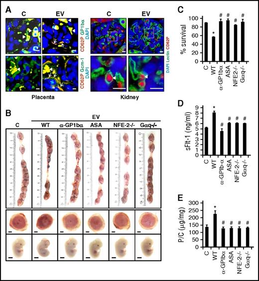
Using a whole-blood clotting assay (rotational thromboelastometry), we established that the EVs used within this study efficiently induced blood clotting (supplemental Figure 3). Yet, detailed analyses of placental sections (H&E, fibrin(ogen) immunostaining) did not reveal an increase in placental thrombosis in EV-injected pregnant mice. However, we detected activated (P-selectin+) platelets within the placenta (but not in other organs such as kidney) of EV-injected, but not of control, pregnant mice (Figure 2A). Activated platelets lined the maternal blood space and were in direct contact with embryonic trophoblast cells. To evaluate whether maternal platelets contribute to EV-induced PE in vivo, we used several complimentary approaches (Figure 2B-E). Thus, immune depletion of maternal platelets with anti-mouse GP1bα antibodies protected pregnant WT mice from EV-induced PE. Likewise, aspirin treatment improved pregnancy outcome in EV-injected pregnant mice. Mice lacking the transcription factor p45-NF-E2 lack functional platelets and Gαq−/− mice have severely impaired platelet activation.21 Pregnant p45-NF-E2−/− or Gαq−/− mice mated with WT males were protected from EV-induced pregnancy loss, intrauterine growth restriction, placental dysfunction, and did not develop signs of PE (Figure 2B-E). This identifies a crucial function of maternal platelet activation for EV-mediated PE in mice independent of blood clot formation.
Maternal platelets mediate EV-induced PE-like phenotype in mice. (A) Double immunofluorescence staining showing activated platelets (CD62P, P-selectin; red) in the placenta (left), but not in the kidney (right), after injections of mouse endothelial cell–derived EVs. CD62P (red) colocalizes (yellow) with GP1bα (green) indicating presence of activated platelets within the placenta (Placenta, top left panel). Colocalization (yellow) of activated platelets (CD62P; red) and Gcm-1+ syncytiotrophoblast (arrows, green) indicates direct contact of activated platelets with syncytiotrophoblasts, which line maternal blood spaces within the placenta (Placenta, bottom left panel); conventional immunofluorescence analyses; arrowheads, autofluorescence of erythrocytes. (B-C) Depletion of maternal platelets using anti-GP1bα antibody, inhibition of maternal platelets using aspirin, genetic platelet deficiency (p45 NF-E2−/− mice), or genetically superimposed diminished platelet activation (Gαq deficiency) protects pregnant female mice from EV-induced impaired pregnancy outcome. Pregnancy outcome at day 12.5 p.c. after EV injection (mouse endothelial cell–derived EVs) at days 10.5 p.c. and 11.5 p.c. into pregnant females mated to WT males. (B) Representative images of uterus (top), placentae (middle), and embryos (bottom) along with (C) bar graphs quantifying embryonic survival. (D-E) Plasma sFlt-1 levels (D) and proteinuria (E, bar graph summarizing data of P/C ratio) are normal despite EV treatment in platelet-depleted, aspirin-treated, NFE-2−/−, or Gαq−/− pregnant female mice. Scale bar represents (A) 80 µm for placenta and 10 µm for kidney, and (B) 1 mm for embryo and placenta. Data represent mean ± SEM of at least 8 placentae or embryos analyzed from at least 3 different litters of each group. Control mice (C) were injected with the supernatant obtained after the last PBS wash during EV isolation. *P < .05 (relative to control, C); #P < .05 (relative to EV). (C-E) ANOVA. ASA, acetylsalicylic acid; DAPI, 4′,6-diamidino-2-phenylindole.
Maternal platelets mediate EV-induced PE-like phenotype in mice. (A) Double immunofluorescence staining showing activated platelets (CD62P, P-selectin; red) in the placenta (left), but not in the kidney (right), after injections of mouse endothelial cell–derived EVs. CD62P (red) colocalizes (yellow) with GP1bα (green) indicating presence of activated platelets within the placenta (Placenta, top left panel). Colocalization (yellow) of activated platelets (CD62P; red) and Gcm-1+ syncytiotrophoblast (arrows, green) indicates direct contact of activated platelets with syncytiotrophoblasts, which line maternal blood spaces within the placenta (Placenta, bottom left panel); conventional immunofluorescence analyses; arrowheads, autofluorescence of erythrocytes. (B-C) Depletion of maternal platelets using anti-GP1bα antibody, inhibition of maternal platelets using aspirin, genetic platelet deficiency (p45 NF-E2−/− mice), or genetically superimposed diminished platelet activation (Gαq deficiency) protects pregnant female mice from EV-induced impaired pregnancy outcome. Pregnancy outcome at day 12.5 p.c. after EV injection (mouse endothelial cell–derived EVs) at days 10.5 p.c. and 11.5 p.c. into pregnant females mated to WT males. (B) Representative images of uterus (top), placentae (middle), and embryos (bottom) along with (C) bar graphs quantifying embryonic survival. (D-E) Plasma sFlt-1 levels (D) and proteinuria (E, bar graph summarizing data of P/C ratio) are normal despite EV treatment in platelet-depleted, aspirin-treated, NFE-2−/−, or Gαq−/− pregnant female mice. Scale bar represents (A) 80 µm for placenta and 10 µm for kidney, and (B) 1 mm for embryo and placenta. Data represent mean ± SEM of at least 8 placentae or embryos analyzed from at least 3 different litters of each group. Control mice (C) were injected with the supernatant obtained after the last PBS wash during EV isolation. *P < .05 (relative to control, C); #P < .05 (relative to EV). (C-E) ANOVA. ASA, acetylsalicylic acid; DAPI, 4′,6-diamidino-2-phenylindole.
Normal pregnancy in NLRP3- or Casp-1–deficient mice despite EV injection
In addition to blood clotting, platelets regulate inflammatory responses, in part through the modulation of the inflammasome.36 Among others, PE is characterized by inflammatory cytokines, including IL-1β, the pivotal cytokine reflecting inflammasome activation.7 To determine whether the EV- and platelet-induced PE-like phenotype is associated with placental inflammasome activation, we determined inflammasome markers (cleaved Casp-1 and IL-1β and NLRP3 expression) in placenta tissues. These markers were elevated in placenta tissues obtained from EV-injected mice compared with controls, establishing placental inflammasome activation by EVs (Figure 3A-B).
EVs cause inflammasome activation in placenta. (A-B) Inflammasome activation in murine placentae after EV injections (mouse endothelial cell–derived EVs). Immunoblots showing increased cleaved Casp-1 and cleaved IL-1β and increased NLRP3 expression in murine placentae after EV injections, analyzed at day 12.5 p.c. (A, representative immunoblots; B, bar graph summarizing results). Arrowheads indicate inactive (proform, white arrowheads) and the active (cleaved form, black arrowheads) form of Casp-1 or IL-1β, respectively (A). Only the active form was quantified (B). (C-D) Representative images of (top) uterus, (middle) placentae, and (bottom) embryos showing protection from mouse endothelial-derived EV-induced pregnancy complications in NLRP3−/− (C) and Casp-1−/− (D, Casp-1) mice. Size bar, 1 mm. (E) Double immunofluorescence staining showing activated platelets (CD62P, P-selectin; red) in the placenta after EV injection in pregnant NLRP3−/− females mated with NLRP3−/− males. Activated platelets are in direct contact with syncytiotrophoblast (Gcm-1; green); yellow indicates colocalization of activated platelets and Gcm-1+ syncytiotrophoblast (arrows); conventional immunofluorescence analyses; arrowheads, autofluorescence of erythrocytes. Scale bar, 20 µm. Control mice (C) were injected with the supernatant obtained after the last PBS wash during EV isolation. Data shown represent mean ± SEM of at least 8 placentae or embryos analyzed from at least 3 different litters of each group *P < .05 (relative to control, C). (B) Student t test. GAPDH, glyceraldehyde 3-phosphate dehydrogenase.
EVs cause inflammasome activation in placenta. (A-B) Inflammasome activation in murine placentae after EV injections (mouse endothelial cell–derived EVs). Immunoblots showing increased cleaved Casp-1 and cleaved IL-1β and increased NLRP3 expression in murine placentae after EV injections, analyzed at day 12.5 p.c. (A, representative immunoblots; B, bar graph summarizing results). Arrowheads indicate inactive (proform, white arrowheads) and the active (cleaved form, black arrowheads) form of Casp-1 or IL-1β, respectively (A). Only the active form was quantified (B). (C-D) Representative images of (top) uterus, (middle) placentae, and (bottom) embryos showing protection from mouse endothelial-derived EV-induced pregnancy complications in NLRP3−/− (C) and Casp-1−/− (D, Casp-1) mice. Size bar, 1 mm. (E) Double immunofluorescence staining showing activated platelets (CD62P, P-selectin; red) in the placenta after EV injection in pregnant NLRP3−/− females mated with NLRP3−/− males. Activated platelets are in direct contact with syncytiotrophoblast (Gcm-1; green); yellow indicates colocalization of activated platelets and Gcm-1+ syncytiotrophoblast (arrows); conventional immunofluorescence analyses; arrowheads, autofluorescence of erythrocytes. Scale bar, 20 µm. Control mice (C) were injected with the supernatant obtained after the last PBS wash during EV isolation. Data shown represent mean ± SEM of at least 8 placentae or embryos analyzed from at least 3 different litters of each group *P < .05 (relative to control, C). (B) Student t test. GAPDH, glyceraldehyde 3-phosphate dehydrogenase.
To ascertain whether the EV- and platelet-induced PE-like phenotype is dependent on inflammasome activation, we injected NLRP3−/− or Casp-1−/− female mice mated with NLRP3−/− or Casp-1−/− male mice, respectively, with EVs. NLRP3 or Casp-1 deficiency prevented EV-induced embryonic death and normalized embryonic and placental development (Figure 3C-D). Furthermore, NLRP3- or Casp-1–deficient pregnant mice lacked hallmarks of PE as described in the first “Results” section (supplemental Figure 4). Hence, inflammasome activation is required for EV-induced PE in mice. Intriguingly, activated platelets were still detectable within the placenta of EV-injected pregnant mice (Figure 3E). This raises the question as to whether inflammasome activation in platelets or a different cell type drives the PE-like phenotype.
EVs and platelets cause inflammasome activation in trophoblast cells
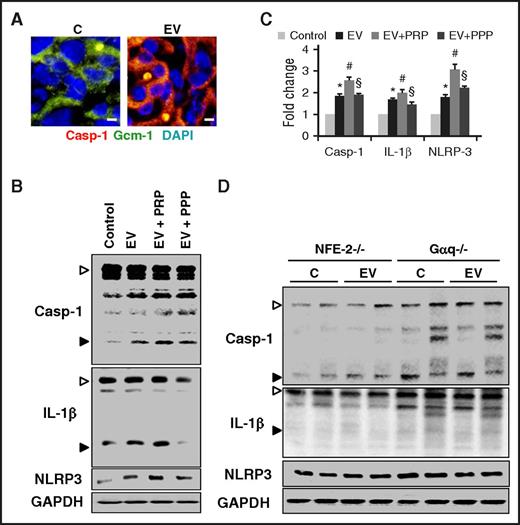
To identify the cells in which EVs induce inflammasome activation, we conducted immunohistochemical analyses. Following EV injection, a marked induction of NLRP3 and cleaved Casp-1 was readily detectable in trophoblast cells (supplemental Figure 5). This was confirmed by immunofluorescent analyses, demonstrating colocalization of cleaved Casp-1 with the syncytiotrophoblast cell marker glial cells missing-1 (Gcm-1) (Figure 4A).
EVs cause platelet-dependent inflammasome activation in trophoblast cells. (A) Representative images (double immunofluorescence staining) showing colocalization (yellow) of cleaved Casp-1 (red) and Gcm1 (green, syncytiotrophoblast marker) indicating inflammasome activation in trophoblasts after EV injections. Size bar represents 80 µm. (B-C) Platelets enhance EV-mediated (mouse endothelial cell–derived EVs) inflammasome activation in murine trophoblast cells. Representative immunoblots showing enhanced EV-mediated inflammasome activation in trophoblast cells in the presence of PRP (EV+PRP) compared with EVs only (EV) or EVs with PPP (EV+PPP). Control cells were exposed to the supernatant obtained after the last PBS wash during EV isolation (B, representative immunoblots; C, bar graph summarizing results). (D) Immunoblots showing no effect on inflammasome activation in p45-NF-E2+/− and Gαq+/− placenta after EV treatment in p45-NF-E2−/− or Gαq−/− pregnant female mice, respectively (representative immunoblots). Arrowheads indicate inactive (proform, white arrowheads) and the active (cleaved form, black arrowheads) form of Casp-1 or IL-1β, respectively (B,D). Only the active form was quantified (C). Data shown represent mean ± SEM from 5 independent experiments. *P < .05 (relative to control, C); #P < .05 (relative to EV); §P < .05 (relative to EV+PRP). (C) ANOVA.
EVs cause platelet-dependent inflammasome activation in trophoblast cells. (A) Representative images (double immunofluorescence staining) showing colocalization (yellow) of cleaved Casp-1 (red) and Gcm1 (green, syncytiotrophoblast marker) indicating inflammasome activation in trophoblasts after EV injections. Size bar represents 80 µm. (B-C) Platelets enhance EV-mediated (mouse endothelial cell–derived EVs) inflammasome activation in murine trophoblast cells. Representative immunoblots showing enhanced EV-mediated inflammasome activation in trophoblast cells in the presence of PRP (EV+PRP) compared with EVs only (EV) or EVs with PPP (EV+PPP). Control cells were exposed to the supernatant obtained after the last PBS wash during EV isolation (B, representative immunoblots; C, bar graph summarizing results). (D) Immunoblots showing no effect on inflammasome activation in p45-NF-E2+/− and Gαq+/− placenta after EV treatment in p45-NF-E2−/− or Gαq−/− pregnant female mice, respectively (representative immunoblots). Arrowheads indicate inactive (proform, white arrowheads) and the active (cleaved form, black arrowheads) form of Casp-1 or IL-1β, respectively (B,D). Only the active form was quantified (C). Data shown represent mean ± SEM from 5 independent experiments. *P < .05 (relative to control, C); #P < .05 (relative to EV); §P < .05 (relative to EV+PRP). (C) ANOVA.
We next assessed the relevance of platelets for EV-dependent inflammasome activation within the trophoblast cells. Exposure of murine trophoblast cells to EVs along with PRP increased levels of cleaved Casp-1 and IL-1β and NLRP3 expression in comparison with trophoblast cells exposed to EVs only or to EVs in the presence of PPP (Figure 4B-C; supplemental Figure 6). This demonstrates that platelets augment EV-induced inflammasome activation in trophoblast cells in vitro. To determine the relevance of platelets for EV-induced inflammasome activation in vivo, we analyzed placenta tissues obtained from pregnant p45-NF-E2−/− and Gαq−/− mice mated with WT mice. In both cases, EVs failed to induce placental inflammasome activation (Figure 4D), which establishes a crucial role of maternal platelets in inducing EV-dependent placental sterile inflammation. This identifies a thromboinflammatory cross-talk at the maternal-embryonic interface.
Placental inflammasome activation causes PE
The findings of the previous 2 sections suggest that inflammasome activation within trophoblast cells may be at the core of PE. To evaluate the mechanistic relevance of trophoblast-specific inflammasome activation in PE, we bred NLRP3+/− females with NLRP3−/− males, which is expected to yield an equal ratio of NLRP3−/− and NLRP3+/− embryos (having the same genotype in their trophoblast cells). EV injection selectively induced intrauterine growth restriction, death, and placental dysfunction of NLRP3-expressing embryos (NLRP3+/−; Figure 5A-B). Conversely, despite maternal NLRP3 expression, the survival rate, size, and development of NLRP3−/− embryos was comparable to that observed in control pregnant mice (Figure 5A-B). As expected, pregnant NLRP3+/− mice carrying both NLRP3-expressing and deficient embryos developed all signs of PE (Figure 5C; supplemental Figure 7A-F). The normal survival and phenotype of NLRP3−/− embryos despite maternal NLRP3 expression establishes that maternal NLRP3 (including NLRP3 in maternal platelets) does not impair placental and embryonic development. Furthermore, despite the maternal PE-like phenotype, the survival and development of NLRP3−/− embryos were normal, demonstrating that impaired embryonic development is not a consequence of the maternal disease.
Placental inflammasome activation causes PE. (A-B) Pregnancy outcome at day 12.5 p.c. after IV. EV (mouse endothelial cell–derived) injection at days 10.5 p.c. and 11.5 p.c. into pregnant NLRP3+/− females mated to NLRP3−/− males. (A) Representative images of uterus (left panel), placenta (right top panel), and embryo (right bottom panel) and (B) bar graph quantifying embryonic survival. (C) Elevated sFlt-1 plasma levels in EV-treated NLRP3+/− pregnant female mice at day 12.5 p.c. following EV injection; bar graphs summarizing results. (D-E) Pregnancy outcome at day 12.5 p.c. after IV EV injection at days 10.5 p.c. and 11.5 p.c. in NLRP3−/− females mated to NLRP3+/+ males. (D) Representative images of uterus (left panel), placenta (right top panel), and embryos (right bottom panel) along with (E) bar graph quantifying embryonic survival. (F) Elevated sFlt-1 plasma levels in EV-injected pregnant mice following mating NLRP3−/− females with NLRP3+/+ males; bar graphs summarizing results. Size bar represents 1 mm for embryo and placenta (A,D). Data shown represent mean ± SEM from at least 8 embryos analyzed from 3 different litters or 5 pregnant females of each group; control mice (C) were injected with the supernatant obtained after the last PBS wash during EV isolation. *P < .05 (relative to control, C). (B) ANOVA; (C-F) Student t test.
Placental inflammasome activation causes PE. (A-B) Pregnancy outcome at day 12.5 p.c. after IV. EV (mouse endothelial cell–derived) injection at days 10.5 p.c. and 11.5 p.c. into pregnant NLRP3+/− females mated to NLRP3−/− males. (A) Representative images of uterus (left panel), placenta (right top panel), and embryo (right bottom panel) and (B) bar graph quantifying embryonic survival. (C) Elevated sFlt-1 plasma levels in EV-treated NLRP3+/− pregnant female mice at day 12.5 p.c. following EV injection; bar graphs summarizing results. (D-E) Pregnancy outcome at day 12.5 p.c. after IV EV injection at days 10.5 p.c. and 11.5 p.c. in NLRP3−/− females mated to NLRP3+/+ males. (D) Representative images of uterus (left panel), placenta (right top panel), and embryos (right bottom panel) along with (E) bar graph quantifying embryonic survival. (F) Elevated sFlt-1 plasma levels in EV-injected pregnant mice following mating NLRP3−/− females with NLRP3+/+ males; bar graphs summarizing results. Size bar represents 1 mm for embryo and placenta (A,D). Data shown represent mean ± SEM from at least 8 embryos analyzed from 3 different litters or 5 pregnant females of each group; control mice (C) were injected with the supernatant obtained after the last PBS wash during EV isolation. *P < .05 (relative to control, C). (B) ANOVA; (C-F) Student t test.
To ascertain whether placental inflammasome activation is sufficient for the EV-induced PE-like phenotype, we mated NLRP3−/− females with WT males resulting in NLRP3+/− embryos and trophoblast cells. Despite maternal NLRP3 deficiency, EVs reduced embryonic survival, impaired the development of surviving NLRP3+/− embryos, and caused placental malperfusion (Figure 5D-E). Hence, embryonic NLRP3 expression within trophoblast cells is sufficient to impair placental and embryonic development in pregnant mice injected with EVs.
Importantly, following EV injection of pregnant NLRP3−/− mice carrying NLRP3-expressing embryos (NLRP3+/− embryos, obtained through mating with male NLRP3+/+ mice), these pregnant mice developed all signs of PE (Figure 5F; supplemental Figure 7G-L). This establishes a central pathogenetic function of the placental inflammasome for the induction of the PE-like phenotype, placental dysfunction, and embryonic demise.
Purinergic and inflammasome signaling induces PE
The data of the previous section establish that inflammasome activation in trophoblast cells induces a PE-like phenotype in mice. Strikingly, maternal platelets induce inflammasome activation within the placenta, whereas the maternal inflammasome, including that in platelets, is dispensable for the PE-like phenotype. To decipher the mechanism underlying this maternal-embryonic thromboinflammatory cross-talk, we considered a potential role of platelet-derived ATP. Upon activation, platelets release ATP, and ATP induces inflammasome activation via purinergic receptors.37 Purinergic receptors, in turn, are expressed by trophoblast cells.38 First, we ascertained that EVs are capable of inducing ATP release from platelets (supplemental Figure 8A). To determine the mechanistic relevance of platelet-derived ATP and the involvement of purinergic receptors, we exposed trophoblast cells to EVs and platelets (PRP) along with apyrase (an ATPase) or oATP (purinergic receptor antagonist). Exposure of trophoblast cells to EVs and platelets resulted in strong inflammasome activation, which was inhibited by treatment with apyrase or oATP (supplemental Figure 8B-C).
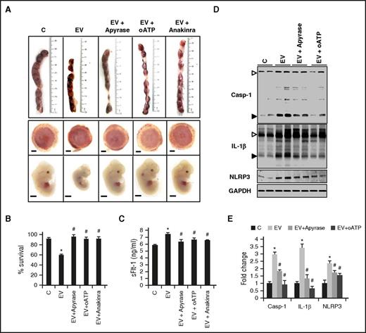
To evaluate the mechanistic relevance of ATP-induced purinergic inflammasome activation in vivo, we treated pregnant mice with apyrase, oATP, or anakinra (IL-1R antagonist). Treatment of EV-injected pregnant mice with either agent markedly improved pregnancy outcome, as reflected by normal embryonic survival, size, and development, and normal placental size and vascularization (Figure 6A-B). Concomitantly, signs of PE were prevented (Figure 6C). Apyrase or oATP treatment furthermore reduced NLRP3 expression and Casp-1 or IL-1β cleavage in the placenta tissues (Figure 6D-E). These results establish that purinergic signaling mediates the EV- and platelet-dependent PE-like phenotype. Inhibition of purinergic or IL-1β signaling protects pregnant mice from the EV- and platelet-dependent induction of PE.
Inhibiting purinergic or inflammasome signaling protects from EV-induced PE. (A-B) Inflammasome inhibition using apyrase, oATP, or anakinra protects mice from EV-induced placental and fetal impairment. Representative images (A) showing uterus (top), placentae (middle), or embryos (bottom) obtained from control (C) or EV-injected (EV) pregnant mice without or with treatment (apyrase, oATP, or anakinra) and quantification of embryonic survival (B). Size bar represents 1 mm. (C) Injections with apyrase, oATP, or anakinra reduce plasma sFlt-1 levels in EV-injected pregnant mice; bar graph summarizing results. (D-E) Immunoblots showing reduced cleaved Casp-1 and IL-1β and NLRP3 expression indicating inhibition of EV-induced inflammasome activation in murine placentae after apyrase or oATP treatment (D, representative immunoblots; E, bar graph summarizing results). Arrowheads indicate inactive (proform, white arrowheads) and the active (cleaved form, black arrowheads) form of Casp-1 or IL-1β, respectively (D). Only the active form was quantified (E). Data shown represent at least 8 placentae or embryos analyzed from 3 different litters or 5 pregnant females of each group; control mice (C) were injected with the supernatant obtained after the last PBS wash during EV isolation. Mean ± SEM. *P < .05 (relative to C); #P < .05 (relative to EV). (E) ANOVA.
Inhibiting purinergic or inflammasome signaling protects from EV-induced PE. (A-B) Inflammasome inhibition using apyrase, oATP, or anakinra protects mice from EV-induced placental and fetal impairment. Representative images (A) showing uterus (top), placentae (middle), or embryos (bottom) obtained from control (C) or EV-injected (EV) pregnant mice without or with treatment (apyrase, oATP, or anakinra) and quantification of embryonic survival (B). Size bar represents 1 mm. (C) Injections with apyrase, oATP, or anakinra reduce plasma sFlt-1 levels in EV-injected pregnant mice; bar graph summarizing results. (D-E) Immunoblots showing reduced cleaved Casp-1 and IL-1β and NLRP3 expression indicating inhibition of EV-induced inflammasome activation in murine placentae after apyrase or oATP treatment (D, representative immunoblots; E, bar graph summarizing results). Arrowheads indicate inactive (proform, white arrowheads) and the active (cleaved form, black arrowheads) form of Casp-1 or IL-1β, respectively (D). Only the active form was quantified (E). Data shown represent at least 8 placentae or embryos analyzed from 3 different litters or 5 pregnant females of each group; control mice (C) were injected with the supernatant obtained after the last PBS wash during EV isolation. Mean ± SEM. *P < .05 (relative to C); #P < .05 (relative to EV). (E) ANOVA.
Placenta-specific inflammasome activation in human PE patients
To ascertain the translational relevance and corroborate the findings obtained in mice with human pathophysiology, we first investigated EV-mediated inflammasome activation in human trophoblast cells. Exposure of Bewo or JEG-3 cells to EVs and platelets (PRP) enhanced inflammasome activation (increased Casp-1 and IL-1β cleavage and NLRP3 expression) in comparison with trophoblast cells exposed to EVs only or to EVs in the presence of PPP (Figure 7A; supplemental Figure 9). Exposure of human trophoblast cells to apyrase or oATP inhibited the EV- and platelet (PRP)-induced inflammasome activation (Figure 7A; supplemental Figure 9). Importantly, the capability of enhancing inflammasome activation was markedly reduced when using platelets obtained from healthy donors who had taken aspirin for 3 consecutive days (Figure 7A; supplemental Figure 9). We next determined placental inflammasome activation in human patients with PE. In placenta tissue samples obtained from women with PE, levels of cleaved Casp-1 and IL-1β were increased in comparison with placenta tissues obtained from healthy pregnant women (supplemental Table 1; Figure 7B; supplemental Figure 10). As in mice, NLRP3 expression was readily detectable at baseline in human placentae, but did not increase in women with PE (Figure 7B; supplemental Figure 10). However, NLRP3 and apoptosis-associated Speck-like protein with a caspase-recruitment domain (ASC) dimerization, reflecting inflammasome activation, was only detectable in placentae obtained from women with PE, but not in control samples (Figure 7C-D). Platelet counts in women with PE were lower compared with controls, compatible with platelet activation and consumption (supplemental Table 1). Furthermore, platelets counts were inversely correlated with placental cleaved IL-1β levels (Figure 7E), corroborating a mechanistic interaction of platelet activation and placental inflammasome activation in humans. Taken together, EVs induce inflammasome activation in human trophoblast cells and in placentae tissue samples obtained from women with PE. These findings substantiate a translational relevance of the PE-inducing mechanism identified within this study.
Inflammasome activation in human trophoblast cells and placenta. (A-E) Platelets enhance ATP-dependent EV-mediated (human umbilical vein endothelial cell–derived EVs) inflammasome activation in human trophoblast cells. (BeWo cells; A, representative immunoblots). EV-mediated inflammasome activation in BeWo cells is enhanced in the presence of PRP (EV+PRP) compared with EVs only (EV) or EVs with PPP (EV+PPP). EV-induced platelet-mediated inflammasome activation (EV+PRP) in BeWo cells is reduced using apyrase (EV+PRP+Apyrase), oATP (EV+PRP+oATP), or when using platelets from donors receiving aspirin (EV+ASA-PRP). (B-D) Inflammasome activation in human placentae obtained from women without pregnancy complications (controls; C) or with PE. Dot plot (B) showing increased cleavage of Casp-1 and IL-1β in PE vs controls, whereas NLRP3 expression does not change. Representative images (C) for proximity ligation assay (PLA) (D, box plot summarizing results) showing an increased frequency of NLRP3-ASC complexes in PE placentae compared with controls. (E) Graph showing an inverse correlation between platelet counts and cleaved IL-1β in human placentae obtained from women without pregnancy complications (controls; C; black) or with PE (gray). Arrowheads indicate inactive (proform, white arrowheads) and the active (cleaved-form, black arrow heads) form of Casp-1 or IL-1β, respectively (A). Only the active form was quantified (B). Size bar represent 20 µm (C). Data shown represent mean ± SEM. Data obtained from 5 independent experiments (A) or from 15 (B) or 5 (D) different placentae per group. *P < .05 (relative to control, C); #P < .05 (relative to EV); **P < .0005 (relative to control placentae, C). (B,D) Nonparametric Mann-Whitney U test; (E) Spearman correlation.
Inflammasome activation in human trophoblast cells and placenta. (A-E) Platelets enhance ATP-dependent EV-mediated (human umbilical vein endothelial cell–derived EVs) inflammasome activation in human trophoblast cells. (BeWo cells; A, representative immunoblots). EV-mediated inflammasome activation in BeWo cells is enhanced in the presence of PRP (EV+PRP) compared with EVs only (EV) or EVs with PPP (EV+PPP). EV-induced platelet-mediated inflammasome activation (EV+PRP) in BeWo cells is reduced using apyrase (EV+PRP+Apyrase), oATP (EV+PRP+oATP), or when using platelets from donors receiving aspirin (EV+ASA-PRP). (B-D) Inflammasome activation in human placentae obtained from women without pregnancy complications (controls; C) or with PE. Dot plot (B) showing increased cleavage of Casp-1 and IL-1β in PE vs controls, whereas NLRP3 expression does not change. Representative images (C) for proximity ligation assay (PLA) (D, box plot summarizing results) showing an increased frequency of NLRP3-ASC complexes in PE placentae compared with controls. (E) Graph showing an inverse correlation between platelet counts and cleaved IL-1β in human placentae obtained from women without pregnancy complications (controls; C; black) or with PE (gray). Arrowheads indicate inactive (proform, white arrowheads) and the active (cleaved-form, black arrow heads) form of Casp-1 or IL-1β, respectively (A). Only the active form was quantified (B). Size bar represent 20 µm (C). Data shown represent mean ± SEM. Data obtained from 5 independent experiments (A) or from 15 (B) or 5 (D) different placentae per group. *P < .05 (relative to control, C); #P < .05 (relative to EV); **P < .0005 (relative to control placentae, C). (B,D) Nonparametric Mann-Whitney U test; (E) Spearman correlation.
Discussion
Due to the lack of sufficient pathophysiologic insights, efficient therapies for PE are lacking. The only “remedy” is delivery of the placenta, which exemplifies that the disease is of placental origin. The current results suggest a new pathomechanism localized to the placenta. Pathogenic EVs induce accumulation of activated maternal platelets in the placental vascular bed, inducing via ATP release and purinergic signaling inflammasome activation in embryonic trophoblast cells. This proposes a novel thromboinflammatory pathway at the maternal-embryonic interface. Importantly, this pathogenetic maternal-embryonic cross-talk may be therapeutically targeted through inhibition of purinergic or inflammasome signaling or by preventing platelet activation. Concurrently, we demonstrate inflammasome activation in trophoblast cells of women with PE, corroborating the disease relevance and the potential translational relevance of the current results.
The relevance of platelets as inflammatory regulators has been increasingly recognized in recent years. Platelet activation has been linked with activation of the complement system or the contact and kallikrein system.11 In addition, platelets contain a functional NLRP3 inflammasome, contributing to sterile and infection-associated inflammation.39 Here, we uncover a novel platelet-related thromboinflammatory mechanism at the maternal-embryonic interface. EVs induce accumulation of activated platelets within the placental vascular bed, resulting in inflammasome activation in trophoblast cells via ATP-purinergic signaling. Importantly, platelets induce sterile inflammation in embryonic trophoblast cells in the absence of blood clots or increased fibrin(ogen) accumulation, demonstrating that platelets directly modulate the innate immune response within the placenta.40 Deficiency of NLRP3 in the embryo and therefore in trophoblast cells prevents the EV-induced systemic PE-like phenotype. Intriguingly, trophoblast-specific inflammasome activation also impaired trophoblast differentiation (both in vitro and in vivo) and compromised placental development, a typical finding in patients with PE. This suggests that the activated inflammasome does not only cause an inflammatory response but in addition directly impedes placental development. The pathomechanism identified within this study is entirely consistent with a disease promoting placental focus. We hypothesize that inflammasome activation within the placenta causes systemic inflammation and endothelial dysfunction and ultimately systemic manifestations of PE through the release of free or EV-bound IL-1β and potentially other disease-promoting mediators, which have been previously linked with PE (eg, soluble endoglin, sFlt-1).3,41,42
While providing a rationale for the placental disease origin and the systemic endothelial dysfunction in PE, the current study has potential limitations and its results raise a number of questions. The murine placenta differs from that in humans in its morphogenesis and endocrine functions.43 Yet, the placenta in both species consists of comparable cell types.44 Importantly, in our model, mice develop major signs of PE as seen in humans, suggesting it to be a suitable model for studying aspects of PE. The anti-inflammatory effect of ADP depletion is well established and the mouse and human placenta expresses several proteins with Ecto-ADPase activity.45 Why and under what conditions these systems fail in the placenta remains to be uncovered. Furthermore, we used EVs after 1 freeze-thawing cycle within the current study. Although EVs for all experiments, including the determination of the procoagulant activity, were prepared following exactly the same protocol, and although we confirmed that freshly prepared EVs and EVs after 1 freeze-thawing cycle had comparable effects in vitro, we cannot entirely exclude that freeze-thawing influenced the properties of EVs. Additionally, we observed accumulation of activated platelets within the placental maternal vascular bed (which is lined by trophoblast cells), but not elsewhere in the maternal vascular system (which is lined by endothelial cells). A direct interaction of platelets with trophoblast cells and spiral arteries has been documented,46 but the mechanism mediating accumulation of activated platelets predominately within the placental vascular bed remains unknown. Furthermore, it remains currently unknown why pathological EV generation is induced in the first place in women, which subsequently develop PE. We speculate that factors such as maternal diabetes, preexisting hypertension, or genetic and epigenetic risk factors (eg, hypomethylation of the thromboxane synthase promoter) predispose certain women to the generation of quantitatively and/or qualitatively different EVs during pregnancy (supplemental Figure 11).47-49 Along the line, thrombophilia has been linked with pregnancy-associated complications such as PE, but given the lack of occluding placental blood clots and frequently inadequately designed and partially contradictory studies, its mechanistic and clinical relevance remains unknown.50,51 Thrombophilia may boost platelet activation and/or EV generation in pregnant women, putting these women at an increased risk of trophoblast-specific inflammasome activation and PE. However, additional risk factors (eg, hypertension or diabetes mellitus) may be required for the manifestation of placental dysfunction (supplemental Figure 11). We expect that answering these questions will provide novel pathogenetic insights into and potential translational targets for PE.
The identified mechanism may initiate a self-perpetuating disease process. Activated platelets, which accumulated within the placenta, will promote IL-1β release from trophoblast cells. IL-1β, in turn, can stimulate platelets,52 resulting in more ATP release and inflammasome activation. Furthermore, ATP can induce the generation of procoagulant EVs via inflammasome signaling,53 further promoting the disease process. Inflammasome inhibition will allow targeting this platelet-dependent self-perpetuating disease mechanism without compromising the hemostatic function of platelets: an important aspect in pregnant women. Platelet inhibition in pregnant women with or at risk of PE is only partially protective,54 potentially reflecting insufficient platelet inhibition (which, in humans, needs to be counterbalanced by the increased risk of hemorrhage). Given the current findings, reports of the safe use of IL-1R antagonists in pregnant women, and development of new drugs targeting the pathomechanisms identified here, we propose that targeting purinergic or inflammasome signaling may constitute an efficacious and possibly safe approach in women with PE.55-57
EVs are not only associated with PE, but also with recurrent miscarriage, intrauterine growth restriction, and preterm delivery58-60 : the current insights may have implications beyond PE. Thus, EV generation, platelet activation, and IL-1β generation are induced in various infectious diseases12,61 and hence the mechanism identified here may induce abortion if maternal health is jeopardized. Intriguingly, as in other cell types, inflammasome regulators are already expressed in trophoblast cells at baseline62 (this study). This may allow prompt activation of a placental inflammatory process and hence abortion while limiting the risk for the mother in the presence of danger. The thromboinflammatory mechanism identified here provides a new conceptual framework linking altered EV quantity and quality, platelet activation, thrombophilia, sterile inflammation, and impaired pregnancy outcome with PE-like signs in pregnant mice that resemble human PE (supplemental Figure 11). Of note, EVs from various sources are increased during normal pregnancy and are thought to have physiological roles, but their frequency, size and content change during PE.19,63-65 EVs are, however, heterogeneous, and the question of whether only certain subtypes of EVs, for example, those providing sufficient procoagulant activity,63 will impair placental function remains. Characterization of the disease-promoting EVs may provide a tool for early diagnosis of PE and potentially other pregnancy complications. Carefully planned preclinical studies and meticulous analyses of observational clinical studies are needed to address the open questions and to evaluate the therapeutic potential of the pathway described here for pregnancy complications.
The online version of this article contains a data supplement.
The publication costs of this article were defrayed in part by page charge payment. Therefore, and solely to indicate this fact, this article is hereby marked “advertisement” in accordance with 18 USC section 1734.
Acknowledgments
The authors thank Kathrin Deneser, Julia Judin, Juliane Friedrich, René Rudat, and Rumiya Makarova for excellent technical support.
This work was supported by grants of the Deutsche Forschungsgemeinschaft (IS-67/4-3, IS-67/5-3, and SFB 854/B26N [B.I.], Me 1365/8-1 and SFB 854/A1 [P.R.M.], SH 849/1-2 [K.S.]), of the German-Israeli Foundation (GIF; 156-202-14/2006) (B.I., A.A., and B.B.), and of the Stiftung Pathobiochemie und Molekulare Diagnostik (B.I.).
Authorship
Contribution: S.K. designed, performed, and interpreted in vivo, in vitro, and ex vivo experiments; J.H. performed rotational thromboelastometry analysis and helped in collection of human placenta samples; M.K., E.A.D., M.M.A.-D., S.R., F.B., S.N., and K.S. supported mouse experiments and ex vivo analysis; P.R.M. and K.-D.F. provided instrumental support and analyzed data; A.C.Z. analyzed the data; H.H. and M.R. provided human placenta samples; S.O. provided Gαq−/− mice and analyzed the data; A.A. and B.B. helped in initial experimental planning and data interpretation; and B.I. designed and interpreted the experimental work and prepared the manuscript.
Conflict-of-interest disclosure: The authors declare no competing financial interests.
Correspondence: Berend Isermann, Institute of Clinical Chemistry and Pathobiochemistry, Otto von Guericke University Magdeburg, Leipziger Str 44, D-39120 Magdeburg, Germany; e-mail: berend.isermann@med.ovgu.de.


This feature is available to Subscribers Only
Sign In or Create an Account Close Modal