Abstract

Introduction:
The revised 2016 WHO classification of MDS has highlighted the value of morphologic evaluation and mutation analysis of bone marrow (BM)/ peripheral blood (PB) to further refine prognostication. These highlights include: (1) increased emphasis on lineage dysplasia compared with cytopenias; (2) objective enumeration of blast % for reproducibility; (3) accurate quantification of ring sideroblasts (RS); and (4) mutation analysis for SF3B1 in cases showing RS >5% and TP53 in MDS with isolated del(5q). Most of the proposed changes are within the categories of low-grade MDS. In this study, we evaluated 264 cases of MDS with diploid karyotype using the 2016 WHO system.
Methods:
We selected consecutive cases of MDS with diploid karyotype with BM morphological evidence of dysplasia and reclassified using the 2016 WHO system. Mutation analysis for SF3B1 (exons 14 and 15), SRSF2 (exon 1) and U2AF1 (exons 2 and 6) was performed using Sanger sequencing. Patient data were collected from the medical record. The Kaplan-Meier method was used to estimate OS and time-to-AML transformation. The associations between outcome and clinical and pathological parameters were determined using univariate and multivariate Cox proportional hazards regression models.
Results:
The study group included 264 MDS patients: 168 (64%) men and 96 (36%) women with a median age of 66.9 years (range, 28.3 - 89.1). The median hemoglobin, absolute neutrophil count (ANC), platelet count, and white blood cell (WBC) count were 10.0 g/dL, 1.9 x 10⁹/L, 114.5 x 10⁹/L, and 3.5 x 10⁹/L, respectively. The median BM blast percentage was 2.5; 74% of the patients had < 5% BM blasts. MDS sub-classification according to the 2008 WHO classification was: RCUD, n=5 (2%); RA, n=9 (3%); RARS, n=16 (6%); RCMD, n=152 (58%); RAEB-1, n=56 (21%); RAEB-2, n=20 (8%), and MDS-U, n=6 (2%). Reclassification using the 2016 WHO classification: MDS with single lineage dysplasia (MDS-SLD, n=14, 5%), MDS with multi-lineage dysplasia (MDS-MLD, n=112, 42%), MDS with RS (including single lineage and multi-lineage dysplasia, MDS-RS, n=56, 21%); MDS-EB1, n=56 (21%), MDS-EB2, n=20 (8%) and MDS-U, n=6 (2%). Grading of fibrosis using reticulin/ trichrome stains showed absent-minimal fibrosis (grades 0-1) in 56/85 (66%) and moderate-severe fibrosis (2-3) in 29/85 (34%) cases. Mutation analysis for splicing factors was performed on 15 cases. Ten cases with 0-5% RS showed 2 cases each with SRSF2 and U2AF1 mutations. No cases had SF3B1 mutation. 5 cases with >5% RS showed SF3B1 mutations in 4 cases and 1 case each with SRSF2 and U2AF1 mutations.
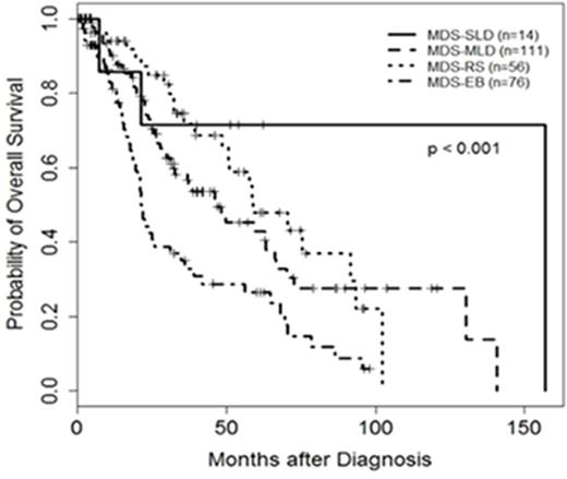
Over a median follow-up duration of 22.4 months (range, 0-156.8), 128 (48%) patients died. The median OS was 46.1 months (95% CI: 32.3, 58.4). Patients categorized as MDS-SLD by 2016 WHO had the best OS (156.8 months), followed by MDS-RS (58.7 months), MDS-MLD (46.3 months) and MDS-EB (21.2 months) (p<0.001). Older age, lower hemoglobin, lower ANC, lower platelet count, ≥5% BM blasts, MDS-EB1 and MDS-EB2 by 2016 WHO were significantly associated with worse OS (≤0.044). Accounting for all significant measures, age, hemoglobin, MDS-EB1 and MDS-EB2 remained significantly associated with OS.Sixteen patients transformed to AML; the median time-to-AML transformation was not reached; 5-year AML transformation rate was 88%. Patients with ≥ 5% BM blasts (p=0.056) and older age (p=0.063) tended to have a higher AML transformation rate.
Conclusions:
Morphological evaluation of BM/PB (for dysplasia, % BM blasts and RS) provides additional prognostic value and continues to be a critical component for evaluation of MDS patients. Molecular studies for splicing factor mutations are ongoing on all samples with >1% RS.
Jabbour:ARIAD: Consultancy, Research Funding; Pfizer: Consultancy, Research Funding; Novartis: Research Funding; BMS: Consultancy.
Author notes
Asterisk with author names denotes non-ASH members.

This icon denotes a clinically relevant abstract


This feature is available to Subscribers Only
Sign In or Create an Account Close Modal