Abstract
INTRODUCTION: Among patients (pts) with chronic lymphocytic leukemia/small lymphocytic lymphoma (CLL/SLL), the indications for liver biopsy are not known. Additionally, the histopathologic findings of CLL infiltration in the liver along with its clinical implications have not been thoroughly described in the literature.
METHODS: We identified all pts with a diagnosis of CLL/SLL from the CLL Clinical Database at Mayo Clinic, Rochester, MN who underwent a liver biopsy. We also cross referenced the Mayo Clinic Hematopathology Database for all pts who underwent a liver biopsy. The indications for biopsy, description of the pathologic findings in the liver biopsy, CLL therapy and outcomes were abstracted from the clinical records. Retrospective pathology review was performed on cases with CLL involvement in which slides were available for review.
RESULTS: Fifty-two CLL/SLL pts who underwent a liver biopsy were identified. The indications for liver biopsy were as follows: liver lesion identified on radiographic imaging in 21 (41%) pts, abnormal liver function tests (LFTs) in 17 (33%) pts, hepatosplenomegaly in 11 (21%) pts, and unknown in 3 (6%) pts. Among all 52 pts, there was concern for Richter transformation at time of biopsy in 7 (13%) pts. Types of biopsy were as follows: percutaneous in 20 (39%) pts, intraoperative in 17 (33%) pts, transjugular in 3 (6%) pts, and unknown in 12 (23%) pts.
CLL/SLL involvement was identified in 38/52 (73%) pts undergoing liver biopsy. Among these, the diagnosis of CLL/SLL was made on liver biopsy in 8 pts, and 7 pts had additional findings on their liver biopsy including metastatic colon cancer (n=3), hemochromatosis (n=2), steatohepatitis (n=1), and cirrhosis (n=1).
Liver biopsy revealed Richter transformation in 4 (8%) pts (3 pts with diffuse large B cell lymphoma (DLBCL) and 1 patient with Hodgkin lymphoma), other malignancy in the absence of CLL in 7 (15%) pts (2 metastatic colorectal, 2 metastatic esophageal, 1 hepatocellular carcinoma, 1 metastatic lung, and 1 neuroendocrine), and 1 patient with steatohepatitis. Two (4%) pts had normal findings on liver biopsy.
Among the 38 pts with liver involvement by CLL, 27 had slides available for retrospective pathology review. CLL involvement ranged from 2-100% of the liver parenchyma. Seven pts (26%) underwent excisional biopsies or resections and 20 (74%) underwent core biopsies. The portal tracts were the most common region of the liver involved (22/26 pts, 85%). Of these, 8/22 (36%) pts had extension into the lobules or sinusoids, as well. Of the 4 cases without portal involvement, 2 showed extensive involvement such that it effaced the liver parenchyma in the region of involvement. One patient (4%) had a predominantly sinusoidal involvement. One patient (4%) showed small nodular infiltrates that were difficult to localize due to their size. Proliferation centers were noted in only 3 pts (11%).
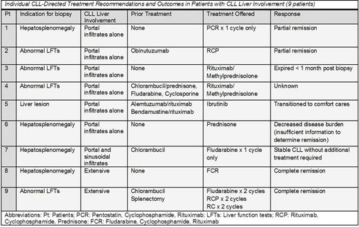
Most pts (29/38; 76%) whose liver biopsy demonstrated involvement by CLL did not receive immediate CLL-specific treatment. Nine of the 38 pts (24%) with CLL involvement on liver biopsy received therapy (Table): purine analog based therapy (n=4), rituximab and steroids (n=2), steroids only (n=1), alkylator and rituximab (n=1), and ibrutinib (n=1). Pts with extensive involvement of the liver by CLL were more likely to receive purine nucleoside analog based therapy compared to pts with portal infiltration by CLL (2/2 pts [100%] vs. 1/6 pts [17%]). The remaining pts were offered no therapy due to incidental finding of CLL in the liver biopsy (n=11), chemotherapy for other cancer (n=5), palliative care (n=3), or unknown (n=10). Pts with Richter's transformation received R-CHOP (n=2) and CHOP (n=1) for DLBCL, and ABVD (n=1) for Hodgkin lymphoma. Pts with other cancers without CLL received appropriate therapy for their underlying malignancy.
CONCLUSION: Abnormal LFTs and radiographic abnormalities were the most common indications for a biopsy in this large cohort of CLL pts. Infiltration of the portal tracts by CLL/SLL was the most common histopathological finding. Proliferation centers were not commonly seen, which may make the diagnosis challenging for the pathologist if there is not a prior history of CLL/SLL. The mere presence of CLL/SLL infiltration did not mandate treatment and many pts were able to continue observation.
Ding:Merck: Research Funding. Kenderian:Novartis: Patents & Royalties, Research Funding. Shanafelt:GlaxoSmithkKine: Research Funding; Celgene: Research Funding; Cephalon: Research Funding; Genentech: Research Funding; Janssen: Research Funding; Pharmacyclics: Research Funding; Hospira: Research Funding. Parikh:Pharmacyclics: Honoraria, Research Funding.
Author notes
Asterisk with author names denotes non-ASH members.


This feature is available to Subscribers Only
Sign In or Create an Account Close Modal