Key Points
Immune reconstitution after CBT is excellent provided ATG exposure is low or absent.
Individualized dosing, or omission of ATG in selected patients, may increase the chance of survival after CBT.
Abstract
Successful immune reconstitution (IR) is associated with improved outcomes following pediatric cord blood transplantation (CBT). Usage and timing of anti–thymocyte globulin (ATG), introduced to the conditioning to prevent graft-versus-host disease and graft failure, negatively influences T-cell IR. We studied the relationships among ATG exposure, IR, and clinical outcomes. All pediatric patients receiving a first CBT between 2004 and 2015 at the University Medical Center Utrecht were included. ATG-exposure measures were determined with a validated pharmacokinetics model. Main outcome of interest was early CD4+ IR, defined as CD4+ T-cell counts >50 × 106/L twice within 100 days after CBT. Other outcomes of interest included event-free survival (EFS). Cox proportional-hazard and Fine-Gray competing-risk models were used. A total of 137 patients, with a median age of 7.4 years (range, 0.2-22.7), were included, of whom 82% received ATG. Area under the curve (AUC) of ATG after infusion of the cord blood transplant predicted successful CD4+ IR. Adjusted probability on CD4+ IR was reduced by 26% for every 10-point increase in AUC after CBT (hazard ratio [HR], 0.974; P < .0001). The chance of EFS was higher in patients with successful CD4+ IR (HR, 0.26; P < .0001) and lower ATG exposure after CBT (HR, 1.005; P = .0071). This study stresses the importance of early CD4+ IR after CBT, which can be achieved by reducing the exposure to ATG after CBT. Individualized dosing of ATG to reach optimal exposure or, in selected patients, omission of ATG may contribute to improved outcomes in pediatric CBT.
Introduction
Allogeneic hematopoietic cell transplantation (HCT) has been introduced as a curative option for several diseases, including malignancies, immune deficiencies, bone marrow failure, and selected metabolic disease. Since the late 1980s, umbilical cord blood has become available as an alternative donor source in pediatric HCT.1 Compared with bone marrow (BM) or peripheral blood stem cells (PBSCs), umbilical cord blood transplantation (CBT) has the advantage of less stringent HLA matching requirements, increasing the chance of finding a suitable donor, and is rapidly available.2 Furthermore, CBT shows at least comparable survival compared with BM and PBSCs3-6 and lower rates of chronic graft-versus-host disease (GVHD), and it has been suggested to have a more potent antileukemic effect compared with traditional stem cell sources.7,8
When focusing on early T-cell immune reconstitution (IR) (ie, within 3 months after HCT), CBT is suggested to have a slower IR compared with non–T-cell-depleted BM or PBSCs.9-11 Anti–thymocyte globulin (ATG), introduced to the conditioning regimen to prevent GVHD and graft failure, may have a more profound effect on IR after CBT than BM and peripheral blood.6,12-14 This more profound in vivo depletion may be due to the fact that ATG is produced in rabbits using infant human thymus, which may result in more antibodies against naive receptors, which are more likely to be present on cord blood T cells.15,16
Successful IR following HCT is associated with improved chance of survival following HCT,9,11,15,17,18 making efforts to improve IR imperative. Strategies to enhance IR may include optimizing the dosing and thereby the exposure to ATG. Using the same linear dosing regimen in milligrams per kilogram for individuals of all sizes, exposure to ATG is highly variable between patients,19-23 especially in pediatric populations. This is mainly due to large variability in clearance, which depends on both body weight and recipient lymphocyte counts.20,23 Individualized dosing of ATG targeted at a predefined optimal exposure, thereby reducing variability in exposure for all patients, may subsequently lead to better predictable and improved IR. However, for individualized dosing of ATG, the optimal target exposure (ie, the therapeutic window) needs to be further investigated in cord blood recipients.
Studies investigating the relationship between ATG exposure and either IR or clinical outcome are scarce, especially for CBT, where it may be most relevant.15,20 We recently described the relationship between ATG exposure and outcome; however, the cord blood patients in this analysis were heterogeneous in terms of conditioning regimens, GVHD prophylaxis, and supportive care. In the current study, these limiting aspects will be overcome by including a large cohort with uniformly treated children receiving a CBT in an experienced CBT center to study the relationships among ATG exposure, IR, and clinical outcomes. An identified therapeutic window may be used as the target for individualized dosing.
Methods
Study design and patients
In this analysis, we included all patients receiving a single-unit CBT treated in the pediatric Blood and Marrow Transplantation Program of the University Medical Center Utrecht, The Netherlands, between December 2004 and October 2015. Cord blood was selected in patients where an identical sibling donor was not available or based on indication (high-risk acute myeloid leukemia [AML], inborn errors of metabolism, or primary immune deficiencies). Only first allogeneic HCTs were included. There was no restriction on the underlying disease or dose of ATG used. Consecutively treated patients were included. Patients developing immunoglobulin G anti-ATG antibodies were excluded.24 Data were collected prospectively in a local clinical database. Blood samples were collected weekly; ATG concentrations were measured in batches. Patient inclusion and data collection started after informed consent was obtained in accordance to the Declaration of Helsinki. For data and sample collection, institutional ethical committee approval was obtained through trial numbers 05/143 and 11/063-K.
Procedures
Umbilical cord blood (UCB) units were obtained from accredited (inter)national cord blood banks. HLA matching was performed on HLA-A, HLA-B, and HLA-DRB1 and had to be at least 4/6 matched to the patient (using intermediate-resolution typing: low resolution on class 1 and high resolution on DR). The minimum total nucleated cell dose required for the cord blood graft was 2.5, 3, or 5 × 10⁷ nucleated cells per kilogram for 6/6, 5/6, or 4/6 HLA-matched UCB units, respectively. In rare cases, a 3/6 matched UCB unit was chosen with a minimum of 5 × 10⁷ nucleated cells per kilogram. Grafts were thawed and diluted 1:1 with NaCl 0.9%/albumin 10% in a laminar flow cabin before infusion; no washing was performed.
Conditioning regimens were selected according to national and international study protocols open in the program and were most frequently busulfan-based myeloablative regimens. Busulfan was given IV with therapeutic drug monitoring aiming for a target cumulative area under the curve (AUC) of 85 to 95 mg*h/L in a myeloablative setting.25,26 ATG (Thymoglobulin; Genzyme, Cambridge, MA, USA) was administered at a dose of 10 mg/kg in 4 consecutive days according to (inter)national protocols. From 2010 onwards, patients with a body weight >40 kg received a reduced dose of 7.5 mg/kg. Additionally, the first infusion of ATG relative to the CBT was moved from day −5 (before 2010) to day −9 (2010 onwards). Starting from 2013, patients treated for AML or high-risk acute lymphoid leukemia (ALL) did not receive ATG as part of the conditioning regimen to ensure early T-cell reconstitution.27 Patients not receiving ATG were included in the analysis to investigate the full spectrum of ATG exposures, including no exposure. GVHD prophylaxis consisted of cyclosporin A with a target trough concentration of 200 to 250 μg/L combined with prednisolone 1 mg/kg. Prednisolone was tapered in 2 weeks starting 4 weeks post-HCT in benign disorders and 1 week after engraftment in malignant disorders. Cyclosporin A was continued until 3 months (malignant disease) or 6 months (benign disorders) after HCT. Patients were prophylactically treated with acyclovir; treatment of viral reactivations of adenovirus, cytomegalovirus (CMV), and Epstein-Barr virus (EBV) was started after reaching 1000 copies/mL. All patients received gut decontamination and Pneumocystis jiroveci prophylaxis according to local protocol as previously described.15 Patients were treated in high-efficiency, positive-pressure, particle-free isolation rooms.
ATG concentrations
Active ATG concentrations were measured in serum every week following the first infusion of ATG.23 Using a validated pediatric population pharmacokinetic model,23 full concentration–time curves could be estimated, and pharmacokinetic exposures measures were calculated. These included the total AUC, the AUC before (defined as the AUC from start of first infusion up to the infusion of the graft) and after (defined as the AUC from start of infusion to infinite time, ie, total elimination of all ATG) CBT, maximum concentration, and concentration at the time of infusion of the graft. ATG exposure measures were calculated using NONMEM 7.3.0 (Icon, Dublin, Ireland).
Lymphocyte subsets
Lymphocyte subsets, consisting of CD3+, CD4+, and CD8+ T cells, B cells, (CD19+), and natural killer cells (CD56+CD3−), were measured after reaching a leukocyte count of >0.4 × 10⁹ cells/L. Cell counts were followed every other week up to 12 weeks after CBT and monthly thereafter.
Outcomes
Main outcome of interest was CD4+ T-cell IR, defined as having >50/μL T cells in 2 consecutive measurements within 100 days. The definition was based on the previously demonstrated association between chance of survival and achieving 50/μL CD4+ T cells within 100 days after transplantation.6,9,12,15 Patients who deceased within 100 days were evaluated until date of death.
Other outcomes of interest were overall survival (OS), defined as days to death of any cause or last follow up; and event-free survival (EFS), defined as days to first event (death due to any cause, relapse, or graft failure) or last follow-up. Nonrelapse mortality (NRM) and relapse-related mortality (RRM) were defined as death due to any cause other than relapse and death due to relapse, respectively, or last follow up. Acute and chronic GVHD were classified according to the Glucksberg28 and Shulman29 criteria. Graft failure was defined as nonengraftment or secondary rejection. Probability on neutrophil reconstitution defined as the first day of achieving a neutrophil count ≥0.5 × 10⁹/L of 3 consecutive days; viral reactivations of CMV, adenovirus, and EBV were defined as the first day having >1000 copies/mL in blood . Both AUC of ATG before and after CBT and CD4+ IR were investigated as predictor for clinical outcomes. Additionally, other IR markers including CD3+, CD4+, B cell, and natural killer cell counts were evaluated as a predictor for outcome. All patients were censored at the date of last contact.
Statistical analyses
Duration of follow-up was defined as the time from CBT to last contact or death. Factors considered as predictors for outcome included patient-related variables (age, sex, CMV serostatus, and EBV serostatus), disease variables (malignancy, primary immune deficiency, bone marrow failure syndromes, or benign non–primary immune deficiency ), donor related variables (HLA-disparity, CMV serostatus, EBV serostatus), treatment period (before or after median year of transplantation), ATG exposure measures (AUC before and after CBT) and CD4+ IR. IR was considered as a time-varying predictor. Variables with a 2-sided P value of < .05 in univariate analysis were considered as a predictor in multivariate analysis. Probabilities of survival were determined using the Kaplan Meier estimation; P values were calculated using a 2-sided log-rank test. Cox proportional hazard and logistic regression models were used. For the end points acute and chronic GVHD, NRM, and RRM, Fine-Gray competing risk models were used. For finding optimal cutoff values for the main outcome of interest, receiving–operator characteristic curves were used. The cutoff with the maximum sum of sensitivity and specificity, and therefore the most accurate, was selected. Statistical analyses were performed using R version 3.2.3, with the packages cmprsk, survival, and ROCR.
Results
Patients
A total of 137 patients with a median age of 7.4 (range, 0.2-22.7) were included in this analysis (Table 1; split for ATG exposure in supplemental Table 1, available on the Blood Web site). Of these patients, 66 patients (48%) were included in a previous analysis.15 Most (82%) patients received ATG as part of their conditioning regimen; ATG was omitted in 17 out of 30 patients with AML and 6 out of 20 patients with ALL. One patient (0.7%) was excluded due to the development of anti-ATG antibodies. A busulfan-based conditioning regimen was the most frequently used (89%) and was combined with either fludarabine alone (67%) or fludarabine and clofarabine (22%). The indication for CBT was a nonmalignant disorder in 58% of the patients. Median follow-up was 44 months (0.2-143).
Patient characteristics
| Characteristic . | Total . |
|---|---|
| Number of patients | 137 |
| Male sex | 82 (60) |
| Age at transplant (y), median (range) | 7.4 (0.2-22.7) |
| Patients receiving ATG | 112 (82) |
| Diagnosis | |
| Malignancy | 56 (41) |
| ALL | 22 (16) |
| AML | 30 (22) |
| Lymphoma | 4 (3) |
| PID | 33 (24) |
| BM failure | 7 (5) |
| Benign non-PID | 41 (30) |
| Conditioning regimen | |
| Bu-Flu | 92 (67) |
| Bu-Flu-Clo | 30 (22) |
| TBI based | 10 (7) |
| Cy-Flu | 5 (4) |
| Match grade | |
| 6/6 matched | 55 (40) |
| 5/6 matched | 63 (46) |
| 4/6 matched | 18 (13) |
| 3/6 matched | 1 (1) |
| Follow-up (mo), median (range) | 44 (0.2-143) |
Values are n (%) of patients unless indicated otherwise.
Bu, busulfan; Clo, clofarabine; Flu, fludarabine; PID, primary immune deficiency; TBI, total body irradiation.
Main outcome of interest
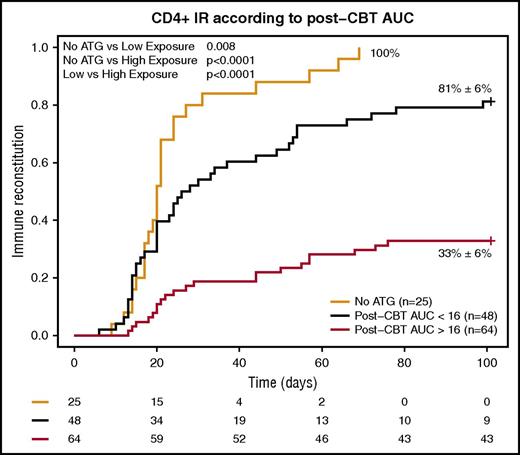
Low exposure to ATG after infusion of the CB transplant was found to be the best predictor for successful CD4+ IR. In multivariate analysis, probability on CD4+ IR was reduced 26% with every 10-point increase in AUC after CBT (hazard ratio [HR], 0.974; 95% confidence interval [CI], 0.962-0.986; P < .0001; Table 2; supplemental Table 2). Next, the optimal cutoff in exposure to ATG after CBT for CD4+ IR was investigated using receiving–operator characteristic curves. The most optimal cutoff was found to be 16 AU*day/mL, with a specificity of 85% with a sensitivity of 65% (supplemental Figure 1). Therefore, the patients were divided in groups of no ATG, low AUC after CBT exposure (<16 AU*day/mL), and high AUC after CBT (≥16 AU*day/mL). Omitting ATG resulted in 100% CD4+ IR, while patients that did receive ATG had a lower chance of reaching IR (81% ± 6%, P = .008 and 33% ± 6%, P < .0001 in no ATG versus low AUC and versus high AUC, respectively; Figure 1). This was also true when malignant and nonmalignant indications were analyzed separately (supplemental Figure 4). No other multivariate predictors for CD4+ IR were found (supplemental Table 1). When investigating whether the optimal ATG exposure differed based on donor, recipient, or transplant characteristics, no variables could be identified, indicating the optimal exposure after CBT is <16 AU*day/mL, irrespective of disease, age, and HLA match grade.
Multivariate analysis
| Variable . | HR . | 95% CI . | P value . | Significance level . |
|---|---|---|---|---|
| Post-HCT AUC of ATG (continuous) | ||||
| CD4+ IR | 0.974 | 0.962-0.986 | <.0001 | **** |
| EFS | 1.005 | 1.001-1.009 | .0071 | ** |
| OS | 1.005 | 1.001-1.009 | .026 | * |
| NRM | 1.005 | 1.001-1.009 | .028 | * |
| Use of ATG (no ATG is reference) | ||||
| Acute GVHD grade 2-4 | 0.878 | 0.353-2.185 | .78 | |
| Acute GVHD grade 3-4 | 0.268 | 0.083-0.862 | .027 | * |
| Chronic extensive GVHD | 1.132 | 0.137-9.383 | .91 | |
| IR (no IR is reference) | ||||
| EFS | 0.264 | 0.156-0.447 | <.0001 | **** |
| OS | 0.516 | 0.279-0.955 | .035 | * |
| NRM | 0.358 | 0.154-0.829 | .017 | * |
| RRM in myeloid leukemia | 0.134 | 0.03-0.595 | .008 | ** |
* <.05; ** <.01; *** <.001; **** <.0001.
Cumulative incidence of CD4+ IR within 100 days according to ATG exposure after CBT. Orange line, no ATG; black line, exposure to ATG after CBT <20 AU*day/mL; red line, exposure to ATG >20 AU*day/mL.
Cumulative incidence of CD4+ IR within 100 days according to ATG exposure after CBT. Orange line, no ATG; black line, exposure to ATG after CBT <20 AU*day/mL; red line, exposure to ATG >20 AU*day/mL.
Other outcomes of interest according to ATG exposure after CBT
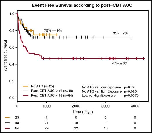
EFS was comparable in patients not receiving ATG and having low ATG exposure after CBT, while those with high AUC after CBT ATG had a significantly lower EFS compared with each of the other 2 groups (Figure 2; supplemental Figure 3). However, patients not receiving ATG are not comparable to those that do, as ATG is omitted only in AML and high-risk ALL. In multivariate analysis, high ATG exposure after CBT (HR, 1.005; 95% CE, 1.001-1.009; P = .0071) and positive recipient CMV serostatus were multivariate predictors for inferior EFS (HR, 1.99; 95% CI, 1.13-3.49; P = .016). Here, ATG exposure after CBT was introduced as a continuous covariate to use the full statistical power of the data and minimize bias. Chance of OS was comparably improved with lower ATG exposure after CBT (HR, 1.005; 95% CI, 1.001-1.009; P = .026). This indicates that every 10-point increase in ATG exposure after CBT results in 5% lower survival probability. RRM and relapse incidence were not impacted by ATG exposure, but the incidence of NRM was significantly reduced in patients with a lower ATG exposure after CBT (HR, 1.005; 95% CI, 1.001-1.009; P = .028). No viral reactivations were observed in the no-ATG group, and a trend toward lower viral reactivations was observed with low ATG exposure when compared with high ATG exposure (supplemental Figure 2). The number of patients at risk were similar in the 3 groups (supplemental Table 1), while the number of infectious deaths was lower in the no-ATG and low-exposure groups (supplemental Table 3). GVHD was not impacted by exposure to ATG after CBT (P = .42, P = .21, and P = .12 for grade 2-4 acute GVHD, grade 3-4 acute GVHD, and extensive chronic GVHD, respectively). No influence of cell dose (total nucleated cells per kilogram) infused was noted on neutrophil reconstitution (supplemental Figure 9), and the impact of ATG exposure on neutrophil engraftment was similar among groups (supplemental Figure 5).
EFS according to ATG exposure after CBT. Orange line, no ATG; black line, exposure to ATG after CBT <20 AU*day/mL; red line, exposure to ATG >20 AU*day/mL.
EFS according to ATG exposure after CBT. Orange line, no ATG; black line, exposure to ATG after CBT <20 AU*day/mL; red line, exposure to ATG >20 AU*day/mL.
Other outcomes of interest according to use of ATG
No difference in GVHD incidence was found based on ATG exposure levels. Therefore, we investigated whether the use of ATG influenced the probability of developing GVHD. Here, we found that although the use of ATG did not predict the incidence of grade 2-4 GVHD (P = .78), patients receiving ATG had a significantly lower incidence of grade 3-4 acute GVHD compared with those not receiving ATG (HR, 0.27; 95% CI, 0.08-0.86; P = .027). No differences were found in incidence of chronic GVHD and graft failure according to the use of ATG, which may be due to the low number of events (5% and 11% for chronic GVHD and graft failure, respectively).
Other outcomes of interest in the context of CD4+ IR
Within the IR markers, we found CD4+ IR to be the strongest and only predictor for the outcomes of EFS, OS, NRM, and RRM.
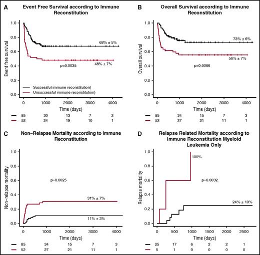
Patients with successful early CD4+ IR at day +100 had a better chance of EFS (HR, 0.26; 95% CI, 0.16-0.45; P < .0001; Figure 3A). This is also true when subdividing in malignant and nonmalignant disease groups (supplemental Figure 7). In addition to CD4+ IR, negative CMV serostatus before transplantation was a multivariate predictor of an improved chance of EFS. OS was comparably improved with successful CD4+ IR (HR, 0.51; 95% CI, 0.28-0.95; P = .035, Figure 3B; supplemental Figure 8). Moreover, successful CD4+ IR lowered the chance of NRM (Figure 3C) and RRM (Figure 3D), the latter only in cases of myeloid leukemia (a small subgroup of patients). In the multivariate models for NRM and RRM, CD4+ IR was the only predictor (NRM: HR, 0.36; 95% CI, 0.15-0.83; P = .0017; RRM: HR, 0.13; 95% CI, 0.03-0.60; P = .008). In patients not reaching CD4+ IR, infectious disease was the most common cause of NRM (11 out of 52 patients compared with 5 out of 85 patients with CD4+ IR). Other causes of NRM were comparable between the groups.
Clinical outcome according to CD4+ immune reconstitution. (A-C) EFS (A), OS (B), and NRM (C) according to successful CD4+ IR in all patients. (D) RRM according to successful CD4+ IR in myeloid leukemia only. Black lines, successful CD4+ IR; red lines, no successful CD4+ T-cell IR.
Clinical outcome according to CD4+ immune reconstitution. (A-C) EFS (A), OS (B), and NRM (C) according to successful CD4+ IR in all patients. (D) RRM according to successful CD4+ IR in myeloid leukemia only. Black lines, successful CD4+ IR; red lines, no successful CD4+ T-cell IR.
Discussion
To our knowledge, this is the largest pediatric single-center study comprehensively investigating the relationships among ATG exposure, CD4+ T-cell IR, and clinical outcomes. We show that even very minimal exposure to ATG after CBT has a detrimental effect on early CD4+ IR. We also found that both ATG exposure and CD4+ IR are predictors of clinical outcome, including EFS, OS, and NRM. The use of ATG was associated with a lower incidence of grade 3-4 acute GVHD, but not grade 2-4 or chronic GVHD, possibly because of the low incidence of chronic GVHD (<5%). While recognizing the limitations of a retrospective cohort study, the strengths of this study include the homogeneous group of patients analyzed (eg, conditioning regimens, GVHD prophylaxis, and supportive care). Additionally, the inclusion of consecutive patients with prospective data collection minimizes the risk of potential bias, and including patients who did not receive ATG (n = 25) provided information regarding the maximum potential of CD4+ reconstitution after CBT. Taken together, these results stress the importance to aim for optimal ATG exposure in pediatric CBT, both for achieving early IR and for ensuring an improved chance of survival.
Outcomes of CBT have improved significantly over the last decade. In the 1990s and early 2000s, lower engraftment rates were a significant disadvantage of CBT; however, the current use of higher cell-dose units has significantly reduced this disadvantage.4 Although NRM due to infectious causes is still reported to be a serious limitation of CBT, this occurs mainly in the context of ATG in the conditioning regimen. In a report on cell-source comparison without ATG, very low incidences of viral reactivations are reported.30 In the absence of ATG, relapse incidence is suggested to be lower with CBT than with other cell sources. More studies are warranted to confirm this finding, as other factors may be influential.4,5 As timely IR seems essential in preventing RRM and NRM, strategies to improve IR to increase protection against viral disease and relapse of malignancy are warrented.7,15,27,30 T-cell IR has been associated with the use, dose, and timing of serotherapy, indicating that a higher dose of serotherapy shortly before infusion of the graft is detrimental for timely and robust IR.6,14,27,31,32 Thus, in vivo exposure of graft-infused T cells to serotherapy results in significant depletion of T cells and limits the exceptional proliferative capacity of cord blood T cells, as recently shown.15 The present study confirms that very low exposure to ATG has a dramatic effect on T-cell reconstitution potential irrespective of disease group, and no exposure to ATG results in exceptional IR potential. The IR potential in CBT is possibly even better than that in transplantation of other stem cell sources, as suggested before.27 In the major landmark studies by Rocha et al and Eapen et al comparing cord blood to other stem cell sources, most cord blood patients received ATG.3,33 Taking the results of the current study into account, when reducing in vivo exposure of ATG to the graft in these studies, the cord blood groups may have performed better. Moreover, the presented results are very likely true in adult CBT as well, potentially at a greater magnitude. Due to the higher body weights in adults compared with children, the number of infused T cells per kilogram is mostly lower. Additionally, from a pharmacokinetic perspective, ATG clearance is markedly higher in smaller (younger) children compared with older children and is likely to be even lower in adults. Therefore, current dosages of ATG in adults may result in very high ATG exposure after infusion of the CBT and fully deplete the even lower amount of graft-infused T cells, subsequently leading to poor IR.
Both successful CD4+ IR and lower ATG exposure after CBT were associated with survival, as they both lowered NRM and RRM in AML. This is in line with previous studies, where CD4+ IR was associated with lower incidence of viral reactivations and relapse and a better chance of survival.6,9,15,27,30 However, few studies have reported on the association between ATG pharmacokinetics (PK) and survival, where PK is evaluated either as single concentration measurements or as exposure. Studies investigating single concentrations at a certain time point could not find a correlation with survival, while actual exposure of ATG before and after transplantation was associated with a variety of outcomes.15,19-21,34 This discrepancy may be due to a lower number of patients included in the concentration studies; however, ATG exposure (AUC) more likely harbors more information compared with single concentration samples and therefore is a stronger predictor.35
In the current study, the use of ATG led to a lower incidence of grade 3-4 acute GVHD, which is in line with previous studies.6,36-38 However, no correlation was found between the use of ATG and grade 2-4 GVHD, extensive chronic GVHD, and graft failure, which in previous studies were related to ATG exposure before transplantation. The absence of a correlation may be due to the relatively low incidence of chronic GVHD and graft failure in this cohort (5% and 11% for extensive chronic GVHD and graft failure, respectively). The incidence of acute GVHD grade 2-4 was relatively low compared with a previous study in which ATG was omitted.27 This difference may be due to different GVHD-prophylaxis regimens: the current study used prednisolone (together with cyclosporine) while Chiesa et al used mycophenolate mofetil (together with cyclosporine).27
Strategies for improved and predictable T-cell IR are warranted and may lead to an improved chance of survival following HCT. This is especially important in CBT, where even very low exposure after CBT has a major impact on the probability of CD4+ IR. Omission of ATG in a CBT setting for all indications may contribute to improved IR but is less feasible in immune-competent recipients, in whom the omission of ATG could potentially lead to more graft failure or GVHD.6,27 All chemo-naive patients (eg, benign disorders) received ATG in the current analysis. Therefore, no inferences can be made on omitting ATG in these patients based on this study. Individualizing ATG exposure is an alternative strategy; dosing is based on patient characteristics (eg, weight and absolute lymphocyte count), with the aim of reaching optimal exposure before and after CBT. The advantages of individualized dosing include optimal IR while protecting against GVHD and graft failure, thereby offering the best of both worlds. This approach is currently being investigated in a prospective clinical trial (Dutch trial register NTR4960). If proven to be effective, an individualized dosing regimen for ATG, based on body weight and absolute lymphocyte count, is relatively easy to implement in all centers, as ATG PK measuring is not required. For more complicated cases (eg, patients at high risk for graft failure), therapeutic drug monitoring may be an additional tool to target the ATG exposure more precisely. This requires some expertise in terms of pharmacological modeling and setting up and validating an assay. Alternatively, samples can be sent to a centralized laboratory with an operational validated active ATG measuring assay.
These data and data from others suggest that ATG can be safely omitted in patients receiving a CBT for a chemotherapy pretreated malignancy.6,27,34 The chance of rejection is low in these patients, making the omission of ATG an easier and safer solution to ensure low or no exposure to ATG after CBT. For nonmalignant and chemo-naive malignant indications, ATG exposure before CBT may help prevent rejection, as previously shown.15 Future studies have to show whether individualized ATG dosing (aiming for sufficiently high exposure before CBT) in malignant indications is of additional value to prevent severe acute GVHD (as suggested in a previous study) while ensuring optimal EFS.15 Furthermore, the broader T-cell repertoire of reconstituting naive CD4+ cells after CBT may have an additional advantage over BM and PBSCs,39 as cord blood cells have an exceptional learning capacity despite their naivety and show a lower incidence of viral reactivation30 and stronger antileukemic activity.4,5,7 The high number of naive cells makes CBT a powerful platform for adjuvant cellular therapies (eg, cell vaccinations),40,41 as the above-mentioned associations suggest that efficient priming of these cells may enhance antileukemic activity.
In conclusion, this study shows the importance of adequate targeting of ATG exposure after CBT to ensure early CD4+ T-cell reconstitution. By omitting ATG or using individualized dosing and timing of ATG aimed at optimal target exposure, T-cell IR will improve. Our data indicate that promoting CD4+ IR will likely improve the chance of survival by lowering both RRM and NRM.
The online version of this article includes a data supplement.
The publication costs of this article were defrayed in part by page charge payment. Therefore, and solely to indicate this fact, this article is hereby marked “advertisement” in accordance with 18 USC section 1734.
Acknowledgments
Amelia Lacna and Lysette Epskamp-van Raaij are acknowledged for measuring the ATG concentrations.
This study was funded by a Rational Pharmacotherapy Program grant (40-41500-98-11044) from the Dutch Organization for Scientific Research.
Authorship
Contribution: R.A. designed the research, analyzed data, and wrote the manuscript; C.A.L. and J.J.B. designed the research, analyzed data, included patients, and wrote the manuscript; S.N. and C.v.K. analyzed data and wrote the manuscript; A.B.V. and M.B.B. included patients and wrote the manuscript; all authors reviewed and approved the final version of the manuscript; and R.A. and J.J.B. had full access to all of the data in the study and take responsibility for the integrity of the data and the accuracy of the data analysis.
Conflict-of-interest disclosure: UMC Utrecht is conducting an investigator-initiated clinical trial to evaluate individualized dosing of ATG, which is supported by an unrestricted research grant from Sanofi. The work was performed independently of all funders.
Correspondence: Jaap J. Boelens, Department of Pediatric Immunology, University Medical Centre Utrecht, PO Box 85500, 3508 AB Utrecht, The Netherlands; e-mail: j.j.boelens@umcutrecht.nl.


