Abstract
Severe hemolysis and associated high levels of hemolytic biomarkers, including LDH and heme, are among major constituents of the pathophysiology of sickle cell disease (SCD). Elevated extracellular heme due to hemolysis overwhelms endogenous detoxification mechanisms and leads to oxidative stress, triggering systemic endothelium activation, vascular dysfunction, and end organ damage. To understand the role of red blood cells (RBC) in this process, we assessed sickle RBC adhesion to heme-activated endothelial cells utilizing an endothelialized microfluidic platform in a clinically diverse patient population.
Human umbilical vein endothelial cells (HUVECs) were seeded into fibronectin (FN) functionalized microfluidic channels and incubated for 4 hours in a 37°C and 5% CO2 environment. Next, the confluent monolayers were loaded and incubated for 45 minutes with fresh culture medium or at one of two concentrations of heme solutions: (1) 20 μM that corresponds to physiological heme levels in SCD patients, and (2) 40 μM. SCD blood samples, collected from 8 patients (7 HbSS and 1 HbS/β0 thal; 3 males and 5 females), were centrifuged to remove the plasma and washed with PBS thrice prior to flow experiments. RBCs were re-suspended in culture medium at a hematocrit of 25%. A total volume of 15 µl RBC suspension was perfused into the microchannels followed by rinsing with fresh culture medium at 1 dyne/cm2, which corresponds to the typical shear stress value observed in post-capillary venules. The fully closed and hermetically sealed microfluidic system design ensured the stability of gas composition in the culture medium during the experiments.
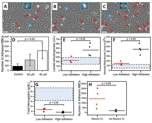
Endothelialized microchannels supported sickle RBC adhesion to non-treated, 20 µM heme-activated, and 40 µM heme-activated HUVECs (Fig. 1A, B, C). Adhesion results suggested that activation of HUVECs by heme mediated RBC adhesion in a concentration-dependent manner, with a significant difference observed at 40 µM (Fig. 1D, paired t-test, p<0.05). Notably, a heterogeneous heme-mediated adhesion profile was seen among patients. Sickle RBC adhesion to 20 μM heme-activated HUVECs was significantly increased in patients with higher LDH levels (Fig. 1E, p=0.024, ANOVA), higher absolute reticulocyte counts (Fig. 1F, p=0.002, ANOVA), and lower total hemoglobin (Fig. 1G, p=0.016, ANOVA), which are indicative of elevated hemolysis. All patients in the high adhesion group (>200 adherent RBCs) had elevations in serum LDH levels and in absolute reticulocyte counts. Moreover, patients with a recent transfusion had higher RBC adhesion to 40 µM heme-activated HUVECs compared to patients with no transfusion in the last 3 months (Fig. 1H, p<0.05, ANOVA).
Here, we report a direct link between heme-driven endothelial activation and RBC adhesion in a patient-specific manner. In patients with a more severe clinical phenotype, as reflected in LDH, total hemoglobin, and absolute reticulocyte or recent blood transfusions, we found greater RBC adhesion to heme-activated HUVECs. These findings suggest that RBCs from those patients most likely to experience hemolysis in vivo may also be those RBCs most likely to adhere to heme-activated endothelium.
Acknowledgments: This work was supported by grants #2013126 and #2015191 from the Doris Duke Charitable Foundation, National Heart Lung and Blood Institute R01HL133574, and National Science Foundation CAREER Award 1552782.
Figure 1: Sickle RBC adhesion to heme-activated HUVECs and clinical associations. Sickle RBCs adherent to immobilized HUVECs in (A) non-activated, (B) 20 µM heme-activated, and (C) 40 µM heme-activated microchannels. The microscope images illustrate a small portion of the full microchannel surface. (D) RBC adhesion to HUVECs increased depending on the heme concentration, with a significant difference at the 40 µM level (paired t test, p<0.05). Patients with higher LDH (E) as well as absolute reticulocyte counts (F), and lower total hemoglobin (G) showed significantly greater RBC adhesion to 20 µM heme-activated HUVECs (ANOVA). (H) Furthermore, patients with a recent transfusion history displayed elevated RBC adhesion to 40 µM heme-activated HUVECs compared to non-transfused patients (ANOVA). The dashed rectangles indicate the normal clinical values for healthy individuals, while all patients had total hemoglobin levels lower than normal. Scale bars represent 30 µm.
Little: Hemex Health: Equity Ownership. Gurkan: Hemex Health: Employment, Equity Ownership.
Author notes
Asterisk with author names denotes non-ASH members.


This feature is available to Subscribers Only
Sign In or Create an Account Close Modal