Abstract
Pediatric myelodysplastic syndromes (MDSs) are a heterogeneous group of clonal disorders with an annual incidence of 1 to 4 cases per million, accounting for less than 5% of childhood hematologic malignancies. MDSs in children often occur in the context of inherited bone marrow failure syndromes, which represent a peculiarity of myelodysplasia diagnosed in pediatric patients. Moreover, germ line syndromes predisposing individuals to develop MDS or acute myeloid leukemia have recently been identified, such as those caused by mutations in GATA2, ETV6, SRP72, and SAMD9/SAMD9-L. Refractory cytopenia of childhood (RCC) is the most frequent pediatric MDS variant, and it has specific histopathologic features. Allogeneic hematopoietic stem cell transplantation (HSCT) is the treatment of choice for many children with MDSs and is routinely offered to all patients with MDS with excess of blasts, to those with MDS secondary to previously administered chemoradiotherapy, and to those with RCC associated with monosomy 7, complex karyotype, severe neutropenia, or transfusion dependence. Immune-suppressive therapy may be a treatment option for RCC patients with hypocellular bone marrow and the absence of monosomy 7 or a complex karyotype, although the response rate is lower than that observed in severe aplastic anemia, and a relevant proportion of these patients will subsequently need HSCT for either nonresponse or relapse.
Introduction
Myelodysplastic syndromes (MDSs) are clonal hematopoietic disorders characterized by peripheral cytopenia, ineffective hematopoiesis, and an increased risk of progression to acute myeloid leukemia (AML). Although sporadic MDS is primarily a disease of the elderly with an incidence of more than 36 per 100 000 in patients age 80 years or older,1 MDS in children (ie, patients age 18 years or younger) is a rare disease with an annual incidence of 1 to 4 cases per million (Table 1).2 Moreover, some of the peculiarities of childhood MDSs are associated with previous exposures to cytotoxic agents, including alkylating agents and topoisomerase inhibitors, inherited bone marrow failure syndromes (IBMFSs), or genetic (ie, germinal) predisposition syndromes (Tables 1 and 2).3-5 Compared with adult MDS, the knowledge of somatic genetic alterations in the pediatric population is still limited, but recent evidence indicates that genes known to be frequently mutated in adult MDSs such as TET2, DNMT3A, and TP53 and the spliceosome complex are not involved in disease pathogenesis in children (Table 1).6-8 By contrast, somatic driver mutations in SETBP1, ASXL1, RUNX1, and RAS oncogenes characterize the genomic landscape of pediatric MDS.8
Differences in MDSs between children and adults
| . | Children (0-18 y) . | Adults (older than age 40 y) . |
|---|---|---|
| Incidence per million | 1-4 | >40 |
| Refractory anemia with ringed sideroblasts (%) | <1 | 25 |
| Associated IBMFSs and predisposition syndromes (%) | >30 | <5 |
| Familial aggregation | Present in a proportion of patients | Uncommon |
| Chromosomal aberrations (%) | ||
| −7/7q− | 25-30 | 10 |
| −5/5q− | 1 | 20 |
| Molecular aberrations | Presence of germ line mutations (eg, GATA2); less frequent somatic mutations; absent or exceptional spliceosomal mutations | Germ line mutations are less common; frequent somatic mutations; spliceosomal mutations are common |
| General aim of treatment | Curative | Often palliative |
Adapted with substantial modifications from Hasle.5
IBMFS, inherited bone marrow failure syndrome.
Classification of syndromes and conditions that predispose children to the development of MDSs
| IBMFSs |
| FA |
| Diamond-Blackfan anemia |
| Shwachman-Diamond syndrome |
| Telomere biology disorders |
| Congenital amegakaryocytic thrombocytopenia |
| Severe congenital neutropenia |
| Germ line predisposition and preexisting platelet disorders |
| Myeloid neoplasms with germ line RUNX1 mutation |
| Myeloid neoplasms with germ line ANKRD26 mutation |
| Myeloid neoplasms with germ line ETV6 mutation |
| Myeloid neoplasms with germ line predisposition and other organ dysfunction |
| Myeloid neoplasms with germ line GATA2 mutation |
| MDS with germ line SAMD9/SAMD9L mutation |
Modified by Arber et al.30
FA, Fanconi anemia.
With these peculiar aspects of childhood MDS and the clinical and biological diversity of the different variants in mind, we discuss 4 paradigmatic cases that exemplify the heterogeneity of both pathophysiology and the treatment approach to be adopted in this rare disease.
Case 1
An 8-year-old boy presented at the hospital emergency room with bruising and hematomas in the legs. Blood cell counts showed a hemoglobin level of 11.1 g/dL, an absolute neutrophil count (ANC) of 1.3 × 10⁹/L, and a platelet (PLT) count of 34 × 109/L. Bone marrow (BM) aspirate was hypoplastic with dysmorphic features involving mainly the myeloid and megakaryocyte lineages; blasts were not detected. Cytogenetic analysis did not show any karyotype abnormality, but marrow trephine confirmed the presence of marked hypocellularity and patchy distribution of erythropoiesis. These evaluations were repeated 3 weeks later and substantially confirmed the initial findings, so a diagnosis of refractory cytopenia of childhood (RCC) was made. Family HLA typing did not show any compatible donor, and a search for an unrelated donor (UD) was initiated. Despite unremarkable physical examination and family history, chromosomal breakage testing and telomere length testing were performed to exclude FA and telomeropathies. With an ANC >1.0 × 10⁹/L and an unsupported PLT count of 34 × 109/L, a watch-and-wait strategy was used. Within 3 months, the ANC dropped below 1.0 × 10⁹/L, and the patient became transfusion-dependent for PLTs. A fully matched (in 10/10 HLA loci by high-resolution techniques) UD was located, which enabled the patient to receive an allogeneic hematopoietic stem cell transplantation (HSCT) after a conditioning regimen consisting of thiotepa and fludarabine and graft-versus-host disease (GVHD) prophylaxis that included anti-T-lymphocyte globulin (ATLG), cyclosporine A, and short-course methotrexate. He had slightly delayed hematologic recovery with full donor hematopoiesis and developed grade 1, skin-only acute GVHD that resolved with topical steroids. Twenty-seven months after HSCT, he is in good clinical condition with normal blood counts and full immune recovery.
Epidemiology and clinical features of RCC
Pancytopenia with a severe decrease of BM cellularity in children may be caused by many different disorders; of these, RCC, acquired severe aplastic anemia (SAA), and IBMFS are the 3 most common hematopoietic diagnoses. Clinical and histopathologic distinction among these 3 groups of disorders is a well-known challenge and has important therapeutic implications.2,9
RCC is the most common subtype of MDS in children.2,10 Boys and girls are equally affected, and median age at diagnosis is 7 to 8 years, although the disease can be diagnosed in all age groups.11,12 In view of the unique clinical and morphologic features of MDS without increase in blasts in children, the diagnosis of RCC was introduced in 2008 as a provisional entity into the World Health Organization (WHO) classification of tumors of the hematopoietic and lymphoid tissues.13 In contrast to adults who usually present with isolated anemia, hematologic manifestations in children frequently include thrombocytopenia and/or neutropenia.2,5,12,14 In addition, an elevated mean corpuscular volume and moderately increased hemoglobin F values are present in a large proportion of patients if age-adjusted reference values are applied.
Diagnostic procedures
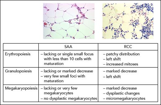
RCC is defined as persistent cytopenia with <5% blasts in BM, <2% blasts in peripheral blood (PB), and dysplastic features, most frequently observed in the erythroid and megakaryocytic lineages (Figure 1).9,13 Notably, peculiar histopathologic findings of childhood RCC consist of islands of immature erythroid precursors accompanied by sparsely distributed granulocytic cells (Figure 1). Megakaryocytes are either decreased or absent; few micromegakaryocytes can be detected through immunohistochemistry. BM cellularity is significantly reduced in up to 80% of patients with RCC.2,9,14 BM morphologic assessment is challenging; it should include evaluation of both aspirates and marrow trephines, including immunostaining for CD61 for detection of micromegakaryocytes.9,13
Pancytopenia in the presence of hypocellular BM with dysplastic features might be indicative of MDS. As already mentioned, in children, the differential diagnosis includes a variety of hematologic as well as nonhematologic diseases,2 the most important being aplastic anemia (AA) and IBMFS. It has been reported that experienced pathologists can reliably distinguish the morphologic pattern associated with RCC from that characteristic of patients with SAA (Figure 1).9 In contrast, RCC and IBMFS share common morphologic features, and their distinction is not possible based on morphology alone. In fact, in about 15% of patients for whom symptoms were suggestive of RCC, FA was diagnosed on the basis of functional testing; moreover, 30% of the patients diagnosed with FA and BM failure had no obvious physical abnormalities.15,16 Similarly, patients with telomere disorders might present with isolated BM failure as one of the first signs of the disease and therefore are not reliably diagnosed on clinical grounds. These observations demonstrate that, together with a medical history that includes an extensive family history and a thorough clinical examination, functional tests such as investigation for increased chromosomal breakage, G2 cell-cycle arrest, western blot or mutational analyses for FA genes, and telomere length are essential before the diagnosis of RCC can be confirmed. More recent studies that use next-generation sequencing (NGS) techniques in patients with IBMFS indicated a low frequency of specific molecular lesions but gave a satisfactory diagnostic yield for the whole group.17-19 It remains to be fully established how these methods can be incorporated into a diagnostic algorithm.
In contrast to advanced MDS in which an abnormal karyotype is found in ∼60% of patients, no chromosomal aberrations are detected in 70% to 80% of patients with RCC.2 Monosomy 7 is the most frequent cytogenetic lesion, occurring in about 11% of patients and being more frequently detected in patients with normo-cellular or hypercellular BM than in children with marrow hypocellularity.
Therapeutic approaches
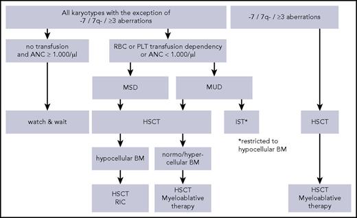
Allo-HSCT is a curative treatment for many patients with MDS and should be considered the treatment of choice if there is an indication for therapy and a suitable donor is identified. Therefore, HLA typing of patient’s family should be performed as soon as the diagnosis of MDS is established; in the absence of a matched family donor, a search for a UD should be initiated. In patients with RCC, the presence of monosomy 7 is correlated with a high risk of progression to more advanced MDS as well as to frank AML, and therefore these patients should receive a transplant as soon as possible (Figure 2).12 The rare RCC patients with a complex karyotype (characterized by ≥3 chromosomal aberrations, including at least 1 structural aberration) should be offered a swift allograft as well, although the presence of these somatic genetic abnormalities portend a dismal prognosis. In contrast, patients with a normal karyotype may have a stable course of disease over a long period, and therefore, in the absence of transfusion dependency and/or marked neutropenia (ie, <1 × 10⁹ or 0.5 × 10⁹/L), we recommend a careful watch-and-wait strategy (Figure 2).11,12 Patients with sustained neutropenia (we recommend a threshold of 1 × 10⁹/L, but 0.5 × 10⁹/L can also be an option) and/or transfusion dependency for PLTs and/or red blood cells have an indication for therapeutic intervention (Figure 2). Historically, HSCT with a myeloablative regimen has resulted in a probability of event-free survival (EFS) of 75%, transplantation-related mortality (TRM) being the major cause of treatment failure.20 In view of these findings, reduced-intensity conditioning might be an attractive approach at least for patients with hypocellular marrow and normal karyotype (Figure 2).21,22 In a pilot study conducted in 19 children treated at centers affiliated with the European Working Group on Childhood MDS (EWOG-MDS), HSCT after a preparative regimen that includes thiotepa and fludarabine has resulted in a probability of overall survival (OS) of 84% and an EFS of 74%.22 These results have considerably improved over time, since the last update of the EWOG-MDS group reported an OS probability of 94% and an EFS probability of 88% in 172 patients with RCC.23 In view of the negligible risk of disease recurrence, there is no benefit for patients to develop even limited-severity GVHD; therefore, BM should be the preferred stem cell source combined with an effective GVHD prophylaxis. We also recommend that patients with RCC and normal or hypercellular BM, monosomy 7, or complex karyotype be offered a fully myeloablative preparation (Figure 2). Results of unrelated cord blood transplantation in pediatric patients with different MDS variants (including RCC) have recently been reported to be inferior to results when using either BM or PB as a source for stem cells.24 Thus, this type of allograft can be recommended only for those patients who lack a matched related or unrelated donor.
Treatment algorithm in RCC. IST, immunosuppressive therapy; MSD, matched sibling donor; MUD, matched unrelated donor; RBC, red blood cell; RIC, reduced intensity conditioning.
Treatment algorithm in RCC. IST, immunosuppressive therapy; MSD, matched sibling donor; MUD, matched unrelated donor; RBC, red blood cell; RIC, reduced intensity conditioning.
In the absence of a suitable donor, IST with ATLG and cyclosporine A may be a therapeutic option in patients with hypocellular RCC and the absence of poor-risk karyotype (Figure 2). The rationale this treatment is based on comes from the observation that BM failure can, at least in part, be mediated by T-cell immunosuppression of hematopoiesis and that there is an overlap in pathophysiology of AA and RCC.25,26 In an initial report, IST resulted in a 6-month complete or partial response in about 75% of 31 patients with RCC.27 However, the failure-free survival rate was only 57%.27 Similar to reports in patients with AA,28 the results are better when patients are given horse ATLG (lymphoglobulin) compared with rabbit antithymocyte globulin (response at 6 months, 74% vs 53%; P = .04).14 The inferior response in the rabbit ATLG group resulted in lower 4-year transplantation-free (46% vs 69%; P = .003) and failure-free (48% vs 58%; P = .04) survival rates in this group compared with those in the horse ATLG group.14 It remains to be tested whether the addition of eltrombopag to IST will be of benefit for patients with RCC, as was recently shown in patients with SAA.29 In contrast to patients who receive HSCT, children with RCC who receive IST remain at risk for relapse and clonal evolution, the risk of the latter complication being low if an accurate diagnostic work-up has been performed.9
Case 2
A 14-year-old girl was admitted to the hospital for moderate-grade fever that had persisted for the previous 15 days. The hematology laboratory test showed reduced PLT count (52 × 109/L) and slightly increased white blood cell (WBC) count (12.3 × 10⁹/L), with 2% blasts in the differential and abnormal hemoglobin level (7.2 g/dL). BM aspirate showed 18% blast infiltrate, with dysplastic changes mainly involving the megakaryocytic and erythroid series. Cytogenetic examination of 21 metaphases exhibited monosomy 7. Screening for recurrent aberrations occurring in AML was negative. A BM aspirate repeated 2 weeks later gave the same results. In view of these findings, the patient was diagnosed with MDS with excess of blasts (MDS-EB). Because an HLA-identical sibling was not available, a search for a suitable HLA-matched UD was started, which led to the identification 6 weeks later of a volunteer identical at the HLA-A, -B, -C, -DRB1, and -DQB1 loci using high-resolution molecular typing. The patient received a transplantation after a fully myeloablative conditioning regimen consisting of busulfan, cyclophosphamide, and melphalan as recommended by the EWOG-MDS group. Fourteen months after HSCT, this patient is alive and in complete remission of her disease.
Advanced MDS in childhood
MDS with ≥2% blasts in PB or ≥5% but <20% blasts in the BM is classified as MDS-EB in the most recent WHO classification of myeloid neoplasms.30 The variant MDS-EB in transformation (MDS-EB-t) is retained in the pediatric classification of MDS13 and is characterized by a PB or BM blast percentage between 20% and 29%. However, it must be emphasized that the blast percentage in a single specimen is in itself not sufficient for differentiating MDS-EB or MDS-EB-t from AML. Exclusion of AML-specific or recurrent translocations, detection of cytogenetic anomalies such as monosomy 7 (more typical of MDS than of AML), and the absence of both organomegaly and rapid progression of the disease can help correctly diagnose advanced MDS.5,31
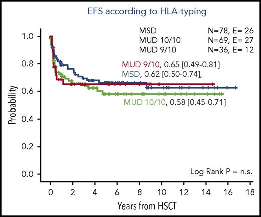
Treatment of patients with MDS-EB with or without signs of transformation and of AML that evolves from MDS (ie, myelodysplasia-related AML [MDR-AML]) remains a major challenge. Therapy for these disorders has been associated with intensive treatment–related toxicities and a high risk of relapse; in particular, conventional AML-type chemotherapy without HSCT resulted in survival rates <30%.32,33 In contrast, it has been shown that a large proportion of children with advanced MDS can be successfully treated with HSCT,34-36 although data are scarce on the outcomes of these children, and previously published reports often included only a limited number of patients who received transplants after heterogeneous regimens. In the largest cohort of children with advanced MDS reported so far, we demonstrated that the 5-year OS probability was 63%, with TRM and disease recurrence contributing equally to treatment failure.36 In that study, all patients received the same fully myeloablative regimen consisting of the combination of busulfan, cyclophosphamide, and melphalan. Age older than 12 years at HSCT, interval between diagnosis and HSCT longer than 4 months, and occurrence of acute or chronic extensive GVHD were associated with increased TRM, whereas the risk of relapse increased with more advanced disease.36 Outcomes for children with MDS-EB and MDS-EB-t were comparable, whereas patients with MDR-AML have an increased risk of relapse. This observation underlines the need to repeat the BM aspirate in patients with MDS-EB or MDS-EB-t to detect any progression of the disease in a timely manner. A more recent update of the EWOG-MDS data in patients with advanced MDS who were given HSCT after myeloablative conditioning regimen including busulfan, cyclophosphamide and melphalan, shows a decrease in TRM, particularly in the adolescent subgroup. The update also shows that the outcome for patients who received a transplant from either an HLA-identical sibling or a UD matched for 9/10 or 10/10 HLA-loci by using high-resolution typing is superimposable (Figure 3). In children with advanced MDS, the presence of structurally complex karyotype was found to be strongly associated with poor prognosis.37
Outcome of patients with advanced MDS given allogeneic HSCT. Probability of EFS according to the type of donor (HLA-identical sibling, 10/10 allelic matched and 9/10 allelic matched unrelated volunteer) used in patients with MDS-EB given an allograft after a myeloablative conditioning regimen that included busulfan, cyclophosphamide, and melphalan. Data from the EWOG-MDS registry. E, events observed; N, number of patients at risk; n.s., not significant.
Outcome of patients with advanced MDS given allogeneic HSCT. Probability of EFS according to the type of donor (HLA-identical sibling, 10/10 allelic matched and 9/10 allelic matched unrelated volunteer) used in patients with MDS-EB given an allograft after a myeloablative conditioning regimen that included busulfan, cyclophosphamide, and melphalan. Data from the EWOG-MDS registry. E, events observed; N, number of patients at risk; n.s., not significant.
One of the most controversial issues in the treatment of children with advanced MDS is the impact of intensive chemotherapy before HSCT. In our original study,36 the use of intensive chemotherapy before the allograft did not improve OS or EFS, because no difference in either relapse incidence or TRM was detected in patients who did or did not receive intensive chemotherapy before HSCT. When the analysis was restricted to children with MDR-AML, there was a significantly decreased risk of relapse in the intensive chemotherapy group that resulted in a nonsignificant advantage in terms of EFS.36 However, the use of intensive chemotherapy was not tested in a systematic way, and therefore, the results must be interpreted with caution. Although the role of intensive chemotherapy before HSCT for patients with advanced MDS remains a matter of debate, we suggest that intensive chemotherapy cannot be routinely recommended for children with MDS-EB, but 1 cycle of cytoreductive chemotherapy should be considered for children with MDR-AML with the aim of reducing the pretransplant blast count. AML-like chemotherapy might also be indicated in patients with ≥20% BM blasts to reduce the risk of relapse. There is a paucity of data to inform the best type of intensive chemotherapy to be utilized.
Case 3
A 13-year-old boy with congenital deafness was observed in another hospital for moderate trilinear cytopenia (WBC count was 3.3 × 10⁹/L, PLT count was 92 × 109/L, and hemoglobin was 10.4 g/dL). The child was admitted to the emergency department of our hospital for high-grade fever and pneumonia. When we were asked to consult, we noted marked bilateral lymphedema of the legs. Immunologic investigations documented marked B-cell lymphopenia and monocytopenia. A BM aspirate showed hypocellularity with 3% blasts, and cytogenetic analysis of marrow cells documented trisomy 8 in the 28 metaphases analyzed. We suggested a diagnosis of RCC with germ line GATA2 mutation; Sanger sequencing documented the presence of a missense mutation in exon 6 of the GATA2 gene. The constitutional nature of the mutation, absent in both parents, was demonstrated. Because the patient was an only child, we started the search for a suitable UD.
A complex interplay exists between IBMFS and other genetic predisposing conditions and development of childhood MDS
Outside the well-known setting of MDS evolving from IBMF disorders and heterozygous germ line RUNX1 and CEBPA mutation causing familial MDS/AML,38-46 little information was available until a few years ago regarding the contribution of hereditary predisposition to the etiology of primary MDS. Recent years have witnessed an increased awareness of non-syndromic familial MDS/AML predisposition syndromes, such as those caused by mutations in GATA2, ETV6, SRP72, SAMD9, and SAMD9L.3,4,47-52 Notably, the discovery that these mutations predispose children to MDS/AML has led to these syndromes being considered as a separate category in the revised 2016 WHO classification of myeloid neoplasms.30
GATA2 germ line mutations leading to haploinsufficiency have been identified as being responsible for a wide spectrum of diseases, including monocytopenia, B-cell lymphopenia, natural killer cell deficiency, congenital deafness, lymphedema (MonoMAC or Emberger syndrome), or cytopenia complicated by systemic infections and an increased risk of developing MDS or AML.5,53-56 Patients with GATA2-related disease present early in life with symptoms that affect the hematopoietic, immune, and lymphatic systems and that are complicated by systemic infections; later in life, these patients have a high risk of developing MDS/AML. Recently, our cooperative EWOG-MDS consortium screened >600 cases of primary or secondary MDS in children and adolescents for the presence of germ line GATA2 mutations.57 We showed that the incidence of GATA2 mutations was significantly higher in patients with advanced MDS (15% [13 of 85]) compared with children affected by low-grade MDS (4% [15 of 341]) (P < .01).57 We also documented that 70% of patients with GATA2 mutations had monosomy 7 compared with only 11% without (P < .01); no mutations were recorded in patients with MDS that evolved from AA or with treatment-related MDS (t-MDS). This latter finding suggests that this gene does not play a relevant role in conferring a risk of developing this severe complication related to childhood cancer treatment. It is also noteworthy that patients with GATA2 mutations were older at diagnosis compared with those who did not carry this genetic aberration (median age, 12.3 vs 10.3 years; P < .01); no child with GATA2 mutation was diagnosed before age 4 years.57 In view of these findings, we propose that GATA2 analysis be included in the workup of all children older than age 4 years and young adults presenting with MDS and monosomy 7. GATA2 mutations do not confer poor prognosis in childhood MDS. However, the high risk for progression to advanced disease should guide decision-making toward timely HSCT, thus avoiding IST. ASXL1 lesions have been reported to recur in one-third of patients with germ line GATA2 lesions, and additional acquired mutations like this can contribute to progression toward more advanced phenotypes.58,59 In addition to clinical indications for an allograft related to the occurrence of severe or life-threatening infections (some occurring in patients with GATA2 mutations), the question of timing of preemptive transplantation is still open. We suggest that the ideal time for HSCT in GATA2-related MDS is during the hypocellular phase of the disease, that is, before severe complications (ie, invasive infections) manifest and before progression to advanced MDS. Although we showed that in a majority of patients, GATA2 deficiency occurred sporadically without preexisting family history of MDS or other GATA2-related symptoms,57 we recommend that potential family donors be tested for GATA2 abnormalities before donating stem cells. Interestingly, increased risk of graft failure and relapse have been reported in 14 patients with GATA2 deficiency who used a nonmyeloablative regimen60 ; this finding suggests that conditioning therapy that can achieve more consistent engraftment and eradication of the malignant myeloid clones should be preferred.
Case 4
A 5-year-old girl was initially diagnosed with abdominal stage IV (due to marrow involvement) Burkitt lymphoma. She was treated with 6 courses of chemotherapy that included cyclophosphamide and etoposide combined with the anti-CD20 monoclonal antibody rituximab. The patient achieved complete remission and was observed in the outpatient unit after she discontinued treatment. At the 6-month control visit, a decline in the PLT count (63 × 109/L) was observed. The child was then retested 2 weeks later, and worsening of thrombocytopenia, together with the appearance of neutropenia (0.9 × 10⁹/L), was found. We decided to perform a BM aspirate, which showed trilinear dysplasia with 12% blasts. Cytogenetic investigations documented a complex karyotype with 3 aberrations, 1 of which involved chromosome 11q23. A diagnosis of therapy-related MDS-EB was then confirmed. Family HLA typing showed that a sister was HLA-compatible with the patient, so HSCT was then performed. The conditioning regimen consisted of busulfan, cyclophosphamide, and melphalan. The patient developed grade 3 acute GVHD involving skin and liver that resolved after treatment with steroids and extra-corporeal photochemotherapy. Immunosuppressive therapy was tapered around day 100 after HSCT. Nine months after HSCT, she developed thrombocytopenia; a BM aspirate was performed that showed 10% blasts with the cytogenetic aberration that had been detected before the HSCT. Three months later, the patient died as a result of disease progression.
Epidemiology and clinical features
In the 2016 WHO classification,30 t-MDS and therapy-related AML (t-AML) are included in the group of therapy-related myeloid neoplasms that occur after exposure to chemotherapy and/or radiation therapy for treatment of a previous unrelated malignancy. Median age at diagnosis of a therapy-related myeloid neoplasm is about 60 years; however, it can occur at any age and is one of the most devastating late complications after treatment of childhood malignancies.61-63 The cumulative incidence of t-MDS and t-AML ranges from 5% to 11% for children treated with standard solid-tumor protocols, including high-dose alkylating agents and topoisomerase II inhibitors64-66 and from <1% to 5% for patients treated for acute lymphoblastic leukemia.61,67,68 The interval from first malignancy to the diagnosis of t-MDS or t-AML is highly variable and may range from several months to many years.63 In adults, 2 distinct patterns have been described: (1) t-MDS or t-AML after treatment with alkylating agents and/or radiotherapy that usually occurs after a latency period of 4 to 7 years and is frequently associated with aberrations of chromosome 5 and 7 and (2) t-MDS or t-AML after topoisomerase II inhibitor therapy, usually occurring after a shorter latency period and frequently associated with aberrations involving MLL.69 Furthermore, these aberrations are frequently part of a complex karyotype.37,69 NGS studies revealed that up to 30% of adult patients with t-MDS or t-AML carry somatic mutations in TP53.70,71 The early acquisition of TP53 mutations in the founding malignant clone probably contributes to the frequent cytogenetic abnormalities and poor responses to chemotherapy that are typical of patients with t-AML or t-MDS.71
Recently, Churpek et al72 investigated the presence of germ line mutations that predispose to malignancy in women who developed t-MDS or t-AML after therapy for breast cancer and identified a cancer predisposition in 21% of patients. This observation indicates the need to consider germ line predisposition syndromes in patients with t-MDS or t-AML.
Treatment approaches
The treatment of patients with t-MDS or t-AML is challenging, and treatment options are limited. Allo-HSCT offers the highest probability of survival, whereas the use of conventional chemotherapy alone has resulted in a high rate of induction failure associated with dismal outcome.73-75 Large series that include pediatric and adult patients with t-MDS or t-AML who received allogeneic HSCT have been reported by the Center for International Blood and Marrow Transplant Research (CIBMTR) and the European Society for Blood and Marrow Transplantation (EBMT).76,77 The OS probability was comparable in both groups (∼35%). Pediatric single-center studies that included a limited number of patients with t-MDS or t-AML who were treated over a long period reported an OS probability ranging between 13% and 20%.75,78,79 Interestingly, Kobos et al80 reported the results of 21 patients who received a transplant over a 15-year period at their institution. The majority of patients (14 of 21) received a cytarabine-based induction before HSCT, and 13 patients reached either complete remission or <5% BM blasts with persistent cytogenetic aberrations. Most patients received conditioning with a busulfan-containing preparative regimen and received a T-cell–depleted allograft. The 5-year OS probability was 61%, and although the analysis had the intrinsic limitations related to the retrospective nature of the study and the long recruitment period, the authors hypothesized that the combination of the cytarabine-based induction with the T-cell–depleted graft may have contributed to the patients’ good outcome which reduced the risk of non-relapse mortality. These encouraging results contrast with those obtained in the series reported by Maher et al81 that included 32 patients with t-MDS or t-AML, of whom 26 received induction chemotherapy with a high rate of non-response (11 of 26). The 5-year OS probability was 36%; patients who achieved complete remission after the first cycle of chemotherapy and patients with MDS without an increase in blasts had a better OS probability compared with patients who achieved remission after multiple therapies or after receiving a transplant in nonremission. However, the only factor associated with better OS in multivariable analysis was a shorter interval from diagnosis of t-MDS or t-AML to HSCT. This observation corroborates our recommendation that patients with t-MDS or t-AML should proceed to HSCT as soon as possible. We do not recommend T-cell depletion of the graft if the donor is immunogenetically compatible with the recipient. But in patients who lack an HLA-identical donor, transplantation from an HLA-disparate donor after innovative approaches of graft manipulation (such as those based on negative depletion of αβ T cells) can be a suitable alternative.82 Even though the role of intensive chemotherapy before the allograft remains unclear in this setting, the results of HSCT in primary advanced childhood MDS indicate that the benefit derived from induction chemotherapy, if any, is limited to patients with >20% BM blasts.36 Disease recurrence after transplantation is the main result of treatment failure, and the prognosis for patients who relapse is dismal. Successful withdrawal of immunosuppressive therapy and donor leukocyte infusions in early relapse have occasionally been reported.83 Close monitoring of chimerism status after HSCT may allow initiation of immunotherapy before overt hematologic recurrence, which potentially translates to a better probability of rescuing patients.84 HSCT can be an option for relapsing patients, although it is associated with a limited chance of success.
In conclusion, significant differences in MDS between children and adults are evident for morphology, cytogenetics, and therapy approaches (Table 1). The somatic mutation landscape also differs between children and adults with myelodysplasia. With the increased use of specific molecular tests and of NGS in clinical practice, more children diagnosed with MDS will be found to have a genetic predisposition syndrome. This will affect genetic counseling for patients and their families; therapeutic decision-making for patients, including HSCT considerations such as indication for and timing of the procedure; the choice of the donor to be used; and optimal graft manipulation in case of a non-HLA–compatible donor. However, careful interpretation of any identified gene mutation will certainly be the great challenge of pediatric hematologists for the next several years.
Acknowledgment
This work was partly supported by Special Grant “5xmille”-9962 and grant IG-17200 from Associazione Italiana Ricerca sul Cancro, grant RF-2010-2316606 from the Ministero della Salute, and grant Finanziaria Laziale di Sviluppo (FILAS) from Regione Lazio (all to F.L.).
Authorship
Contribution: F.L. and B.S. contributed equally to conceiving, writing, and reviewing the manuscript.
Conflict-of-interest disclosure: The authors declare no competing financial interests.
Correspondence: Franco Locatelli, University of Pavia, Dipartimento di Oncoematologia Pediatrica, Istituto di Ricovero e Cura a Carattere Scientifico Ospedale Pediatrico Bambino Gesù, Piazza Sant’Onofrio 4, 00165 Rome, Italy; e-mail: franco.locatelli@opbg.net.


