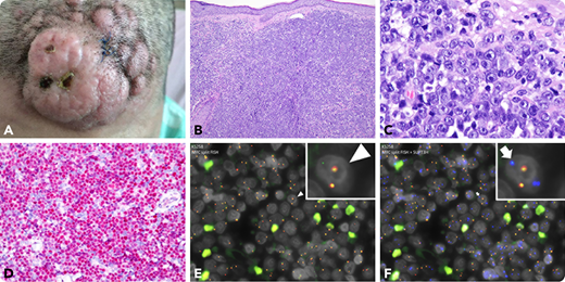
A 53-year-old woman presented with 1 month of a rapidly growing tumor on the forehead that measured 4 × 3.5 cm and had nodular and erythematous appearance (panel A; gross, ×1). A skin biopsy showed the dermis was diffusely infiltrated by a blue cell tumor with a focally starry-sky pattern (panel B; hematoxylin and eosin, 10× objective, total magnification ×100). The tumor cells possessed a moderate amount of cytoplasm and medium-sized to large nuclei with central prominent nucleoli, reminiscent of immunoblasts (panel C; hematoxylin and eosin, 100× objective, total magnification ×1000). Immunohistochemical analysis showed the tumor cells were positive for LCA, CD4, TCL1, and CD123, and negative for CD3, CD20, CD56, CD68, CD138, ALK1, CD30, and MPO, consistent with a diagnosis of blastic plasmacytoid dendritic cell neoplasm (BPDCN). c-MYC was overexpressed (panel D; 40× objective, total magnification ×400). Fluorescence in situ hybridization analysis identified MYC break apart (panel E, arrowhead; green signals, ×600; inset, ×1000) and MYC-SUPT3H fusion (panel F, arrow, blue-green, ×600; inset, ×1000) in more than 70% of the cells, consistent with t(6;8)(p21;q24), which involves MYC but also RUNX2, close to SUPT3H. The patient has completed induction chemotherapy (idarubicin and cytarabine) with tumor regression.
Most cases of BPDCN have a lymphoblast- or myeloblast-like appearance, whereas 10% to 30% of BPDCN cases show immunoblast-like morphology, which are all positive for MYC rearrangement and MYC overexpression, and are associated with older age and a poorer outcome.
For additional images, visit the ASH Image Bank, a reference and teaching tool that is continually updated with new atlas and case study images. For more information, visit http://imagebank.hematology.org.


This feature is available to Subscribers Only
Sign In or Create an Account Close Modal