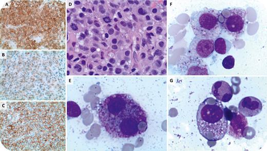
A 63-year-old woman presented with a destructive lesion of L2 with soft tissue extension. Biopsy revealed sheets of cells with abundant eosinophilic cytoplasm, moderate nuclear pleomorphism, and frequent mitoses (panel D, 50× objective, ×500 magnification). Malignant cells were positive for CD45, CD117, tryptase, CD4, and CD30 and negative for CD2, CD25, CD34, and myeloperoxidase by immunohistochemistry, consistent with mast cell sarcoma (MCS) (panels A [CD117], B [tryptase], and C [CD4]; 20× objective, ×200 magnification). Serum tryptase was elevated (132 ng/mL). Iliac crest bone marrow aspiration showed mast cell leukemia (MCL) with 41% malignant mast cells; morphologic features included abnormal granulation, binucleation, vacuolization, and frequent hemophagocytosis (panels E-G, 100× objective, ×1000 magnification). Features of an associated hematologic neoplasm were absent. A KIT mutation was not detected by next-generation sequencing on the aspirate; conventional cytogenetics was normal. Due to poor functional status, the patient received single-agent midostaurin but died 2 months after diagnosis.
MCL is an aggressive form of systemic mastocytosis defined by ≥20% immature mast cells in the bone marrow, which occasionally represents the disseminated phase of MCS. It frequently lacks the KIT D816V mutation but may harbor other KIT mutations. The typical immunophenotype includes CD117, tryptase, and frequent aberrant expression of CD2, CD25, and CD30; CD4 expression is uncommon. This case highlights a rare MCL/MCS with unusual morphologic and immunophenotypic findings.
For additional images, visit the ASH Image Bank, a reference and teaching tool that is continually updated with new atlas and case study images. For more information, visit http://imagebank.hematology.org.


This feature is available to Subscribers Only
Sign In or Create an Account Close Modal