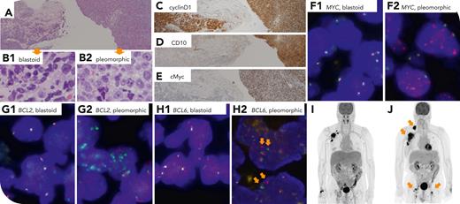
A 72-year-old man had stage IV blastoid mantle cell lymphoma and responded to induction rituximab, bendamustine, and cytarabine therapy initially. He had disease progression 18 months after completing front-line treatment. The biopsy revealed 2 distinct cell populations: blastoid cells (panel A, left side, original magnification ×40; panel B1, original magnification ×400) and pleomorphic cells (panel A, right side, original magnification ×40; panel B2, original magnification ×400). The blastoid cells displayed typical markers like CD20+, CD5+, and cyclin D1+ (panel C; original magnification ×40), SOX11+, CD10− (panel D; original magnification ×40), and cMyc− (panel E; original magnification ×40). In contrast, the pleomorphic cells showed CD5+, cyclin D1+ (panel C) and SOX11+, but CD10+ (panel D), BCL6+, and cMyc+ (panel E). No aberrant p53 staining pattern indicating TP53 alteration was seen. Fluorescence in situ hybridization of MYC, BCL2, and BCL6 break-apart probes revealed no rearrangement in the blastoid area (panels F1-H1; original magnification ×1000), whereas the pleomorphic area exhibited amplification of rearranged MYC (numerous isolated red signals; panel F2) and BCL2 (numerous isolated green signals; panel G2) along with single-copy rearranged BCL6 (arrow: split signals; panel H2). The lymphoma progressed despite treatment with ibrutinib, 560 mg/d, for 6 months (positron emission tomography before/after ibrutinib; panel I/J).
This unique case demonstrates that mantle cell lymphoma can acquire MYC, BCL2, and BCL6 rearrangements with or without amplifications and can be resistant to a Bruton tyrosine kinase inhibitor. Morphologic change and unusual germinal center B phenotypes may unveil such transformation.
For additional images, visit the ASH Image Bank, a reference and teaching tool that is continually updated with new atlas and case study images. For more information, visit https://imagebank.hematology.org.


This feature is available to Subscribers Only
Sign In or Create an Account Close Modal