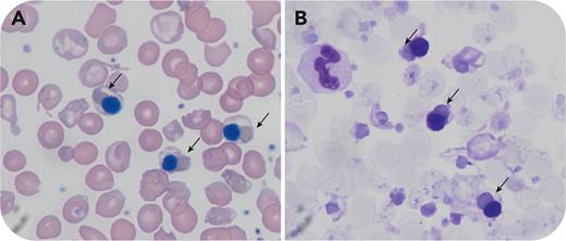
A splenectomized 28-year-old man with transfusion-dependent (ie, receiving transfusion every 4-6 weeks) β-thalassemia is described. The complete blood count showed a hemoglobin of 10.4 g/dL and elevated nucleated red cell count (109 × 109/L). The blood film appeared dimorphic with a subset of cells showing marked anisopoikilocytosis and hypochromia (panel A; May-Grünwald Giemsa stain, 100× lens objective). Some red cells showed lakes of pooled hemoglobin surrounded by very pale areas of cytoplasm (indicated by arrows in panel A, May-Grünwald Giemsa stain, ×100 original magnification). These inclusions were highlighted by supravital staining (panel B; methyl violet, 100× lens objective, arrows).
Globin chain imbalance is a major contributor to anemia in thalassemias. In β-thalassemia, excess α chains form α4 tetramers that aggregate and cause significant oxidative stress/red cell damage leading to hemolysis. These aggregates are known as Fessas bodies in honor of Phaedon Fessas, who reported them in Blood as black-and-white images in 1963. Unlike H-bodies (β4 tetramers seen as small inclusions showing “golf ball” appearance with supravital staining), Fessas bodies form much larger aggregates with prominence in normoblasts. These are removed by the spleen and are therefore not often found circulating in significant numbers. However, they can be uncovered following splenectomy, providing a rare visual glimpse into the pathophysiology of β-thalassemia.
For additional images, visit the ASH Image Bank, a reference and teaching tool that is continually updated with new atlas and case study images. For more information, visit https://imagebank.hematology.org.


This feature is available to Subscribers Only
Sign In or Create an Account Close Modal