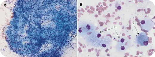
A 53-year-old man was diagnosed with type 1 Gaucher disease (genotype: N370S/L444P) with splenomegaly extending 16 cm below the rib margin, hemoglobin 11.2 g/dL, severe thrombocytopenia (25 × 109/L), and occasional hemorrhagic events. After 7 years of treatment with velaglucerase (50 U/kg and then 60 U/kg every 14 days), his platelet count remained at ∼40 × 109/L. Bone marrow (BM) examination (panel A, May-Grünwald-Giemsa stain, original magnification ×20; panel B, May-Grünwald-Giemsa stain, original magnification ×60) revealed many Gaucher cells (GCs) and very few hematopoietic cells. The usual hematons, elementary units of BM, contained almost exclusively GCs clustered into structures that resembled Gaucheromas (panel A). The cytological appearance of GCs was more (panel B, a) or less (panel B, b) typical. Magnetic resonance imaging showed BM infiltration and bone infarctions, but no bone pseudotumoral features. However, 1 splenic Gaucheroma (diameter: 16 mm) and 1 hepatic Gaucheroma (diameter: 12 mm) were visible on magnetic resonance imaging. Velaglucerase was increased to 95 U/kg every 2 weeks. Splenomegaly was partially reduced. Gaucher disease biomarkers decreased with platelet counts fluctuating between 60 and 80 × 109/L, and bleeding events were observed despite normal coagulatpion parameters.
This exceptional case of BM Gaucheromas, defined as nonmalignant pseudotumors composed of GCs, highlights the need to perform a BM aspiration in rare cases of treatment-resistant thrombocytopenia to identify the mechanism of resistance.
For additional images, visit the ASH Image Bank, a reference and teaching tool that is continually updated with new atlas and case study images. For more information, visit https://imagebank.hematology.org.


This feature is available to Subscribers Only
Sign In or Create an Account Close Modal