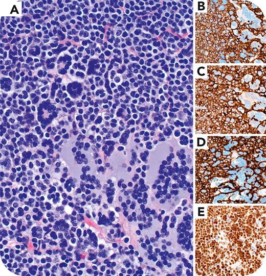
A 77-year-old man with significant smoking history and chronic lymphocytic leukemia/small lymphocytic lymphoma (CLL/SLL) was found to have a right lower lobe lung mass and mediastinal lymphadenopathy on imaging. Subsequent lobectomy showed an invasive squamous cell carcinoma, and all mediastinal lymph nodes were involved by CLL/SLL without large-cell transformation. One lymph node contained large multinucleated cells, either with nuclei arranged in a wreathlike configuration or with abundant amphophilic cytoplasm, with identical nuclear features to the adjacent CLL/SLL cells (panel A, hematoxylin and eosin, 40× lens objective). Immunohistochemistry showed that the multinucleated cells had an identical immunophenotype to the CLL/SLL, positive for CD20 (panel B), CD5 (panel C), CD23 (panel D), and LEF1 (panel E) (all images, 40× lens objective) and negative for pan-cytokeratin (not shown). These results supported Warthin-Finkeldey cells in CLL/SLL and no metastatic carcinoma.
Warthin-Finkeldey cells were first described in the tonsils of children with measles and are a hallmark of measles infection. Nodal Warthin-Finkeldey cells can be found incidentally in other viral infections, Kimura disease, or lymphomas. Their origin is unclear in reactive conditions, showing variable expression of B-cell, macrophage, or follicular dendritic-cell markers. In lymphomas they usually represent a morphologic variation of the neoplastic cells. Warthin-Finkeldey cells should not be confused with metastatic carcinoma, which could result in a wrongful staging.
For additional images, visit the ASH Image Bank, a reference and teaching tool that is continually updated with new atlas and case study images. For more information, visit https://imagebank.hematology.org.


This feature is available to Subscribers Only
Sign In or Create an Account Close Modal