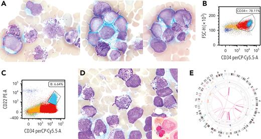
A woman, aged 62 years, with a 7.5-year history of ovarian cancer and on chemotherapy, presented with anemia/circulating blasts. Peripheral blood examination showed a leukocyte count of 31.3 × 10⁹/L, neutrophil count of 3.8 ×10⁹/L, hemoglobin level of 7.9 g/dL, platelet count of 55 ×109/L, and blast count of 55.6%, as well as increased basophils/basophilic myeloid precursors (panel A; Wright-Giemsa stain, 100× lens objective). Flow cytometry analysis of aspirate revealed 78% of blasts, which were CD34+/CD117+(variable)/HLA-DR+(variable)/CD13+/CD33+/CD38+/CD123+/cMPO(variable)/CD7+(aberrant) (panel B). A subset of blasts (6.64%) was CD22+ /CD11b+/CD13+/CD33+/CD123+(bright)/CD15–/CD19–/CD10–/CD79–/TdT– (panel C). Marrow aspirate showed hypercellularity with increased blasts, some with basophilic granules, 15% basophilic myeloid precursors/basophils (panel D; May-Grünwald Giemsa stain, 100× lens objective), megakaryocytic/erythroid dysplasia, and 18% ring sideroblasts (RS) (panel D, inset; Perls stain, 100× lens objective). Optical genome mapping showed a highly complex karyotype (CK) including del(5q) and del(13q) (panel E). Next-generation sequencing showed biallelic TP53 (variant allele frequency: 49.6%; 42.5%) and PRPF40B/TET2 mutations. A diagnosis of acute myeloid leukemia (AML) with mutated TP53, therapy-related was rendered per International Consensus Classification criteria. The patient died 3 months later.
AML with basophilic differentiation is often associated with t(6;9)(p22.3;q34.1)/DEK::NUP214 and only rarely reported in AML with myelodysplasia-related cytogenetic abnormalities. CD22, a B-lineage marker, can be moderately expressed in basophils. Its positivity, along with CD123+(bright)/CD13+/ CD33+/CD11b+/CD15–/basophilic granules, supports basophilic differentiation. RS in AML is associated with CK/TP53 mutations. AML with mutated TP53, therapy-related with basophilic differentiation, partial CD22/RS is rare, reflecting an aggressive disease with poor outcomes.
For additional images, visit the ASH Image Bank, a reference and teaching tool that is continually updated with new atlas and case study images. For more information, visit https://imagebank.hematology.org.


This feature is available to Subscribers Only
Sign In or Create an Account Close Modal