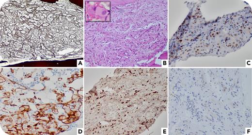
A 61-year-old woman with essential thrombocythemia (ET) presented with anemia necessitating cytoreduction cessation. Bone marrow biopsy was performed to exclude fibrotic progression. Full blood count results demonstrated a hemoglobin level of 8.8 g/dL, white blood cell count of 4.1 × 10⁹/L, and platelet count of 535 × 109/L. Bone marrow trephine results showed marked reticulin fibrosis (MF-3) (panel A, 10× objective; silver stain). Careful examination revealed small nests and cords of epithelioid cells (panel B, 10× objective; hematoxylin and eosin [H&E] stain) with round to oval nuclei, fine chromatin, distinct nucleoli, and abundant pale eosinophilic cytoplasm (panel B inset, 100× objective; H&E stain). The abnormal cells were positive for ERG (panel C, 20× objective), CD34 (panel D, 20× objective), CD31, and factor VIII; diffusively positive for TFE3 (panel E, 10× objective); and negative for CAMTA1 (panel F, 20× objective). The uninvolved areas showed megakaryocytic hyperplasia consistent with underlying ET. Dual-color break-apart fluorescence in situ hybridization analysis confirmed TFE3 rearrangement. A diagnosis of epithelioid hemangioendothelioma (EHE) was established. Imaging studies confirmed disseminated metastases. Further biopsies were conducted, but no notable findings were revealed.
EHE is a malignant endothelial neoplasm with heterogeneous presentation and prognosis, most commonly involving the soft tissue, bone, lung, skin, and liver. However, bone marrow metastasis is extremely rare. CAMTA1 rearrangement is highly specific for EHE, which can be detected by immunohistochemistry and molecular studies. In CAMTA1-negative cases, TFE3 rearrangement should be tested.
For additional images, visit the ASH Image Bank, a reference and teaching tool that is continually updated with new atlas and case study images. For more information, visit https://imagebank.hematology.org.


This feature is available to Subscribers Only
Sign In or Create an Account Close Modal