Abstract
The adhesion of leukocytes to platelets deposited at the site of vascular injury may represent an important mechanism by which leukocytes contribute to hemostasis and thrombosis. In this study, we examined whether, in comparison with their distribution in circulating blood, certain leukocyte types are enriched at sites of platelet deposition. We used an experimental vascular injury model, in which human fibrillar collagen was exposed to anticoagulated human whole blood flowing through parallel-plate chambers (venous shear rate, 65/s). The platelet-adherent leukocytes were detached by EDTA treatment and analyzed by flow cytometry using cell-type–specific antibodies. The predominant leukocytes found in platelet thrombi were polymorphonuclear leukocytes, accounting for 76% of bound leukocytes (62% in circulating blood), whereas T and B lymphocytes did not significantly accumulate on thrombi, comprising a fraction of less than 5% (32% in circulating blood). Monocytes constituted 16% of platelet thrombus-bound leukocytes, which represents an almost fourfold enrichment as compared with their proportion in circulating blood. Almost identical results were obtained when we analyzed leukocytes adhering to platelet monolayers, which were formed by blocking glycoprotein IIb-IIIa, thus preventing platelet aggregation on top of the collagen-adherent platelets. Furthermore, leukocyte adhesion to platelet monolayers was completely inhibited by an anti-P-selectin antibody (50% inhibitory concentration, 0.3 μg/mL), whereas it reached a plateau at about 70% inhibition on platelet thrombi. This difference could be explained by a possible function of glycoprotein IIb-IIIa in leukocyte immobilization to thrombi or by the high local concentration of P-selectin in the growing thrombi. The results suggest that, because of their known abilities to promote coagulation and thrombolysis, the monocytes and polymorphonuclear leukocytes accumulating on forming platelet thrombi could play an important role in modulating thrombotic and hemostatic processes.
DAMAGE TO A BLOOD VESSEL results in the adhesion of platelets to the site of injury and the subsequent formation of a platelet thrombus. Leukocytes comprise an additional cellular component of thrombi1 and are able to adhere to platelets even under high shear stress conditions.2 The leukocytes immobilized on a platelet thrombus may contribute to the localized generation of thrombin leading to fibrin formation and further platelet activation. The procoagulant activity of thrombus-bound leukocytes was shown in an arteriovenous shunt model in baboons.3 All leukocyte types, polymorphonuclear leukocytes (PMNs), monocytes, and lymphocytes, are able to promote coagulation by providing assembly sites for the prothrombinase complex.4,5 Monocytes have additional procoagulant capabilities in that they can express tissue factor on stimulation by appropriate agonists,6-8 provide assembly sites for the intrinsic tenase complex,9 and directly activate Factor X (FX) by a CD116/CD18-dependent mechanism.10
In vitro studies showed that many different leukocyte types can bind to activated platelets through the leukocyte adhesion molecule P-selectin,11-16 a constituent of the platelet α-granules that is transported to the platelet surface on stimulation with agonists.17-19 P-selectin is a member of the selectins, a family of structurally related adhesion molecules that function as leukocyte rolling receptors.20-22 Under physiological shear stress, leukocyte interaction with surface-bound P-selectin or activated platelet layers leads to leukocyte rolling23-25 and the subsequent firm attachment26 through additional adhesion mechanisms21 involving the β2-integrin CD11b/CD18.27 28
Although the importance of P-selectin for leukocyte immobilization to thrombi was shown in vivo,3 it remained unclear which types of leukocytes actually accumulate on thrombi and potentially contribute to the procoagulant activity. To examine whether certain leukocyte types become enriched at vascular injury sites under blood flow conditions, we determined the distribution pattern of leukocyte types bound to deposited platelets and compared it with their proportion in circulating blood. For that purpose, we used a human ex-vivo blood flow system29 30 in which fibrillar collagen, a thrombogenic constituent of the vessel wall, was exposed to human whole blood at venous shear stress leading to platelet deposition and subsequent platelet activation. The results of the experiments provide evidence for an almost exclusive accumulation of monocytes and PMNs on forming platelet thrombi and on collagen-adherent platelet monolayers. P-selectin was strongly expressed on both the platelet monolayers and platelet thrombi, and functional studies indicate that P-selectin plays an important role for immobilizing the leukocytes to the platelets. The implications of this local enrichment of monocytes and PMNs for thrombosis and hemostasis are discussed.
MATERIALS AND METHODS
Antibodies, inhibitors, and proteins.Fluorescence-activated cell sorting (FACS) lysing solution and monoclonal antibodies directed against different leukocyte types used for FACScan analysis were obtained from Becton Dickinson (San Jose, CA): phycoerythrin (PE)-labeled anti-CD20 (B lymphocytes), PE-labeled anti-CD3 (T lymphocytes), PE-labeled anti-CD14 (monocytes), fluorescein isothiocyanate (FITC)-labeled anti-CD15 (PMNs), PE-labeled antiCD62 (P-selectin), and FITC-labeled anti-CD61 (glycoprotein IIb-IIIa [GPIIb-IIIa]; ie, platelets). PE-labeled anti-CD45 and FITC-labeled anti-CD45 (all leukocytes) were purchased from DAKO (DAKO Diagnostics AG, Zug, Switzerland). The monoclonal anti-P-selectin antibodies were WAPS12.2 (Endogen Inc, Cambridge, MA) and anti-CD62, clone AK-6 (Serotec Ltd, Kidlington, UK). As a control antibody, we used the monoclonal anti-GPIIb antibody pl48, which does not interfere with the function of the GPIIb-IIIa complex on platelets (from Beat Steiner, F. Hoffmann-La Roche). The thrombin inhibitor Ro 46-6240 (napsagatran) was used as an anticoagulant,31,32 and the GPIIb-IIIa antagonist Ro 44-9883 (lamifiban) was used as an inhibitor of platelet aggregation.33,34 The thrombin receptor activation peptide (TRAP peptide) with the amino acid sequence Ser-Phe-Leu-Leu-Arg was synthesized by solid-phase peptide synthesis using a p-alkoxybenzyl alcohol polystyrene resin.35
Human collagen type III was purified from lyophilized pepsin-extracted placenta by salt precipitation as described previously.36 Fibril formation was induced by dialyzing a solution of 1 mg/mL of collagen type III in 0.1 mol/L acetic acid against 20 mmol/L Na2HPO4 (pH, 7.5) at 4°C for 24 hours. The activity of the preparations was tested in the aggregometer using human plasma, and they were stored at 4°C until used in the perfusion experiments.
Human blood flow system.Human collagen type III was sprayed in fibrillar form onto Thermanox plastic coverslips (Miles Lab, Naperville, IL) at a concentration of 20 μg/cm2 using an air-brush (model 100G; Badger Air-Brush Co, Franklin Park, IL). The collagen-coated coverslips were dried for several hours at room temperature, washed with 0.9% NaCl solution over a period of 1 hour, and kept in NaCl solution containing 0.1% bovine serum albumin (BSA) until they were used for the perfusion experiments. The coverslips were positioned in three parallel-plate perfusion chambers as described recently.29,30 Blood was drawn from the antecubital vein of a healthy donor directly into a Plexiglas distribution block, where the blood was separated into three tubings. The blood flow of 1 mL/min was controlled by three individual roller pumps positioned distal to the parallel-plate perfusion devices. Immediately before entering the mixing chambers, the flowing blood was anticoagulated by infusion of 1 μmol/L (blood concentration) thrombin inhibitor Ro 46-6240 via an infusion pump (Infu 362; Datex AG, Uhwiesen, Switzerland) at a rate of 50 μL/min. To examine leukocyte adhesion to platelet monolayers, we additionally infused 0.5 μmol/L of GPIIb-IIIa antagonist lamifiban (control) and different concentrations of the anti–P-selectin antibody WAPS12.2 or control antibody pl48. The same protocol was used to examine leukocyte adhesion to platelet thrombi, except that infusion of the GPIIb-IIIa antagonist lamifiban was omitted. The infusion solutions were mixed with the blood in parallel in the three mixing chambers.29 30 The blood-inhibitor mixture then entered the three parallel-plate perfusion devices containing the collagen-coated coverslips. The blood flow of 1 mL/min resulted in a shear rate of 65/s on the collagen surface, which corresponded to venous blood flow conditions. After a 5.5-minute perfusion period, the wash solution (phosphate-buffered saline [PBS]) was connected to the distribution block without interrupting the flow. During the 3-minute washing period, the inhibitor infusion of 50 μL/min was maintained, except in experiments in which leukocyte adhesion to platelet thrombi was determined by infusion of a high concentration of WAPS12.2 (blood concentration, 50 μg/mL). In these experiments, infusion of antibody was stopped after 1 minute. Then, the mixing device was disconnected from the parallel-plate devices, which were subsequently perfused at 1 mL/min for 2 minutes with 3% paraformaldehyde in PBS after a brief interruption of flow (≈5 seconds). For morphometrical examinations, we used 2.5% glutaraldehyde in 0.1% cacodylate buffer (pH, 7.4) containing 2.5 mmol/L CaCl2 and 0.9 mmol/L MgCl2 instead of paraformaldehyde. Then, the coverslips were removed from the chambers, incubated in fresh fixative for an additional 1 hour, and stored in PBS-0.03% azide. For morphometrical analysis, the fixative was cacodylate buffer containing 7% sucrose.
Quantification of leukocyte adhesion and thrombus dimensions on cross sections.After the perfusion experiments, the coverslips were embedded in Epon (Fluka Chemie, Buchs, Switzerland) and cross sections 1 mm downstream to the flow entrance were prepared as described previously.37 The cross sections were stained with 0.01% toluidine blue and 0.01% fuchsin and were analyzed.37 The number of leukocytes bound to platelet monolayers (infusion of GPIIb-IIIa antagonist) and to platelet thrombi (no infusion of GPIIb-IIIa antagonist) was determined on cross sections (width, 8 mm). The dimensions of the deposited platelet thrombi, such as the thrombus area and the average thrombus height, were measured by computer-assisted morphometry (DIASYS II; Meier, Thoerigen, Switzerland) using a Zeiss Standard microscope (Carl Zeiss AG, Zurich, Switzerland).37
Double staining for leukocytes and platelet-expressed P-selectin.Coverslips were incubated with 10 μg/mL of monoclonal anti-P-selectin antibody (anti-CD62, clone AK-6; Serotec) in PBS-0.1% BSA. After washing with PBS, the coverslips were incubated at room temperature for 30 minutes with 5 nm gold-labeled antimouse antibody (Auro Probe LM; Amersham, Buckinghamshire, UK) diluted 1:50 in PBS-0.1% BSA. Then, the coverslips were washed with PBS, treated for 10 minutes with 2% glutaraldehyde in PBS, and then washed with PBS and distilled water. After incubation with silver enhancer for 10 to 15 minutes (IntenSEM; Amersham), the coverslips were fixed with Rapidfix (Eastman-Kodak, Rochester, NY) and thoroughly washed in distilled water. After air-drying, the adherent leukocytes were stained with Diff-Quick solution (Baxter-Dade AG Düdingen, Switzerland). The coverslips were embedded in Merckoglass (Merck, Darmstadt, Germany) and examined under the microscope (Zeiss Axiophot).
Measurement of P-selectin expression and platelet binding to leukocytes in post–chamber blood.To measure platelet activation in the entire perfusion system, a tubing was positioned between the parallel-plate perfusion chamber containing a noncoated plastic coverslip and the roller pumps. The perfusion conditions were the same as those for the leukocyte adhesion experiments including infusion of 1 μmol/L thrombin inhibitor and 0.5 μmol/L GPIIb-IIIa antagonist or infusion of PBS-0.1% BSA buffer. After the tubing was perfused for 5 minutes, the blood content of the tubing (120 mm in length and 2.0-mm inner diameter; ≈350 μL vol) was collected into an Eppendorf tube (Sarstedt AG, Sevelen, Switzerland) containing 40 μL of 108-mmol/L citrate buffer. During this perfusion period, the mixing device and the parallel-plate chambers have been exposed to the blood for at least 5.5 minutes, which is the standard perfusion time in all experiments. A total of 100 μL of the anticoagulated blood sample was pipetted into 1 mL of cold 0.5% paraformaldehyde fixation solution and was incubated for 1 hour at 4°C. Control blood samples were first activated with 10 μmol/L of the TRAP peptide or 10 μmol/L adenosine diphosphate (ADP) before fixation. After fixation, 10 μL of the samples were directly added to 90 μL of modified Tyrode buffer (133 mmol/L NaCl, 2.7 mmol/L KCl, 12 mmol/L Na2CO3 , 0.4 mmol/L NaH2PO4 , 0.4 mmol/L MgCl2 , 5 mmol/L glucose, 0.2% BSA, 10 mmol/L HEPES [pH, 7.4], and 50 μmol/L CaCl2 ). To determine the fraction of activated platelets, 3 μL of FITC–anti-CD61 (GPIIb-IIIa) and PE–anti-CD62 (P-selectin) was added. The CD61+ platelet population was analyzed with a forward-scatter (FSC) versus side-scatter (SSC) dot blot, encircled with a gate, and analyzed for the presence of a CD62 signal indicative of platelet activation. To detect leukocytes with bound platelets, 3 μL of FITC–anti-CD45 and 3 μL of PE–anti-CD62 antibodies were added before incubation at room temperature for 15 minutes. FACScan analysis was performed in an SSC versus FITC signal (FL1) dot plot with both photomultipliers set on logarithmic gain. The FL1-positive cells (FITC–anti-CD45; leukocytes) were encircled with a gate, and 1,000 events were acquired. In the analysis mode, the CD45+ population was then analyzed in an FL1 versus FL2 dot plot for the presence of the CD62 signal (FL2), indicative of leukocyte binding to one or more activated platelets.
Quantification of leukocyte types by flow cytometry.After the 3-minute washing period with PBS, the coverslips were removed from the parallel-plate chambers, positioned into 6-well Costar plates (Costar Corp, Cambridge, MA), and incubated twice for 5 minutes in 1 mL of 5 mmol/L EDTA in PBS. After each 5-minute interval, the detached leukocytes were transferred into full medium (M199 supplemented with 10% fetal bovine serum). After centrifugation at 400g for 5 minutes, the leukocytes collected from three coverslips were resuspended in 0.2 mL full medium and were pooled. After fixation in 0.5% paraformaldehyde-PBS on ice for 1 hour, the cell suspension was centrifuged and the leukocytes were resuspended in 0.2 mL of 0.5% paraformaldehyde-PBS for flow cytometry. The total number of detached leukocytes was quantified by use of a hemocytometer. The viability of the cells, as assessed by the Trypan-Blue exclusion test, was more than 95%. The leukocytes that still bound to collagen-adherent platelets on the coverslips after the EDTA treatment were stained with Diff-Quick solution (Baxter Dade AG), and they were quantified on the imaging analysis system. To determine the leukocyte type distribution in the donor blood, a small volume of blood was supplemented with 10 μmol/L thrombin inhibitor and 5 μmol/L GPIIb-IIIa antagonist before the start of the perfusion experiment, and the distribution of leukocyte types was analyzed on the FACScan.
FACScan analysis was performed with greater than 10,000 detached leukocytes or with 5 μL anticoagulated blood in 80 μL of modified Tyrode buffer. The volume of antibody solution required for saturation binding was determined to be 3 μL for each antibody (data not shown). The following antibody combinations were used: (1) FITC–anti-CD45 + PE–anti-CD14 (for monocytes); (2) FITC–anti-CD45 + PE–anti-CD3 (for T lymphocytes); (3) FITC–anti-CD45 + PE–anti-CD20 (for B lymphocytes); and (4) PE–anti-CD45 + FITC–anti-CD15 (for PMNs). After incubation for 10 minutes, the blood samples (85 μL) were mixed with 500 μL of FACS lysing solution (Becton Dickinson) to lyse erythrocytes and were incubated for 5 minutes before analysis. The leukocyte suspension derived from a total of three coverslips was diluted with 500 μL of modified Tyrode buffer before analysis. Data (1,000 events) were acquired on an FSC versus SSC dot plot, and the leukocyte population was encircled by a gate. The leukocyte types within the CD45+ leukocyte population were quantified by FL1 (FITC signal) versus FL2 (PE signal) dot plot analysis.
Analysis of leukocyte subtypes by differential staining on coverslips.For the differentiation of leukocytes adhering to platelet monolayers, the coverslips were washed with PBS in the parallel-plate chambers for 1.5 minutes. Then, the coverslips were fixed immediately with 3% paraformaldehyde in PBS for 1 minute, rinsed with PBS, and air-dried. Leukocytes were then stained with Diff-Quick solution (Baxter Dade AG). Lymphocytes could not unambiguously be differentiated from monocytes; therefore, these two leukocyte types were combined as mononuclear cells. The other leukocyte types determined were neutrophils and eosinophils. At least 200 leukocytes were analyzed per coverslip and on stained blood smears of the same donor.
Quantification of adherent leukocytes by an image analysis system.After perfusion experiments, the unfixed coverslips were rinsed with PBS, air-dried, and stained with the leukocyte-staining solution Contrast Blue (Kirkegaard and Perry Laboratories, Inc, Gaithersburg, MD). The coverslips were then air-dried and embedded in Merckoglass (Merck). The number of adherent leukocytes per square millimeter was measured on an image analysis system using OPTIMAS version 4.10 software (BioScan, Edmonds, WA) on a Philips PC (486 DX-25) connected to a JVC video camera (model KY-M280E, Basel, Switzerland) that was installed on a Zeiss microscope. The values obtained were the average of five determinations taken at a distance of 1 mm from the blood entrance of the coverslip.
RESULTS
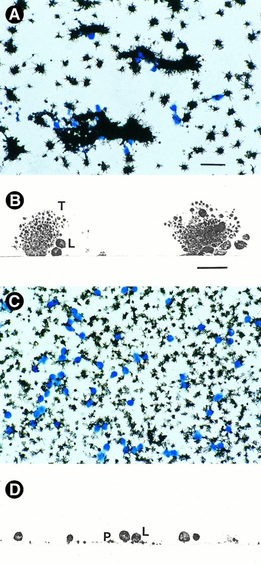
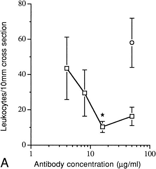
Role of P-selectin in the adhesion of leukocytes to platelet thrombi and to platelet monolayers.Exposure of collagen-coated coverslips to flowing anticoagulated human blood (venous shear rate, 65/s corresponding to a shear stress of about 2 dynes/cm2 ) resulted in the deposition of fibrin-free platelet thrombi to which numerous leukocytes were bound (Fig 1B). As shown in Figs 1A and B, platelet thrombi were formed despite anticoagulation with thrombin inhibitor. Similar results were obtained previously30 and are consistent with results of other blood flow systems, which showed that the influence of the coagulation system on platelet thrombus formation appears more pronounced at arterial shear rates of 650/s, than at venous shear rates.38-41 Immunochemical staining with a monoclonal P-selectin antibody showed a strong expression of P-selectin by platelet thrombi (Fig 1A). Moreover, infusion of the blocking anti–P-selectin antibody WAPS12.2 inhibited leukocyte adhesion and reached a plateau at about 70% inhibition between 16 to 50 μg/mL, as determined by morphometry on cross sections (Fig 2A). A control antibody (pl48) had no inhibitory effect (Fig 2A). To exclude the possibility that the effect of the anti–P-selectin antibody was caused by inhibition of platelet thrombus formation, the thrombus dimensions were analyzed by computer-assisted morphometry. The results showed that neither the P-selectin antibody nor the control antibody pl48 reduced the thrombus area or the average height of the formed platelet thrombi (Table 1).
Leukocyte adhesion to platelet thrombi and platelet monolayers. Collagen-coated surfaces were exposed for 5.5 minutes to flowing human blood (shear rate, 65/s) that was anticoagulated by infusion of thrombin inhibitor. To obtain collagen-adherent platelet monolayers, platelet aggregation on top of the adherent platelets was prevented by additional infusion of the GPIIb-IIIa antagonist. (A and C) “En face” view of coverslips with platelet thrombi (A) and platelet monolayers (C). P-selectin–expressing platelets (black) were visualized by an immunogold-silver staining using a monoclonal anti–P-selectin antibody, and the adherent leukocytes (blue) were stained with Diff-Quick solution. (B and D) Cross sections of coverslips with platelet thrombi (B) and platelet monolayers (D). Leukocytes can be seen to adhere to the deposited platelet thrombi (in B) and to single collagen-adherent platelets (in D). L, leukocytes; P, single adherent platelet; T, platelet thrombus.
Leukocyte adhesion to platelet thrombi and platelet monolayers. Collagen-coated surfaces were exposed for 5.5 minutes to flowing human blood (shear rate, 65/s) that was anticoagulated by infusion of thrombin inhibitor. To obtain collagen-adherent platelet monolayers, platelet aggregation on top of the adherent platelets was prevented by additional infusion of the GPIIb-IIIa antagonist. (A and C) “En face” view of coverslips with platelet thrombi (A) and platelet monolayers (C). P-selectin–expressing platelets (black) were visualized by an immunogold-silver staining using a monoclonal anti–P-selectin antibody, and the adherent leukocytes (blue) were stained with Diff-Quick solution. (B and D) Cross sections of coverslips with platelet thrombi (B) and platelet monolayers (D). Leukocytes can be seen to adhere to the deposited platelet thrombi (in B) and to single collagen-adherent platelets (in D). L, leukocytes; P, single adherent platelet; T, platelet thrombus.
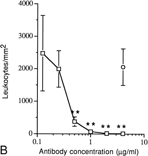
Inhibition of leukocyte adhesion by infusion of the anti–P-selectin antibody (WAPS12.2) to the flowing blood. Experimental conditions are described in the legend to Fig 1, except that the infusion solutions also contained anti–P-selectin antibody or the control antibody pl48 (nonblocking antibody directed against the GPIIb subunit) or buffer alone (controls). (A) Leukocyte adhesion to platelet thrombi as determined by morphometrical analysis of cross sections. (B) Leukocyte adhesion to platelet monolayers, quantified on an image analysis system after leukocyte staining. (□), anti-P-selectin antibody WAPS12.2; (○), control antibody pl48. The control value (buffer alone) for platelet thrombi was 42.6 ± 9.8 leukocytes/10 mm cross section, and that for platelet monolayers was 2,570 ± 311 leukocytes/mm2. The values are the mean ± SEM of 4 to 19 coverslips. Comparison with control values (buffer alone) by unpaired Student's t-test: *, P < .02; **, P < .001.
Inhibition of leukocyte adhesion by infusion of the anti–P-selectin antibody (WAPS12.2) to the flowing blood. Experimental conditions are described in the legend to Fig 1, except that the infusion solutions also contained anti–P-selectin antibody or the control antibody pl48 (nonblocking antibody directed against the GPIIb subunit) or buffer alone (controls). (A) Leukocyte adhesion to platelet thrombi as determined by morphometrical analysis of cross sections. (B) Leukocyte adhesion to platelet monolayers, quantified on an image analysis system after leukocyte staining. (□), anti-P-selectin antibody WAPS12.2; (○), control antibody pl48. The control value (buffer alone) for platelet thrombi was 42.6 ± 9.8 leukocytes/10 mm cross section, and that for platelet monolayers was 2,570 ± 311 leukocytes/mm2. The values are the mean ± SEM of 4 to 19 coverslips. Comparison with control values (buffer alone) by unpaired Student's t-test: *, P < .02; **, P < .001.
Effect of Anti–P-Selectin Antibody on Platelet Thrombus Dimensions
| . | Thrombus Area . | Average Thrombus Height (μm ± SEM) . |
|---|---|---|
| . | (μm2/μm ± SEM) . | . |
| Buffer | 2.25 ± 0.35 | 10.16 ± 0.73 |
| Anti–P-selectin Ab* | 2.28 ± 0.48 | 10.35 ± 1.08 |
| Control Ab† | 2.98 ± 0.73 | 14.85 ± 1.85 |
| . | Thrombus Area . | Average Thrombus Height (μm ± SEM) . |
|---|---|---|
| . | (μm2/μm ± SEM) . | . |
| Buffer | 2.25 ± 0.35 | 10.16 ± 0.73 |
| Anti–P-selectin Ab* | 2.28 ± 0.48 | 10.35 ± 1.08 |
| Control Ab† | 2.98 ± 0.73 | 14.85 ± 1.85 |
Collagen-coated coverslips were exposed to flowing human blood (shear rate, 65/s) for 5.5 minutes. The blood was supplemented via a mixing device with an anticoagulant (thrombin inhibitor Ro 46-6240) together with P-selectin antibody WAPS12.2 (6 donors) or control antibody pl48 (4 donors) or with buffer (13 donors). The coverslips were washed, fixed, and embedded in Epon resin. Cross sections 1.5 mm from the flow entrance were stained, and thrombus dimensions were measured by computer-assisted morphometry as described in Materials and Methods.
Abbreviation: Ab, antibody.
50 μg/mL WAPS12.2.
50 μg/mL pl48 (nonblocking anti–GPIIb-IIIa antibody).
Next, we examined the role of P-selectin in leukocyte adhesion to platelet monolayers. For that purpose platelet aggregation on top of the collagen-adherent platelets was prevented by the additional infusion of the selective GPIIb-IIIa antagonist lamifiban. This resulted in the formation of a collagen-adherent platelet monolayer in the complete absence of platelet thrombus formation as shown previously.42 Most of the adherent platelets were spread on collagen (Figs 1C and D) and were fully activated, as indicated by the presence of immunostainable P-selectin on the platelet surface (Fig 1C). In contrast to experiments with platelet thrombi, the leukocytes that bound to the collagen-adherent platelet monolayer were lying in an approximate planar layer; therefore, it was possible to quantify the density of stained leukocytes by use of an image analysis system. Figure 2B shows that infusion of P-selectin antibody completely inhibited leukocyte adhesion from 2,570 ± 311 leukocytes/mm2 (mean ± SEM) to 9 ± 6 leukocytes/mm2 at 4 μg/mL antibody (50% inhibitory concentration [IC50], 0.3 μg/mL), whereas a control antibody (pl48) had no inhibitory effect.
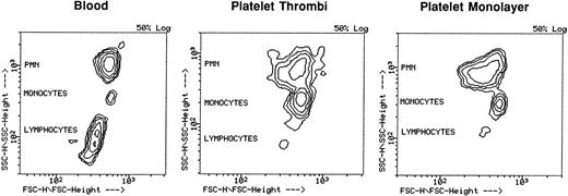
Differentiation of leukocyte types by flow cytometry.For analysis by flow cytometry, the leukocytes were detached from the platelet thrombi and from the platelet monolayers after a short incubation with 5 mmol/L EDTA. The efficiency of leukocyte detachment was in the average of 97% for platelet thrombi and 90% for the platelet monolayers. In experiments with platelet monolayers, the nondetached leukocytes were examined by a differential staining technique. This analysis showed that the leukocyte type distribution pattern of nondetached leukocyte types was similar to that of adherent ones, indicating that the detachment procedure did not lead to the enrichment of a particular leukocyte type (data not shown). After detaching the leukocytes from the platelet thrombi and platelet monolayers, they were first analyzed according to their size/granularity distribution using a 2-parameter correlation display (Fig 3). The results clearly show that monocytes and PMNs were enriched on both platelet thrombi and platelet monolayers, as compared with their proportion in whole blood. In contrast, lymphocytes constituted a marginal portion of platelet-adherent leukocytes (Fig 3).
Leukocyte type distribution in blood, on platelet thrombi, and on platelet monolayers. The experimental conditions for the whole blood perfusions are described in the legend to Fig 1. The platelet-adherent leukocytes were detached with EDTA, fixed in 0.5% paraformaldehyde-PBS solution, diluted in modified Tyrode buffer, and analyzed on the FACScan. A fresh blood sample was taken before the perfusion experiments and treated with FACS lysing solution. The leukocyte distributions of a single blood donor are shown as FSC (size) versus SSC (granularity) contour plots. Shown are the contour plots of fresh blood sample (left), leukocytes adhering to platelet thrombi (center), and leukocytes adhering to platelet monolayers (right).
Leukocyte type distribution in blood, on platelet thrombi, and on platelet monolayers. The experimental conditions for the whole blood perfusions are described in the legend to Fig 1. The platelet-adherent leukocytes were detached with EDTA, fixed in 0.5% paraformaldehyde-PBS solution, diluted in modified Tyrode buffer, and analyzed on the FACScan. A fresh blood sample was taken before the perfusion experiments and treated with FACS lysing solution. The leukocyte distributions of a single blood donor are shown as FSC (size) versus SSC (granularity) contour plots. Shown are the contour plots of fresh blood sample (left), leukocytes adhering to platelet thrombi (center), and leukocytes adhering to platelet monolayers (right).
The different leukocyte types were then quantified by using leukocyte type-specific fluorescent antibodies in the 2-parameter dot plot mode. The results are summarized in Fig 4 and show that monocytes and PMNs comprised 16% to 18% and 73% to 76%, respectively, of all leukocytes adhering to platelet thrombi and platelet monolayers. In contrast, adhesion of T and B lymphocytes to platelet thrombi and to platelet monolayers was less than 5% and less than 8%, respectively (Fig 4). Leukocyte counts of all donors were within the normal range, as was the leukocyte type distribution in whole blood, in accord with the analysis of the individual blood smears (data not shown).
Quantification of monocytes, PMNs, and T and B lymphocytes adhering to platelet thrombi and platelet monolayers. The experimental conditions for the whole blood perfusions are described in the legend to Fig 1. The leukocytes adhering to platelets were detached with EDTA and fixed in 0.5% paraformaldehyde-PBS solution. The leukocyte suspension was then diluted in modified Tyrode buffer before incubation with leukocyte type-specific antibodies (see Materials and Methods). In addition, a fresh blood sample was taken at the start of the perfusion experiments and treated with FACS lysing solution before the addition of antibodies. Data (1,000 events) were acquired on an FSC versus SSC dot plot, in which the leukocyte population was gated. Using a panel of PE- and FITC-labeled leukocyte type-specific antibodies (see Materials and Methods), the leukocyte types were quantified in a FL1 (FITC signal) versus FL2 (PE signal) dot plot mode. Shown is the distribution of leukocyte types adhering to (A) platelet thrombi () and (B) platelet monolayers () in comparison with the leukocyte distribution in fresh blood samples (□). The data represent the average values ± SD of 7 donors (monolayers) and 13 donors (thrombi). Statistical analysis by paired Student's t-test: *, P < .01; **, P < .0001.
Quantification of monocytes, PMNs, and T and B lymphocytes adhering to platelet thrombi and platelet monolayers. The experimental conditions for the whole blood perfusions are described in the legend to Fig 1. The leukocytes adhering to platelets were detached with EDTA and fixed in 0.5% paraformaldehyde-PBS solution. The leukocyte suspension was then diluted in modified Tyrode buffer before incubation with leukocyte type-specific antibodies (see Materials and Methods). In addition, a fresh blood sample was taken at the start of the perfusion experiments and treated with FACS lysing solution before the addition of antibodies. Data (1,000 events) were acquired on an FSC versus SSC dot plot, in which the leukocyte population was gated. Using a panel of PE- and FITC-labeled leukocyte type-specific antibodies (see Materials and Methods), the leukocyte types were quantified in a FL1 (FITC signal) versus FL2 (PE signal) dot plot mode. Shown is the distribution of leukocyte types adhering to (A) platelet thrombi () and (B) platelet monolayers () in comparison with the leukocyte distribution in fresh blood samples (□). The data represent the average values ± SD of 7 donors (monolayers) and 13 donors (thrombi). Statistical analysis by paired Student's t-test: *, P < .01; **, P < .0001.
By use of a differential leukocyte staining, it was possible to analyze the leukocyte type distribution on platelet monolayers (but not on platelet thrombi) without detaching the leukocytes. Because the staining did not allow us to differentiate between monocytes and lymphocytes, both cell types were categorized together as mononuclear cells. We found the following distribution of leukocyte types: 25.2% ± 1.8% mononuclear cells (13 donors), 73.0% ± 1.8% neutrophils (13 donors), and 2.3% ± 0.5% eosinophils (13 donors). This result is in good agreement with the distribution pattern obtained by FACS analysis that gave 25.5% mononuclear cells (monocytes + T lymphocytes + B lymphocytes) and 73.1% PMNs.
P-selectin expression and leukocyte-platelet binding during the passage of the blood through the perfusion system.The contact of the flowing blood with the Plexiglas components and tubings of the perfusion system leads to some activation of the coagulation system and platelets, as shown previously.30 To exclude that significant P-selectin expression and leukocyte adhesion to platelets occurred during the passage of the blood through the system, we analyzed blood samples taken at the exit of the parallel-plate chambers at the end of the 5.5-minute perfusion period. For these experiments, noncoated instead of collagen-coated coverslips were exposed to the flowing blood with the infusion of both lamifiban (GPIIb-IIIa antagonist) and Ro46-6240 (anticoagulant) or just with buffer. Analysis of the blood samples by flow cytometry showed some minimal P-selectin expression on platelets because of the passage of the blood through the perfusion system (Table 2). This agrees with the observed small increases of the platelet activation marker β-thromboglobulin in the same system.30 In comparison, fresh blood that was treated with the weak platelet agonist ADP and the strong agonist TRAP peptide resulted in the expression of P-selectin on 38.2% and 97.7% of the platelets, respectively. Similarly, both ADP and TRAP peptide significantly increased the percentage of leukocytes with bound platelets (13.1% and 37.3%, respectively), whereas the passage of blood through the perfusion system resulted only in a minimal increase (Table 2).
P-Selectin Expression and Leukocyte-Platelet Binding During the Passage of the Blood Through the Perfusion System
| Experimental Condition . | P-selectin Expression . | Leukocytes With Bound Platelets (% of leukocytes) . |
|---|---|---|
| . | (% of platelets) . | . |
| Untreated blood sample* | 0.5 ± 0.3 | 3.9 ± 3.2 |
| 25 μmol/L TRAP peptide* | 97.7 ± 1.0 | 37.3 ± 7.6 |
| 10 μmol/L ADP* | 38.2 ± 7.8 | 13.1 ± 4.1 |
| Infusion of buffer | 1.7 ± 0.9† | 3.8 ± 1.5 (NS) |
| Infusion of anticoagulant + GPIIb-IIIa antagonist | 1.5 ± 0.7‡ | 6.1 ± 4.8 (NS) |
Blood was flowing through the entire perfusion system consisting of distribution block, mixing device, and parallel-plate chambers containing noncoated coverslips. Blood samples were taken at the exit of the chambers (see Materials and Methods) and analyzed for P-selectin expression on platelets using an anti-CD62 (P-selectin) and an anti-CD61 (platelets) antibody. Incubation of the blood samples with anti-CD62 (P-selectin) and anti-CD45 (leukocytes) antibodies allowed the identification of leukocytes with bound platelets, which were quantified by dot plot analysis. For comparison, blood samples taken before the blood perfusions, were incubated with a weak (ADP) and a strong (TRAP peptide) platelet agonist to give intermediate and maximal values of P-selectin expression and leukocyte-platelet binding. The results of five experiments, expressed in percentage of total analyzed cells, is presented as the mean ± SD.
Abbreviation: NS, not significant (P > .05).
Blood samples taken before the perfusion.
Comparison with untreated blood sample by paired Student's t-test; P < .05.
Comparison with untreated blood sample by paired Student's t-test; P < .002.
DISCUSSION
The study presented here shows that circulating monocytes and PMNs specifically localized to deposited platelet thrombi and platelet monolayers under whole blood flow conditions, whereas T and B lymphocytes apparently lacked the ability to significantly adhere to platelets. Moreover, both the platelet thrombi and platelet monolayers showed strong expression of the leukocyte adhesion molecule P-selectin, which was found to play an important role in immobilizing monocytes and PMNs to deposited platelets, using a monoclonal anti–P-selectin antibody.
Analysis by flow cytometry showed that the vast majority of leukocytes immobilized to platelet thrombi and platelet monolayers were monocytes and PMNs (>90%). Examination of postchamber blood showed that the binding of leukocytes to deposited platelets was specific and was not caused by unspecific upregulation of P-selectin or the preformation of leukocyte-platelet aggregates during the passage of the blood through the perfusion devices. Our results agree well with the observed accumulation of PMNs and monocytes in mural platelet thrombi in a rat venous thrombosis model1 and are also consistent with the results of in vitro studies showing PMN and monocyte adhesion to activated platelets.11-14,24,26 The almost exclusive presence of PMNs and monocytes in platelet thrombi together with their ability to develop prothrombotic as well as thrombolytic activities suggests that these two leukocyte types may be involved in the localized modulation of thrombotic and hemostatic processes. Stimulated PMNs generate reactive oxygen metabolites and HOCl, which, in concert with the tissue-degrading proteinases released from their granules, may have an important function in thrombolysis.43 In particular, elastase that is released by PMNs during blood clotting44,45 is a potent fibrinolytic enzyme with a wide range of substrate specificity, including plasminogen and various coagulation factors.46 Conversely, PMNs may also exert prothrombotic activities47 by activating platelets via cathepsin-G release48 and by providing assembly sites for cofactor:enzyme complexes.4,5 More likely sources of procoagulant activity are the monocytes, which can upregulate tissue factor, the essential cofactor for factor VIIa and initiator of blood coagulation.49,50 We find monocytes to accumulate on platelet thrombi with an almost fourfold higher proportion as compared with their distribution in circulating blood. This strong monocyte enrichment indicates that the platelet thrombi formed under the experimental low-flow conditions possess a high procoagulant potential. Several possible mechanisms may elicit monocyte tissue factor expression within the context of a platelet thrombus, such as stimulation by platelet release products51 and monocyte binding to P-selectin,52 which we find expressed abundantly in platelet thrombi. However, it remains to be shown under which conditions tissue factor expression within a platelet thrombus will occur and how this might bear consequences for further thrombus growth and stability.
In contrast to PMNs and monocytes, T and B lymphocytes did not appreciably accumulate on platelet thrombi. This finding was not unexpected because lymphocytes do not substantially adhere to platelets in vitro under static conditions,11,13,14 and it also agrees with the reported minor association of lymphocytes with platelet thrombi in a rat venous thrombosis model.1 It is possible that, in our study, the small population of lymphocytes found in platelet thrombi and on monolayers are natural killer cells and subsets of T lymphocytes, which were shown to adhere to activated platelets and P-selectin.14,15 Although lymphocytes show prothrombinase activity4 5 and, thus, are endowed to mediate coagulation, it seems that, because of their inability to significantly accumulate on platelet thrombi, they may not contribute to thrombus formation, at least under our experimental conditions.
In addition, using a differential leukocyte staining, we found that eosinophils adhered to platelet monolayers in a P-selectin–dependent manner and at a similar proportion as that in circulating blood (≈2%). This finding is consistent with the reported binding of eosinophils to activated platelets and P-selectin under static conditions.14,16 Although activated eosinophils release granule proteins that can activate platelets,53 their contribution to thrombus formation may be limited because of their low numbers. However, the P-selectin–dependent immobilization to vessel wall-adherent platelets may represent a mechanism by which eosinophils contribute to the pathogenesis of other diseases, such as asthma54,55 and eosinophilic endomyocardial disease.56
Inhibition studies with an anti–P-selectin antibody indicated that adhesion of PMNs and monocytes to platelet monolayers was dependent on P-selectin expression by the collagen-adherent platelets. Surprisingly, even 50-fold higher antibody concentrations did not fully prevent leukocyte accumulation on platelet thrombi. A main difference between the two experimental settings was the presence and absence of the GPIIb-IIIa antagonist. The platelet fibrinogen receptor GPIIb-IIIa was recently suggested to be involved in the binding of leukocytes to platelets.57 Therefore, it could be argued that even though functional GPIIb-IIIa may not be an absolute prerequisite for leukocyte-platelet interaction, as suggested by the observed leukocyte adhesion to platelet monolayers, GPIIb-IIIa might still contribute to the leukocyte accumulation on platelet thrombi. Furthermore, it is possible that, in a growing platelet thrombus, the binding affinity of P-selectin might be significantly increased, because the aggregated P-selectin–expressing platelets form a dense and compact structure, leading to a high local P-selectin density. Moreover, unlike the situation for platelet monolayers, the continuous new expression of P-selectin in the growing thrombus by newly arriving activated platelets may further decrease the efficiency of the anti–P-selectin antibody. Therefore, higher antibody concentrations than those used in our experiments may be necessary for efficient inhibition. Alternatively, the persistent leukocyte accumulation on platelet thrombi might involve additional P-selectin–independent adhesion mechanisms yet to be defined.
In conclusion, our findings show that PMNs and monocytes become immoblized at sites of platelet deposition under whole blood flow conditions, whereas T and B lymphocytes apparently lack this ability. The observed preferential accumulation of monocytes on deposited platelets may suggest that the immoblized monocytes participate in the localized initiation and promotion of coagulation, which may further thrombus growth and stability. It is conceivable that, in certain pathological situations, such as venous thrombosis, the procoagulant activity of platelet-bound monocytes may constitute a critical factor in thrombus development.
ACKNOWLEDGMENT
We would like to thank H. Mösch, I. Guillaumat, C. Prümm, A. Allemann, and O. Kuster for technical assistance with the perfusion experiments and B. Wessner and I. Pracht for support with the image analysis system and leukocyte differentiation, respectively.
Address reprint requests to Daniel Kirchhofer, PhD, Pharma Division, Preclinical Research, F. Hoffmann-La Roche Ltd, Grenzacherstrasse 124, CH-4002 Basel, Switzerland.


This feature is available to Subscribers Only
Sign In or Create an Account Close Modal