Abstract
The feature of intact human erythrocytes and erythrocyte white ghosts is a unique sialidase activity with acidic optimal pH (acidic sialidase). The treatment of white ghosts with mildly alkaline isotonic solutions at 37°C, like that used to produce resealed ghosts, is accompanied by the expression, together with the acidic sialidase, of a novel sialidase with a pH optimum of 7.2 (neutral sialidase) that remained masked in the inside-out vesicles prepared from white ghosts. Exhaustive treatment of resealed ghosts with Bacillus Thuringiensis phosphatidylinositol-specific phospholipase C causes an almost complete release of the acidic sialidase, with the neutral enzyme remaining totally unaffected. The treatment of resealed ghosts with 1.2% Triton X-100 resulted in the solubilization of only the neutral sialidase, whereas 3.6% octylglucoside also solubilized the acidic sialidase. The neutral enzyme affected not only the artificial substrate but also any sialoderivatives of a ganglioside, glycoprotein, and oligosaccharide nature; the acidic enzyme did not affect sialoglycoproteins. Erythrocyte endogenous gangliosides were hydrolyzed by both sialidases, whereas the endogenous sialoglycoproteins responded to only the neutral enzyme. It was definitely proved that the acidic sialidase is located on the outer erythrocyte membrane surface, so presumably the neutral enzyme has the same location. It could be that the newly discovered neutral sialidase has a physiologic role in the releasing of sialic acid from erythrocytes during the erythrocyte aging process, leading to eventual phagocytosis by macrophages.
THE MOLECULAR MECHANISM by which circulating human erythrocytes, after an average life span of 120 days, are captured and endocytosed by macrophages is still poorly known and continues to be the object of intensive investigations.1-4 However, it has become more and more evident that partial desialylation of erythrocyte membrane sialoglycoconjugates5-9 constitutes a primary or preliminary signal for erythrophagocytosis, followed by steps in which autoimmune reactions,10-12 membrane vesiculation processes,13,14 and selective proteolysis of membrane components15,16 may play important roles. The removal of sialic acid from the sialo-glycoconjugates (sialoglycoproteins; gangliosides) of the erythrocyte membrane is assumed to be promoted by sialidases, which are known to be located in the membrane of human erythrocytes17-20 and therefore are the best candidates for this role, although the involvement of surface cell sialidases (granulocytes, macrophages, and endothelial cells) that are in contact with red blood cells during circulation15 cannot be excluded. At least one form of sialidase is located in the outer surface of human erythrocyte membrane and appears to be linked by a glycosylphosphatidylinositol (GPI) anchor.20 This makes the hypothesis of self-desialylation of endogenous membrane-bound sialo-glycoconjugates (that are constituents of the cell glycocalix) acceptable, because the flexibility of the GPI arm allows sialidase to extend over a large area of the membrane and reach the sialyl residues. However, when attempting to attribute a physiologic role to sialidase in erythrocyte desialylation, there is the difficulty that the erythrocyte sialidases studied till now (of human, rat, and rabbit origin) have an optimal pH in the acidic range (4.2 to 4.7) and display practically no activity at neutral pH.19-21
On the basis of previous, positive experiences on brain membrane-bound sialidases,22 we performed a systematic study aimed at ascertaining the possible presence of a neutral sialidase in human erythrocyte membranes. We report here the occurrence in these membranes of a sialidase with optimal pH 7.2, active on both exogenous and endogenous gangliosides and sialoglycoproteins, and resistant to the action of bacterial phosphatidylinositol phospholipase C.
MATERIALS AND METHODS
Materials.Commercial chemicals were of the highest grade available. Recombinant phosphatidylinositol-specific phospholipase C (PIPLC) from Bacillus Thuringiensis (2,000 U/mg) was purchased from Oxford GlycoSystem (Abingdon, UK). Ceramide glycanase (14 mU/mg protein, from leech ) was from Boehringer Mannheim (Mannheim, Germany). N-acetyl-D-neuraminic acid (NeuAc), 4-methyl umbelliferyl α-NeuAc (MU-NeuAc), 4-methyl-umbelliferone (MU), human fetuin, human α1-acid glycoprotein, human transferrin, NeuAc-α2-3-lactose, sodium salt (purity >98%), crystalline bovine serum albumin, ovalbumin, Triton X-100, Triton CF-54, octyl-glucoside, and sodium cholate were from Sigma Chemical Co (St Louis, MO). [3H] borohydride (6,500 Ci mol−1) and [3H] acetic anhydride (500 mCi/mmol) were from Amersham International (Amersham, UK). Precoated silica gel high performance thin-layer (HPTLC) plates (Kiesel gel 60; 250-μm thickness; 10 × 20 cm) and precoated cellulose gel thin-layer plates (Cellulose; 100-μm thickness; 10 × 20 cm) were from Merck GmbH (Darmstadt, Germany). Dowex 2 × 8 resin (200 to 400 mesh); prepared in acetate form.23 Bio-Gel P-2 (extra fine), and Bio-Beads SM-2 (300 to 1,180 μm) were from Bio-Rad Laboratories (Hercules, CA). Concanavalin A-Sepharose and diethyl aminoethyl (DEAE)-Sephadex A-25 were from Pharmacia LKB Biotechnology (Uppsala, Sweden). Water was doubly distilled in a glass apparatus and used to prepare the different solutions.
Preparation of gangliosides and ganglioside micelles.Gangliosides GM1* and GD1a* from calf brain and GM3* (NeuAc) from human spleen were prepared and structurally analyzed as described.25,26 GM1 and GD1a were [3H]-labeled at C-3 of the sphingosine moiety27 and the erythro forms, separated and purified,28 were used. GM3 was [3H]-labeled at the level of the N-acetyl group of sialic acid ([NeuAc-3H]GM3) according to Chigorno et al.29 The radiochemical purity of [3H]GM1, [3H]GD1a, and [NeuAc-3H]GM3 was greater than 99%, and the specific radioactivity was 1.25, 0.96, and 0.30 Ci mmol−1, respectively.
[3H]-labeled gangliosides were stored at −20°C in n-propanol water (4:1, by volume). Micellar dispersions of gangliosides, used as substrates for sialidase, were obtained as described.30
Preparation of radiolabeled glycoproteins and oligosaccharides. Glycoproteins (fetuin, α1-acid glycoprotein, and transferrin) were [3H]-labeled in the C-8 position of NeuAc under optimal conditions and purified as described by Veh et al.31 To assess the intramolecular location of the label, the thus formed NeuAc analog was released by heating 1 mg of each of the treated glycoproteins in 1 mL of 0.1 N HCl at 80°C for 1 hour and separated from the glycoprotein by dialysis, followed by purification on a Dowex 2 × 8 (H-C00− form) column.23 After lyophilization, the NeuAc analog was identified by thin-layer chromatography on a cellulose plate31 using n-butanol/n-propanol/aqueous 0.1 N HCl (1:2:1, by volume) as the solvent system and quantified by radiochromatoscanning using a Berthold automatic TLC linear analyzer (model MB 2832; Laboratorium Berthold, Wildbad, Germany) equipped with an M290 Olivetti computer (Olivetti, Ivrea, Italy). More than 98% of the glycoprotein-bound radioactivity proved to be bound to released C-8 sialic acid. The specific radioactivity was 0.45, 0.60, and 0.52 mCi mmol−1 bound NeuAc for fetuin, α1-acid glycoprotein, and transferrin, respectively. NeuAc-α2,3 lactose, [3H]-labeled at the acetyl group of sialic acid, was obtained after enzymatic hydrolysis of [NeuAc-3H]GM3 with ceramide glycanase, according to Li et al.32 Briefly, 100 × 106 dpm of [NeuAc-3H]GM3 was dissolved with 1 mL of chloroform/methanol (2:1, by volume), added to 375 μg sodium cholate, and dissolved in chloroform/methanol (2:1 vol/vol), with the solvent then being completely removed by a gentle flow of N2 . The residue, which was kept under vacuum over P2O5 until processed, was dissolved with 400 μL of bidistilled water and left overnight at room temperature to obtain stable mixed micelles. The reaction mixture was completed by adding 12.5 μL of 1 mol/L sodium acetate buffer, pH 5.0, and 50 μL (5 mU) of ceramide glycanase (an equal portion of enzyme was added after 12 hours of incubation) and then incubated at 37°C for 24 hours. At the end of the incubation, the hydrolysis of [NeuAc-3H]GM3 was greater than 94%. The final mixture was extracted repeatedly with chloroform (2 vol each time), and the obtained aqueous phase contained only [NeuAc-3H]α2-3 lactose. Cholate was removed by successive treatment with Bio-Beads SM-2 (300 to 1,180 μm), and the preparation of [NeuAc-3H]α2,3 lactose was desalted by column chromatography on Bio-Gel P2 (extra fine). The radiochemical purity, assessed by HPTLC in chloroform/methanol/aqueous 0.2% CaCl2 (50:42:11, by volume) followed by autoradiography,33 was greater than 99%, with the specific radioactivity being 0.25 Ci/mmol.
Blood collection and isolation of red blood cells from blood samples.After we obtained informed consent, blood samples were collected in heparinized tubes by veinpuncture from healthy adult donors and used immediately. Erythrocytes were separated from leukocytes and platelets by filtration through a column of α-cellulose and microcrystalline cellulose (2:1, by weight), according to the method of Beutler et al,34 in phosphate-buffered saline (PBS)-buffered isotonic saline solution. When sialidase was directly assayed on intact erythrocytes, a suspension containing 8.0 to 8.2 × 10⁶ cells/mL (counted in a Burker chamber; PBI, International, Milan, Italy) was used.
Preparation of erythrocyte ghosts, resealed ghosts, and inside-out vesicles.Unsealed ghost membranes were prepared at 4°C following the method of Steck and Kant35 that makes use of a hypotonic treatment (5.0 to 1.25 mmol/L PBS, pH 7.2). The ghost membrane yield, expressed as protein, was 1 mg per 5.36 to 5.50 × 10⁶ starting erythrocytes. Resealed ghosts and inside-out vesicles were prepared according to Steck et al.36 Notably, resealed ghosts were obtained by simply dispersing white ghosts with 40 vol of 5 mmol/L PBS-0.15 mol/L NaCl buffer, pH 8.0, and allowing the dispersion to stand at 37°C for 40 minutes. The inside-out vesicles, which were prepared using the same dispersing solution as for resealed ghosts but operating at 4°C, were further purified by passing through a Concanavalin A-Sepharose column to remove right-side-out vesicles,37 with unabsorbed vesicles from this column being used as pure inside-out vesicles. In some experiments, inside-out vesicles were lysed by incubation at 37°C for 3 hours in 50 mmol/L citric acid sodium phosphate buffer, pH 7.4 (no variations in the degree of lysis were observed in the pH range 4.7 to 8.0). Assessment of the resealed ghosts and sealed inside-out vesicle purity and verification of efficacy of the treatment for inside-out vesicle lysis were performed by determining the activity of acetylcholinesterase and NADH-cytochrome c oxidoreductase.
Release of membrane-bound sialidase by treatment with PIPLC. Erythrocyte membranes (unsealed ghosts), resealed ghosts, and inside-out vesicles were treated with PIPLC according to the procedure described by Chiarini et al,20 with some modifications. Briefly, the membrane preparations (2.5 to 7.0 mg, as protein) were washed with 2.0 mL of 25 mmol/L Tris-acetate buffer-0.15 mol/L NaCl, pH 7.4, and centrifuged (10,000g for 15 minutes), and the pellets were dissolved in 0.7 mL of the same buffer. PIPLC (ratio of protein/enzyme units, 7 mg/0.5 U) was added and the mixture was incubated at 37°C for 30 minutes. An additional aliquot of PIPLC (equal to that used in the first treatment) was then added and the incubation continued for an additional 30 minutes. This addition of PIPLC and prolongation of incubation was repeated four more times. At the end, the mixtures were centrifuged (28,000g for 20 minutes) and the supernatant was recentrifuged at 4°C (150,000g for 10 minutes). The final supernatant and the pellet were submitted to analysis. In all of the experiments, a blank mixture without PIPLC was run concomitantly.
Solubilization of membrane-bound sialidase using different detergents.Resealed ghosts (approximately 2.5 mg protein/mL) were incubated (1 hour at 4°C) in 10 mmol/L phosphate buffer, pH 7.0, with different concentrations of Triton X-100 (0.4%, 0.8%, and 1.2%) or octyl-glucoside (0.9%, 1.8%, and 3.6%), according to Hooper and Turner.38 After incubation, the mixtures were centrifuged (28,000g for 20 minutes) and the supernatant was cleared by further centrifugation (150,000g for 10 minutes). The final supernatant and the pellet were analyzed. The activity present in the supernatant (solubilized enzyme) was expressed as a percentage of the total starting activity, stated as 100%. In all of the experiments, a blank mixture without detergents was run concomitantly.
Action of membrane-bound sialidases on endogenous substrates.Mixtures of 0.5 mL of resealed ghosts (2 to 4 mg, as protein), 0.1 mL of 0.5 mol/L citric acid-sodium phosphate buffer at the established optimal pH (4.7 or 7.2), and 0.4 mL of redistilled water were incubated at 37°C for 18 hours with gentle shaking. At the end, the mixtures were brought to pH 2.5 by the slow drop-wise addition of 1 N HCl and centrifuged at 4°C for 10 minutes at 150,000g. The clear supernatant was quantitatively transferred to the top of a Dowex 2 × 8 column (0.25 × 0.5 cm) in acetate form and NeuAc was eluted39 and determined.40 In the blank mixtures, the resealed ghosts were first inactivated by immersion in a boiling water bath for 10 minutes. Their content of total NeuAc, ganglioside-bound NeuAc, and protein-bound NeuAc was determined before and after sialidase action according to Tettamanti et al.25
Sialidase assay.The assay of membrane-bound and PIPLC-released sialidases in erythrocyte white ghosts, resealed ghosts, and inside-out vesicles was accomplished by fluorimetric or radiochemical methods. In the fluorimetric assay,41 MU-NeuAc was used as the substrate and the assay mixtures, containing, in a final volume of 0.1 mL, 50 mmol/L citric acid-sodium phosphate buffer (at the established optimal pHs), 0.02 to 0.6 mmol/L MU-NeuAc, 2 to 60 μg of enzyme preparation as protein, and 0.5 mg of albumin, were incubated for up to 30 minutes at 37°C. The blank mixtures were incubation mixtures lacking enzyme preparation.
Sialidase assays with [3H]-labeled GD1a, GM3, or NeuAc α2-3 lactose were made according to Chigorno et al.42 The assay mixtures contained, in a final volume of 0.1 mL, 88 nmol [3H] GD1a, 80.8 nmol [3H] GM3, or 79 nmol [NeuAc-3H] α2-3-lactose (each carrying 2.5 × 10⁵ dpm), 50 mmol/L citric acid-sodium phosphate buffer (at the established optimal pHs), and 2 to 60 μg of erythrocyte preparation as protein (in the case of GM3, 10 μL of 1%, by volume, aqueous Triton CF-54 was added43 ) and were incubated for 1 hour at 37°C, with gentle shaking. Blank mixtures were prepared with boiled (for 10 minutes) enzyme preparations. Residual [3H]GD1a and formed [3H]GM1 were separated by HPTLC on silica gel plates with chloroform/methanol/aqueous 0.2% CaCl2 (50:42:11, by volume). Residual [3H]GM3 and formed [3H] NeuAc, as well as residual [NeuAc-3H]α2-3 lactose and formed [3H] NeuAc, were separated by HPTLC on cellulose plates with n-butanol/n-propanol/aqueous 0.1N HCl (1:2:1, by vol) and quantified by radiochromatoscanning using a Berthold automatic TLC linear analyzer (model MB 2832) equipped with an Apple II Europlus computer. On the basis of these determinations, the enzyme activity was calculated according to the above-mentioned procedure.42 In sialidase assays with [3H] labeled glycoproteins, mixtures containing 100 nmol of substrate as bound NeuAc ([3H]-fetuin, [3H] α1-acid glycoprotein, or [3H]-transferrin, carrying 5 × 10⁵ dpm), 50 mmol/L citric acid-sodium phosphate buffer (at the established optimal pH), and 2 to 60 μg of enzyme preparation as protein were in a final volume of 0.1 mL incubated at 37°C for 1 hour. The reaction was stopped by adding 0.1 mL of ice-cold ovalbumin solution (80 mg/mL, in sodium phosphate buffer, pH 7.0) under stirring, followed by 0.5 mL of a solution containing 5% (wt/vol) phosphotungstic acid and 15% (wt/vol) trichloroacetic acid to precipitate unreacted substrate. After mixing and standing at 4°C for 20 minutes, the tubes were centrifuged (14,000g for 5 minutes), the radioactivity was determined on 0.5 mL of supernatant by adding 5 mL of scintillation cocktail (Ultima Gold; Packard Instrument, Groningen, The Netherlands) and counting in a liquid scintillation counter (Packard Tri-Carb 460). In the sialidase radiochemical assays, substrate concentrations yielding optimal activities were used on the basis of ad hoc preliminary experiments. The assay of sialidase on intact erythrocytes was performed with the radiochemical method described above, which was adapted for this purpose. The assay mixtures containing, in a final volume of 0.1 mL, 25 to 40 μL erythrocyte suspension in PBS-buffered isotonic saline solution, 21.5 nmol [3H] GD1a, or 31.8 nmol [3H]GM3 (each carrying 2.5 × 10⁵ dpm), 50 mmol/L citric acid-sodium phosphate buffer-0.15 mol/L NaCl at pH 4.7 or 7.2 were incubated at 37°C for 5 hours under gentle shaking. Incubation was terminated by immersion in an ice-water bath. An aliquot of the refrigerated mixture was withdrawn and centrifuged (2,000g for 5 minutes), and the supernatant was used for hemoglobin determination (as a marker of hemolysis). The remainder was lyophilized and the residue was submitted to total lipid extraction.25 The extract was evaporated to dryness under N2 flow and the residue was suspended with chloroform/methanol, 2:1, by volume (when GD1a was used as substrate), or with ethanol/water, 7:3, by volume (when GM3 was used as substrate). The separation and radiochromatoscanning determination of radiolabeled compounds were accomplished as described above. Blank mixtures were prepared with a boiled (for 10 minutes) erythrocyte preparation.
Enzyme activities were expressed as units, with 1 U being the amount of enzyme that liberates 1 μmol/L of product per minute at 37°C under optimal conditions.
Other methods.Protein content was determined by the method of Lowry et al,44 using crystalline bovine serum albumin as the standard; when Tris was present in buffers, the Coomassie Brillant Blue method45 was used. Acetylcholinesterase activity was determined by the method of Ellman et al,46 and NADH-cythochrome c oxidoreductase was determined by the method reported by Steck and Kant.35 Total phospholipids were extracted according to Ladisch and Gillard47 and determined as phosphorus (P) by the method of Bartlett.48 Hemoglobin was assayed as reported by Taguchi et al.49
RESULTS
Treatment of erythrocyte white ghosts to produce resealed ghosts: Appearance of a sialidase activity at neutral pH.Following the reported procedure, resealed erythrocyte ghosts were obtained by dispersing white ghosts with 40 vol of 5 mmol/L phosphate-0.15 NaCl buffer at pH 8.0 and maintaining the dispersion for 40 minutes at 37°C under gentle shaking. As shown in Fig 1, this treatment unmasked a sialidase activity that acted optimally (on Mu-NeuAc) at pH 7.2. In fact, the white ghosts exhibited a single, broad peak of sialidase activity, with highest activity (23.0 ± 0.9 [SD] μU/mg protein) in the pH range 4.2 to 4.7 and low activity (1.2 ± 0.5 [SD] μU/mg protein) at neutral pH values, whereas the resealed ghosts showed two peaks of activity, one at pH 4.7 and a second at pH 7.2. Note that the specific activity was much greater at pH 7.2 than at 4.7 (56.9 ± 2.3 [SD] v 14.3 ± 0.6 [SD] μU/mg protein). Of the three main parameters characterizing the procedure used to prepare resealed ghosts (pH, incubation time, and dispersion of the ghost preparation) the most effective in inducing the appearance of neutral sialidase (pH 7.2 sialidase) at the temperature adopted (37°C) was dispersion (Fig 2). Furthermore, such treatment did not appreciably influence the activity of the acidic sialidase (pH 4.7 sialidase). Dispersion of the ghost preparation with a 40-fold volume of buffered isotonic solution pH at 8.0, which was suggested by the original procedure, provided the highest value of neutral sialidase activity. Interestingly, no appreciable sialidase activity was measurable at pH 7.2 when the resealed vesicle preparation was accomplished at 4°C, indicating that temperature played a major role in unmasking the neutral sialidase activity.
Effect of pH on the sialidase activity contained in human erythrocyte white ghosts and resealed ghosts. For details, see Materials and Methods.
Effect of pH on the sialidase activity contained in human erythrocyte white ghosts and resealed ghosts. For details, see Materials and Methods.
Effect of pH, time, and dispersion on the appearance of neutral sialidase activity on treatment (40 minutes at 37°C) of erythrocyte white ghosts with the dispersing solution (5 mmol/L buffered PBS-0.15 mol/L NaCl, pH 8.0). The white ghost preparation used was the final pellet after erythrocyte lysis and washing, performed as described by Steck and Kant.35 The preparation of erythrocyte white ghosts was dispersed with 1, 5, 10, and 40 vol with the above dispersing solution. (□) neutral sialidase; (▪) acidic sialidase.
Effect of pH, time, and dispersion on the appearance of neutral sialidase activity on treatment (40 minutes at 37°C) of erythrocyte white ghosts with the dispersing solution (5 mmol/L buffered PBS-0.15 mol/L NaCl, pH 8.0). The white ghost preparation used was the final pellet after erythrocyte lysis and washing, performed as described by Steck and Kant.35 The preparation of erythrocyte white ghosts was dispersed with 1, 5, 10, and 40 vol with the above dispersing solution. (□) neutral sialidase; (▪) acidic sialidase.
When resealed ghosts prepared under optimal conditions were submitted to centrifugation (20,000g for 30 minutes at 4°C), all of the neutral sialidase was recovered in the pellet, whereas the supernatant contained some released proteins (0.84% of the starting protein content). The addition of increasing amounts of this supernatant (or of its concentrate) to the resealed ghost preparation mixture, thus supplementing it with released proteins in quantities equal to or exceeding 0.84% of the starting protein content, did not cause any modification of the neutral sialidase activity (Table 1). Thus, it can be excluded that a putative inhibitor of neutral sialidase is released during the resealed ghost preparation. Table 1 also shows that the treatment of resealed vesicles in hypoosmotic conditions, which is used to prepare white ghosts, did not modify the neutral sialidase activity and that this sialidase activity was not affected by the hypoosmotic conditions originally used to prepare the resealed vesicles. This indicates that, once the neutral sialidase is unmasked, it remains fully active.
Influence on the Neutral Sialidase Activity Contained in Resealed Ghosts by (A) Proteins Released From White Ghosts During the Procedure Used to Prepare Resealed Ghosts, (B) Hypoosmotic Treatment of Resealed Ghosts, and (C) Isoosmotic Treatment of Hypoosmotically Treated Resealed Ghosts
| Enzyme Source and Treatment . | Sialidase Activity at pH 7.2 ( μU/mg protein) . |
|---|---|
| White ghosts | 1.2 ± 0.05 |
| RG | 56.9 ± 2.3 |
| (A)RG plus 0.5 μg supernatant proteins | 56.1 ± 2.5 |
| RG plus 1 μg supernatant proteins | 55.4 ± 1.9 |
| RG plus 2.5 μg concentrate supernatant proteins | 56.5 ± 2.1 |
| RG plus 8 μg concentrate supernatant proteins | 57.2 ± 1.8 |
| (B)RG after hypoosmotic treatment | 53.2 ± 1.3 |
| (C)Hypoosmotically treated RG after isoosmotic treatment | 54.8 ± 1.8 |
All assays were performed with 30 μg, as protein, of resealed ghosts using the fluorimetric method (for details, see Materials and Methods). Proteins released from white ghosts were added using adequate aliquots of the supernatant obtained after treatment of white ghosts followed by centrifugation (for details, see Materials and Methods). Alternatively, and to concentrate released proteins, the supernatant was lyophilised, suspended with distilled water, and desalted. Hypoosmotic treatment of resealed ghosts was accomplished with 5 to 1.25 mmol/L PBS, pH 7.2, at 4°C according to Steck and Kant35; isoosmotic treatment of hypoosmotically treated resealed ghosts was accomplished with 5 mmol/L PBS-0.15 mol/L NaCl buffer, pH 8.0, at 37°C for 40 minutes according to Steck et al.36 The data are the mean values of four to five experiments ± SD values.
Abbreviation: RG, resealed ghosts.
No appreciable sialidase activity (with Mu-NeuAc) at pH 7.2 was exhibited by the inside-out vesicles even after lysis (see Table 7), indicating that, in these vesicles, the enzyme remained masked.
Orientation of Sialidase in Human Erythrocyte Membranes
| Enzyme Source . | Sialidase ( μU/mg protein) . | Achesterase (U/mg protein) . | NADH-Cyt.C Ox/Red (mU/mg protein) . | |
|---|---|---|---|---|
| . | pH 4.7 . | pH 7.2 . | . | . |
| White ghosts | 23.0 ± 0.9 | 1.2 ± 0.05 | 2.86 ± 0.1 | 1.8 ± 0.04 |
| Resealed ghosts | 14.3 ± 0.7 | 56.9 ± 2.3 | 2.42 ± 0.1 | 1.8 ± 0.05 |
| Inside-out vesicles | ND | ND | 0.09 ± 0 | 20.0 ± 0.53 |
| Lysed inside-out vesicles | 16.4 ± 0.5 | 0.5 ± 0.02 | 1.95 ± 0.08 | 5.7 ± 0.23 |
All enzyme activities are expressed as international units. For details, see Materials and Methods. The data are the mean values of four to five experiments ± SD values.
Abbreviation: ND, not detectable.
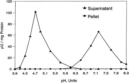
Release of neutral and acidic sialidases from erythrocyte resealed ghosts.The Materials and Methods described conditions for the treatment of various ghost preparations with B Thuringiensis PIPLC. The optimum conditions needed for PIPLC treatment were established through the preparation of resealed ghosts that maintained the sidedness of sialidases and presented the neutral enzyme in the unmasked form. As shown in Table 2, this treatment led to the release of 11.6% protein and 84% acidic sialidase into the supernatant, whereas the neutral sialidase remained totally in the pellet: no acidic sialidase could be measured in the pellet, and no neutral sialidase could be measured in the supernatant. This acidic sialidase in the supernatant was then enriched more than sevenfold, and the neutral sialidase in the pellet was enriched 1.2-fold. Figure 3 shows that, after treatment with PIPLC, the supernatant contained only the acidic sialidase, with a sharp pH optimum at 4.7, and that the pellet contained only the neutral sialidase, with optimal pH 7.2. Thus, the supernatant could be considered a crude preparation of acidic sialidase, and the pellet could be considered a crude preparation of the neutral enzyme. PIPLC treatment, followed by centrifugation, was also applied to white ghosts under the same conditions used for resealed ghosts (Table 3). As shown, the acidic sialidase was measurable only in the supernatant, where it reached a specific activity similar to that obtained with resealed ghosts. The pellet contained the neutral sialidase with a specific activity (52.8 ± 2.7 μU/mg protein) close to that displayed by resealed vesicles (56.9 ± 2.3 μU/mg protein). When the same pellet was dispersed with 40 vol of 5 mmol/L PBS-0.15 mol/L NaCl, pH 8.0, and incubated at 37°C for 40 minutes (the treatment used to prepare resealed ghosts), the specific activity of neutral sialidase increased to 63.9 ± 2.6 μU/mg protein. Interestingly, the appearance of neutral sialidase in the pellet (specific activity, 53.2 ± 3.1 μU/mg protein) was also observed in the blank mixture incubated in the absence of PIPLC, indicating that the unmasking of the neutral sialidase was presumably due to the incubation conditions (30°C at pH 7.4 for a total of 3 hours) and not to any PIPLC action.
Release of Sialidase From Human Erythrocyte Resealed Vesicles by Exhaustive Treatment With PIPLC Followed by Centrifugation
| . | Resealed Vesicles . | Supernatant . | Pellet . |
|---|---|---|---|
| Total protein (mg) | 2.31 | 0.27 | 1.87 |
| % | 100 | 11.6 | 80.8 |
| Acidic sialidase | |||
| μU/mg protein | 14.3 ± 0.7 | 103.7 ± 5.5 | ND |
| Total μU | 33.1 | 27.8 | ND |
| % | 100 | 84 | ND |
| Neutral sialidase | |||
| μU/mg protein | 56.9 ± 2.3 | ND | 66.6 ± 4.6 |
| Total μU | 131.7 | ND | 124.5 |
| % | 100 | 0 | 94.5 |
For details, see Materials and Methods. The data are the mean values of five experiments ± SD values.
Abbreviation: ND, not detectable.
Effect of pH on the sialidase activity present in the supernatant and pellet obtained after exhaustive treatment of human erythrocyte resealed ghosts with PIPLC. For details, see Materials and Methods.
Effect of pH on the sialidase activity present in the supernatant and pellet obtained after exhaustive treatment of human erythrocyte resealed ghosts with PIPLC. For details, see Materials and Methods.
Effect of Exhaustive Treatment of Human Erythrocyte White Gosts With PIPLC on the Activity of Acidic and Neutral Sialidases
| Enzyme Source and Treatment . | Acidic Sialidase ( μU/mg protein) . | Neutral Sialidase ( μU/mg protein) . |
|---|---|---|
| WG | 23.0 ± 0.9 | 1.2 ± 0.5 |
| PIPLC-treated | ||
| WG supernatant | 101.7 ± 4.7 | ND |
| WGP | ND | 52.8 ± 2.7 |
| Blank of PIPLC-treated | ||
| WG supernatant | ND | ND |
| WGP | 22.3 ± 1.1 | 53.2 ± 3.1 |
| WGP submitted to isoosmotic treatment | — | 63.9 ± 2.6 |
PIPLC treatment was followed by centrifugation. The blank mixtures of PIPLC-treated white ghosts, in which PIPLC was not present, were also analyzed. The pellet obtained after white ghosts treatment with PIPLC was submitted to isoosmotic treatment, as reported in the legend to Table 1. For details, see Materials and Methods. The data are the mean values of four to five experiments ± SD values.
Abbreviations: WG, white ghosts; WGP, white ghost pellet; ND, not detectable.
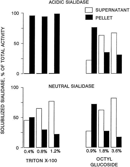
PIPLC treatment (under the conditions given above) of inside-out vesicles was not followed by any appreciable release of acidic sialidase. Conversely, PIPLC treatment of the same preparation after vesicle lysis caused the liberation of acidic sialidase. The possible efficacy of detergent treatment in releasing neutral and acidic sialidases from erythrocyte resealed ghosts was also studied following the strategy of Hooper and Turner.38 As shown in Fig 4, Triton X-100 treatment caused a substantial (72%) solubilization of neutral sialidase, with practically no release of the acidic enzyme; the best solubilizing effect was obtained at 1.2% detergent concentration. Conversely, octyl-glucoside treatment led to partial release of both sialidases; maximal release (67% of the acidic and 82% of the neutral enzyme) was obtained at the 3.6% detergent concentration.
Behavior of acidic and neutral sialidases on treatment of resealed vesicles with different detergents and subsequent centrifugation. For details, see Materials and Methods.
Behavior of acidic and neutral sialidases on treatment of resealed vesicles with different detergents and subsequent centrifugation. For details, see Materials and Methods.
Substrate specificities of neutral and acidic sialidases.Substrate specificity studies were accomplished with the crude preparations of neutral (pellet after PIPLC treatment of resealed ghosts) and acidic (supernatant after PIPLC treatment of resealed ghosts) sialidases. As shown in Table 4, the acidic sialidase acted on MU-NeuAc, GD1a, GM3, and NeuAc α2-3 lactose, but did not affect the glycoproteic substrates; conversely, the neutral enzyme affected all substrates. Acidic sialidase exhibited the highest activity on GM3 (380 μU/mg protein), followed by GD1a, MU-NeuAc, and NeuAc α2-3 lactose; the neutral sialidase was also most active on GM3 (650 μU/mg protein) and the least active on substrates of a glycoproteic nature (25 to 30 μU/mg protein).
Substrate Specificity of Acidic and Neutral Sialidases Obtained From Human Erythrocyte Membranes
| Substrate . | Apparent Vmax ( μU/mg protein) . | |
|---|---|---|
| . | Acidic Sialidase . | Neutral Sialidase . |
| Ganglioside | ||
| GD1a | 170 | 420 |
| GM3 (+0.1% Triton CF-54) | 380 | 650 |
| Glycoprotein | ||
| Fetuin | ND | 24.8 |
| α1-Acidic glycoprotein | ND | 29.8 |
| Transferrin | ND | 28.0 |
| Oligosaccharide | ||
| α-2-3 Sialyllactose | 37.1 | 33.0 |
| Artificial substrate | ||
| MU-NeuAc | 104 | 66.6 |
Enzyme source of acidic sialidase was supernatant obtained after exhaustive PIPLC treatment of resealed vesicles and centrifugation. Enzyme source of neutral sialidase was pellet obtained after exhaustive PIPLC treatment of resealed vesicles and centrifugation. For details, see Materials and Methods. The data are the mean values of five experiments ± SD values.
Abbreviation: ND, not detectable.
The action of neutral and acidic sialidases on endogenous substrates (sialoglycoproteins and gangliosides) was explored using the resealed ghosts preparation, in which both enzymes were expressed (Table 5). This preparation contained a total of 147.8 ± 8.2 (SD) nmol bound NeuAc, of which only 9.7 ± 0.5 (SD) nmol (representing approximately 6.6% of the total) was ganglioside-bound NeuAc, with the remainder being of a glycoproteic nature. After incubation under the conditions specified in the Materials and Methods, both the acidic and the neutral sialidases were able to release a substantial portion (70% and 67%, respectively) of ganglioside-bound NeuAc, whereas only the neutral enzyme appreciably (14%) degraded the sialoglycoproteins. No activity of the acidic enzyme on endogenous sialoglycoprotein could be detected.
Action of Acidic and Neutral Sialidases on Endogenous Substrates (Glycolipids and Glycoproteins)
| . | Before Incubation . | After Incubation . | |
|---|---|---|---|
| . | . | pH 4.7 . | pH 7.2 . |
| Ganglioside-bound sialic acid | 9.7 ± 0.5 | 2.9 ± 0.2 | 3.2 ± 0.1 |
| Released sialic acid (%) | 0 | 70.1 | 67 |
| Protein-bound sialic acid | 134.6 ± 7.5 | 134.8 ± 5.4 | 115.7 ± 4.2 |
| Released sialic acid (%) | 0 | 0 | 14 |
| Total sialic acid | 147.8 ± 8.2 | 141.6 ± 8.2 | 122.4 ± 6.1 |
| Released sialic acid (%) | 0 | 4.2 | 17 |
Enzyme preparation was resealed vesicles from human erythrocyte membranes. Incubation conditions was 50 mmol/L citric acid-sodium phosphate-0.15 mol/L NaCl, pH 4.7 and 7.2 (for the acidic and neutral sialidase, respectively), for 18 hours at 37°C. Data are expressed as nanomoles of total-bound, ganglioside-bound, and glycoprotein-bound sialic acid per milligram protein. The data are the mean values of five experiments ± SD values.
Measurement of sialidase activity in intact erythrocytes.The direct measurement of sialidase activity on intact erythrocytes is not an easy task, owing to the need to avoid cell lysis and the abundance of hemoglobin that can greatly affect many of the approaches used in the enzyme reaction. We circumvented most of these difficulties on the evidence that both acidic and neutral sialidases are much more active on gangliosidic substrates. On this basis, we used the very sensitive radiochemical assay method with [3H]-labeled GD1a and GM3 as substrates and, using an isoosmotic buffer, were able to reduce the incubation time to 5 hours, thus limiting erythrocyte lysis (determined as hemoglobin release in the assay mixture) to less than 2%. As shown in Table 6, sialidase activity on intact erythrocytes was measurable with both GD1a and GM3. It was twofold to threefold higher on GM3 than on GD1a and more than sevenfold higher at pH 4.7 than 7.2. It should be noted that, in the resealed ghosts, the specific activity of sialidase was markedly higher at pH 7.2 than 4.7.
Direct Assay of Sialidase on Intact Human Erythrocytes
| Substrate . | pH . | Sialidase Activity . | |||
|---|---|---|---|---|---|
| . | . | nmol Released Sialic Acid/h/μmol P . | μU/μmol P . | nmol Released Sialic Acid/1 × 107 Cells . | μU/1 × 107 Cells . |
| GD1a | 4.7 | 0.74 ± 0.05 | 12.3 ± 0.83 | 1.23 ± 0.09 | 20.5 ± 1.5 |
| GM3 | 4.7 | 1.85 ± 0.2 | 30.8 ± 3.3 | 3.0 ± 0.21 | 50.0 ± 3.3 |
| GD1a | 7.2 | 0.14 ± 0.02 | 2.3 ± 0.33 | 0.17 ± 0.02 | 2.8 ± 0.33 |
| GM3 | 7.2 | 0.39 ± 0.04 | 6.5 ± 0.66 | 0.64 ± 0.08 | 10.6 ± 1.3 |
Assays were performed at pH 4.7 and 7.2 using 25 to 40 μL of an erythrocyte suspension carrying 8.08 × 10⁶ cells/mL. The radiochemical assay method was used with [3H]GD1a and [3H]GM3 as substrates. At the end of incubation, an aliquot of the mixture was withdrawn and centrifuged, and the hemoglobin content was determined in the supernatant. The degree of hemolysis was in the range of 1.83% to 1.96%. For details, see Materials and Methods. The data are the mean values of five experiments ± SD values.
Orientation of the acidic sialidase in human erythrocyte membrane.As shown in Table 7, Ach-esterase, which is known as a typical enzyme of the external membrane surface, was, as expected, (1) fairly measurable on white and resealed ghosts, (2) present in trace amounts in inside-out vesicles, and (3) easily measurable in lysed inside-out vesicles. Furthermore, NADH-cytochrome C oxidoreductase, which is known to expose its active site on the internal membrane surface, appeared to be much more abundant in inside-out vesicles than in white and resealed ghosts and lysed inside-out vesicles. The acidic sialidase was easily measured in white and resealed ghosts, could not be detected in inside-out vesicles, and reappeared in lysed inside-out vesicles; thus, its behavior was essentially identical to that of the marker enzyme located in the external membrane surface. As already mentioned, the neutral sialidase did not appear to be expressed in the used preparation of inside-out vesicles or in the same vesicles after lysis. Of course, the content of neutral sialidase was much higher in the resealed ghosts than in the white, in which the enzyme is masked.
DISCUSSION
This report provides evidence of the occurrence of a neutral sialidase (optimum pH 7.2) in human erythrocyte plasma membrane. This new finding widens the physiologic implications of mammalian sialidases, enzymes known to occur at different subcellular levels (plasma membrane, lysosomes, and cytosol) and characterized by an acidic pH optimum.50 The data reported here (Table 6) show that intact human erythrocytes feature a major sialidase activity (assayed on gangliosides GD1a and GM3) at pH 4.7 and some minor activity, possibly the residual activity of the acidic enzyme, at pH 7.2. Also, the neutral sialidase is practically undetectable in the unsealed (white) erythrocyte ghosts that carry, as expected,17-21 the acidic sialidase. However, our treatment, which was devised to reseal ghosts,35 unmasked the neutral enzyme. In fact, it became the preponderant sialidase in the resealing preparation (Fig 1). According to the original procedure,35 the optimum conditions for resealing ghosts (proper dispersion in an isoosmotic buffer; pH 8.0; 40 minutes of incubation at 37°C) were also optimum for the full expression of neutral sialidase activity (Fig 2). It is to be noted that the conditions used to prepare inside-out vesicles from erythrocyte white ghosts did not lead to the unmasking of the neutral sialidase, even though such conditions differed from those used to prepare resealed ghosts only in temperature (4°C instead of 37°C). Instead, in the conditions used to treat white ghosts with PIPLC (incubation at 30°C, pH 7.4), the neutral sialidase was unmasked, regardless of the presence of PIPLC. This indicates that temperature is a critical factor for removing neutral sialidase crypticity. During incubation under conditions appropriate to producing resealed ghosts, small, but definite amounts of protein were released into the medium. However, when added to the formed resealed ghosts, these proteins did not influence in any ways the neutral sialidase activity (Table 1), proving that no sialidase inhibitor was released during incubation. Moreover, once the neutral sialidase had been unmasked, it remained fully active (Table 1), indicating that the enzyme gained a stable conformation. It cannot be excluded that autolysis processes occurred during incubation, leading to degradation of a putative physiologic inhibitor of the neutral sialidase, or that there was the transformation of an inactive enzyme precursor into the active enzyme.
The substrate specificity of the neutral and acidic sialidases from erythrocyte membrane was studied using the pellet and supernatant, obtained after exhaustive PIPLC treatment of resealed ghosts, as the respective sources of the enzymes (Fig 3). The specific activity enrichment was about sixfold greater for the acidic enzyme. Instead, the neutral sialidase showed broader specificity, working on the artificial substrate MU-NeuAc and sialoderivatives of a glycolipid, glycoprotein, and oligosaccharide nature; conversely, there was no measurable activity by the acidic sialidase on the sialoglycoproteins (Table 4). On the basis of the recorded values of apparent Vmax and in consideration of the higher purity of the acid sialidase preparation, the neutral sialidase appeared to be much more potent than the acidic enzyme, especially on gangliosides. By incubation under the conditions described in the experimental section (37°C for 18 hours), both the neutral and acidic sialidases were able to release about 70% of NeuAc bound to endogenous ganglioside; conversely, only the neutral enzyme affected endogenous sialoglycoproteins, causing a 14% release of protein-bound NeuAc (Table 4). Because the membrane bound endogenous gangliosides and sialoglycoproteins are known to be located in the outer membrane leaflet, the finding that they can be affected by membrane-bound sialidases strongly supports the hypothesis that these enzymes have the same topological localization as their endogenous substrates.
Indeed, the acidic sialidase was shown to expose its catalytic site on the external surface of the erythrocyte membrane (Table 7). In fact, like Ach-esterase, which is known to be located in the external membrane surface,35 the acidic sialidase was fairly measurable in the white ghosts and resealed ghosts preparations, but not in intact inside-out vesicles, although its activity became measurable after inside-out vesicle lysis. Unfortunately, this methodological approach could not be applied to directly assess the sidedness of neutral sialidase in the erythrocyte plasma membrane, because the enzyme appeared to maintain its crypticity throughout treatment. Therefore, the outer location can be cautiously suggested for the neutral sialidase on the basis of its activity on endogenous gangliosides and sialoglycoproteins. Thus, it would seem that the outer membrane surface location of human erythrocyte sialidase(s) differs from the inner surface location of the rabbit erythrocyte sialidase, as reported by Chen et al.21 This difference may well reflect species differences or may depend on the analytical conditions used (the 3-hour incubation period at pH 4.5 used by Chen et al21 to assay sialidase activity may have produced partial lysis of inside-out vesicles, thus causing exposure of sialidase). The neutral sialidase was unaffected by bacterial PIPLC (Table 2) and was efficiently solubilized by resealed ghosts treatment with different detergents (namely 1.2% Triton X-100 and 3.6% octylglucoside; Fig 4), whereas the acidic enzyme was released by PIPLC action and solubilized only by octylglucoside treatment (Fig 4), as predicted38 for protein linked to membrane by a GPI anchor. These data, together with the evidence of the different pH optima, are consistent with the notion that erythrocyte acidic and neutral sialidases are distinct entities, residing in different membrane domains possibly being of different composition and having different connections with the cytoskeleton.
On the basis of the overall evidence provided in this report, the neutral sialidase may be considered the candidate enzyme responsible for the release of sialic acid from all sialoderivatives present in the erythrocyte membrane during the physiologic aging of human erythrocytes in circulating blood. The finding that to become active the neutral sialidase requires the removal of a putative inhibitor or the occurrence of modifications in certain domains of the erythrocyte membrane focuses attention on an important, presently unknown, point: what is the molecular event that triggers the activation of neutral sialidase, thus initiating the cascade of events that leads to erythrocyte aging and eventual phagocytosis by macrophages? It could be interesting, for instance, to ascertain whether the perturbation of the erythrocyte membrane linked to the process of erythrocyte membrane vesiculation, known to involve well-defined membrane domains carrying GPI-anchored proteins and to accompany erythrocyte aging,51-53 plays a role in neutral sialidase activation.
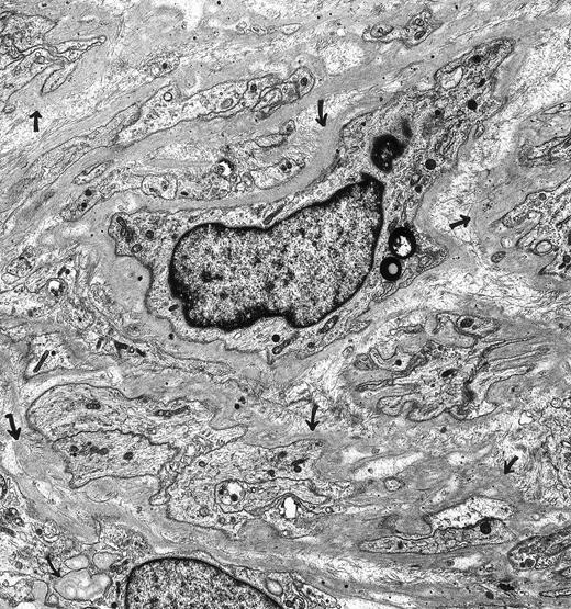
Schwannoma. A 66-year-oldman had a nonenlarging retroperitoneal mass for 3 years that was composed of Schwann cells. The spindle cells and their extensive, narrow cellular processes were encased in basal lamina (arrows). The tumor cells lacked surface vesicles, bundles of microfilaments, dense bodies and dense surface plaques—features characteristic for tumors of smooth muscle cell origin. Immunoperoxidase stains were positive for vimentin and S-100 and negative for muscle-specific actin. Original magnification × 7,500. (Courtesy of Ann M. Dvorak, MD, Department of Pathology, Beth Israel Deaconess Medical Center, 330 Brookline Ave, Boston, MA 02215.)
Schwannoma. A 66-year-oldman had a nonenlarging retroperitoneal mass for 3 years that was composed of Schwann cells. The spindle cells and their extensive, narrow cellular processes were encased in basal lamina (arrows). The tumor cells lacked surface vesicles, bundles of microfilaments, dense bodies and dense surface plaques—features characteristic for tumors of smooth muscle cell origin. Immunoperoxidase stains were positive for vimentin and S-100 and negative for muscle-specific actin. Original magnification × 7,500. (Courtesy of Ann M. Dvorak, MD, Department of Pathology, Beth Israel Deaconess Medical Center, 330 Brookline Ave, Boston, MA 02215.)
*The ganglioside nomenclature proposed by Svennerholm24was followed.
Supported in part by grants from the Italian Research Council (CNR), Target Project “Genetic Engineering” (Grant No. 95. 00666.PF99), and Strategic Project for Mezzogiorno (Grant No. CT95.04684.ST75).
Address reprint requests to Guido Tettamanti, MD, PhD, Department of Medical Chemistry and Biochemistry, The Medical School, University of Milan, Via Saldini 50, 20133 Milan, Italy.


This feature is available to Subscribers Only
Sign In or Create an Account Close Modal