Abstract
Platelet factor XI is an alternatively spliced product of the factor XI gene expressed specifically within megakaryocytes and platelets as an approximately 1.9-kb mRNA transcript (compared with ∼2.1 kb in liver cells) lacking exon V. Flow cytometry with an affinity-purified factor XI antibody, with PAC1 antibody (to the GPIIb/IIIa complex on activated platelets), and with S12 antibody (to P-selectin, an α-granule membrane protein expressed on the platelet surface during secretion) on platelets activated with ADP, thrombin, thrombin receptor peptide (SFLLRN amide), or collagen at various concentrations exposed platelet factor XI and PAC1 antibody binding in parallel. Unactivated platelets expressed approximately 40% of total platelet factor XI but no PAC1 binding sites. Enhanced membrane exposure of platelet factor XI is independent of α-granule secretion, because ADP and collagen exposed platelet factor XI but no S12 binding sites. Platelets from four patients with plasma factor XI deficiency (<0.04 U/mL) had normal constitutive and activation-dependent expression of platelet factor XI. Well-washed platelets from normal and from factor XI-deficient donors incubated with low concentrations of thrombin (0.05 to 0.1 U/mL) corrected the clotting defect observed with factor XI-deficient plasma. Thus, functionally active platelet factor XI is differentially expressed on platelet membranes in a tissue-specific manner both constitutively and in a concentration-dependent fashion by various agonists in the absence of detectable plasma factor XI.
COAGULATION FACTOR XI is a glycoprotein (GP) present in plasma as a zymogen and is required for normal hemostasis.1,2 Factor XI circulates in plasma noncovalently bound to high molecular weight kininogen.1 When a surface-associated complex of factor XI, high molecular weight kininogen, and factor XIIa is formed, it results in the formation of factor XIa.2 Human platelets participate in the interaction of intrinsic coagulation proteins, including factor XII, high molecular weight kininogen, and factor XI.3-5 However, in addition to providing a surface that promotes coagulation, platelets also contain a number of coagulation proteins, including fibrinogen, factor V, high molecular weight kininogen, and factor XI.6
Previous studies have demonstrated that factor XI is present in platelets3,4,7-10 in a form that is structurally different from plasma factor XI and that is referred to as platelet factor XI. Platelet factor XI migrates by sodium dodecyl sulfate-polyacrylamide gel electrophoresis (SDS-PAGE) with an apparent molecular weight (Mr) of 220 kD (55 kD after reduction),7-9 compared with plasma factor XI, which has a Mr of 160 kD and a subunit with an Mr of 80 kD. Factor XI coagulant activity and antigen in well-washed platelet suspensions constitutes about 0.5% of the factor XI activity in normal plasma.4,7-9,11,12 Assuming a specific activity similar to that of plasma factor XI, this represents approximately 300 molecules of platelet factor XI per platelet. The physiologic role and importance of platelet factor XI is unknown, but the fact that factor XI activity is associated with the platelet plasma membrane7 suggests the possibility that the protein can participate in blood coagulation. Moreover, the observation that a few patients with severe plasma factor XI deficiency but no evidence of hemostatic abnormality have normal amounts of platelet factor XI suggests that platelet factor XI may substitute for plasma factor XI in hemostasis.7-10 This suggestion is supported by reports of several hemostatically abnormal patients with no detectable factor XI either in plasma or platelets.4,13 Confirmation of the existence and biochemical nature of platelet factor XI has come from recent studies in our laboratory (Hsu et al, manuscript submitted) that demonstrate that platelet factor XI is an alternatively spliced form of the factor XI gene, lacking exon V and expressed specifically within megakaryocytes.14
To determine the subcellular localization of expressed platelet factor XI and to analyze the mechanisms of exposure of platelet factor XI in response to a variety of agonists, we have developed a flow cytometric assay using an affinity-purified antihuman plasma factor XI antibody. For these studies, we have used two additional antibodies, PAC1 and S12. PAC1 is a monoclonal IgM antibody that binds only to the activated form of the GPIIb/IIIa complex and recognizes an epitope on the GPIIb/IIIa complex critical for fibrinogen binding.15 The S12 antibody recognizes a 140-kD α-granule membrane protein16 P-selectin17 (previously known as GMP-140 or PADGEM) that becomes associated with the platelet surface during secretion. Our studies indicate that platelet factor XI is expressed constitutively on unactivated platelets from both normal donors and donors with severe plasma factor XI deficiency and that the exposure of platelet factor XI is increased by various platelet agonists in a concentration-dependent manner.
MATERIALS AND METHODS
Materials.
Fluorescein-5-isothiocyanate (FITC), Sephadex G-25 (PD10) gel filtration resin, CNBr-activated Sepharose 4B, p-nitrophenylphosphate, alkaline-phosphatase–conjugated rabbit antigoat IgG, apyrase, prostaglandin E1, and bovine thrombin were purchased from Sigma Chemical Co (St Louis, MO). S12 antibody was purchased from Centocor Inc (Malvern, PA). FITC-labeled PAC1 antibody was purchased from the Cell Center of the University of Pennsylvania (Philadelphia, PA). Plasma factor XI was purchased from Enzyme Research Laboratories (South Bend, IN). Nitro-blue tetrazolium chloride and 5-bromo-4-chloro-3′-indolylphosphate p-toluidine salt (NBT/BCIT) phosphatase substrate were purchased from Kirkegaard & Perry Laboratories, Inc (Gaithersburg, MD). D-Phe-Pro-Arg chloromethyl ketone (PPACK) was purchased from Calbiochem-Novabiochem Corp (La Jolla, CA). Collagen (type 1) was purchased from Chrono-Log Corp (Havertown, PA). FITC-conjugated F(ab′)2 fragment rabbit antigoat IgG antibody was purchased from Accurate Chemical & Scientific Corp (Westbury, NY). Thrombin receptor peptide (TRP; SFLLRN amide) was synthesized using 9-fluorenylmethyloxycarbonyl (FMOC) chemistry on a Applied Biosystems 430A Synthesizer (Applied Biosystems, Foster City, CA) and reverse-phase high-performance liquid chromatography (HPLC) purified to greater than 99.9% homogeneity.
Fluorescence labeling of S12 antibody.
The labeling procedure was modified according to the method of Dachary-Prigent et al.16 In brief, the purified S12 antibody was dialyzed overnight at 4°C against 20 mmol/L borate buffer, pH 9.5, containing 150 mmol/L NaCl. S12 was incubated at room temperature for 4.5 hours with FITC at a 15 to 1 molar ratio of FITC to antibody. FITC-labeled S12 was separated from unbound FITC by gel filtration on a Sephadex G-25 gel filtration column.
Affinity purification of goat antihuman plasma factor XI antibody.
Antihuman plasma factor XI antibody was prepared by Hugh Hoogendoorn and Alan R. Giles (Queen's University, Kingston, Ontario, Canada) in a goat injected with highly purified human plasma factor XI prepared in our laboratory as previously described.18 The antibody was partially purified by caprylic acid precipitation and ammonium sulfate precipitation. The antibody was then affinity-purified using a column of human factor XI coupled to CNBr-activated Sepharose 4B, according to the procedure described by the manufacturer (Pharmacia LKB Biotechnology, Uppsala, Sweden).
Titration of factor XI antibody using enzyme-linked immunosorbent assay (ELISA).
A 96-well microtiter plate (Falcon, Lincoln Park, NJ) was coated with purified plasma factor XI in 15 mmol/L Na2CO3, 35 mmol/L NaHCO3 buffer, pH 9.6. The wells were then incubated overnight at 4°C with 3% bovine serum albumin–phosphate-buffered saline (BSA-PBS) to block nonspecific binding. After incubation with various concentrations of either preaffinity-purified antibody, affinity-purified antibody, or preimmune goat IgG, the plate was washed with PBS containing 0.02% Tween 20. Alkaline phosphatase-conjugated rabbit antigoat IgG was placed in each well. After incubation and three washes, substrate solution was added. After incubation at room temperature for a suitable time period, the reaction was stopped by the addition of 3 mol/L NaOH, and absorption at 405 nm was read on a microplate reader (Thermo Max; Molecular Devices, Menlo Park, CA).
SDS-PAGE and Western blot.
PAGE of plasma was performed according to the procedure of Laemmli.19 The electrophoretic transfer to polyvinylidene difluoride (PVDF; Millipore Corp, Bedford, MA) membrane was performed using a Transphor apparatus (Model TE 50; Hoefer Scientific Instruments, San Francisco, CA). The membrane strips were incubated with anti-factor XI antibody, washed, incubated with alkaline phosphatase-conjugated rabbit antigoat IgG, and developed with NBT/BCIT substrate.
Blood collection and preparation of platelets.
Fresh blood was obtained from healthy volunteers or factor XI-deficient patients and anticoagulated with acid-citrate-dextrose (ACD) solution (2.5% trisodium citrate, 1.5% citric acid, 2.0% dextrose; 1 part anticoagulant:6 parts blood). Platelet-rich plasma (PRP) was prepared by centrifugation at 180g for 20 minutes at room temperature. Apyrase (1 U/mL), PPACK (20 nmol/L), and prostaglandin E1 (PGE1; 1 μmol/L) were added to PRP that was then incubated at room temperature for 15 minutes. PRP was gel-filtered on Sepharose CL-2B (Sigma Chemical Corp) equilibrated in HEPES-Tyrode's buffer (126 mmol/L NaCl, 2.7 mmol/L KCl, 1 mmol/L MgCl2, 0.38 mmol/L NaH2PO4, 5.6 mmol/L dextrose, 15 mmol/L HEPES, pH 7.4) containing 2 mg of BSA per milliliter. Platelets eluting in the void volume were adjusted to 2 × 10⁸/mL in the same buffer.
Platelet activation and fixation.
Gel-filtered platelet suspensions containing 2 mmol/L CaCl2were incubated without stirring for 3 minutes at 37°C in the presence of one of the following agonists: thrombin (0.2 to 10 nmol/L), SFLLRN amide (0.2 to 10 μmol/L), ADP (1 to 15 μmol/L), and collagen (2.5 to 50 μg/mL). Activated platelets were then added to an equal volume of 2% formalin buffered with PBS and incubated for 30 minutes at 37°C. Fixed platelets were washed three times with PBS-1% BSA and stored in the same buffer at 4°C until assay. The concentration of fixed platelets was adjusted to 1 × 10⁸/mL.
The measurement of platelet factor XI on platelets and the binding of FITC-S12, FITC-PAC1 to platelets using flow cytometric assay.
Twenty-five microliters of fixed platelets was mixed with 25 μL of affinity-purified goat antihuman factor XI antibody or preimmune goat IgG, respectively, at a final concentration of 200 μg/mL and incubated for 60 minutes at 4°C. After being washed twice, platelets were incubated with FITC-conjugated F(ab′)2fragment rabbit antigoat IgG at a final concentration of 5 μg/mL for 60 minutes at 4°C. Platelets were then washed twice and suspended in 0.4 mL of 1% BSA-PBS for flow cytometry.
Ten microliters of a fixed platelet suspension containing 1 × 10⁶ platelets was incubated at room temperature with 40 μL of FITC-S12 (20 μg/mL) or 40 μL of FITC-PAC1 (100 μg/mL) for 30 minutes and then diluted with 0.35 mL of 1% BSA-PBS for flow cytometry.
The platelet samples were analyzed in a Coulter Epics Elite Flow Cytometer (Coulter Corp, Miami, FL) equipped with a Coherent Innova 300 Argon laser that was calibrated daily with 2-μm calibrate beads (Coulter Corp).
Coagulation assays.
One hundred microliters of factor XI-deficient plasma or normal plasma was incubated with either 50 μL of 30 μmol/L phosphatidylserine and L-α-dioleoyl-phosphatidylcholine (1:3 molar ratio) from bovine brain in 20 mmol/L PBS or washed platelets (50 μL, 2 × 10⁸ platelets/mL) and 50 μL of CaCl2 (25 mmol/L). In some assays, goat antihuman factor XI was added (14.6 μmol/L) without preincubation. Thereafter, thrombin at various concentrations was added to initiate clot formation. In the absence of added thrombin, the clotting times of all samples were greater than 5 minutes.
Patients and normal donors.
Four hemostatically normal donors and four patients with moderately severe plasma factor XI deficiency (<0.04 U/mL) were studied, none of whom had received drugs known to affect platelet function for 2 weeks before blood donation. Clinical data were obtained and evaluations of factor XI-deficient patients were performed by an individual (B.A.K.) without knowledge of the results of the studies. The studies were performed without knowledge of the clinical data or evaluations. The factor XI-deficient patients were females of Ashkenazi Jewish extraction; were 41, 77, 37, and 59 years of age; had plasma factor XI levels of 0.02, <0.01, 0.02, and 0.03 U/mL; and had plasma partial thromboplastin times (normal, 25 to 35 seconds) of 68.2, 91.1, 70.5, and 55 seconds, respectively. All were documented to have no evidence of a factor XI inhibitor, and prothrombin times and other coagulation assays and platelet counts were normal. Two of the four patients gave a history of easy bruising and two gave a history of melena (1 on one occasion and 1 recurrently) in the past, but none had experienced epistaxis, hematuria, menorrhagia, hematemesis, muscle hematomata, central nervous system bleeding, or hemarthrosis. Each of the four factor XI-deficient patients had experienced surgical operations and/or traumatic injury: one had abdominal surgery with prophylactic factor XI replacement (plasma) without excessive bleeding; one had multiple dental extractions without prophylaxis or excessive bleeding and both a hysterectomy and surgical repair of facial trauma, both with prophylaxis and both without excessive bleeding; one had experienced mild excessive bleeding, in no instance requiring either prophylactic or therapeutic factor XI or blood replacement therapy, after a hysterectomy, after a breast biopsy and after trauma associated with an automobile accident; and one had experienced no excessive bleeding and no prophylactic factor XI replacement therapy after a tonsillectomy and after a breast biopsy.
RESULTS
Characterization of affinity-purified goat anti-factor XI antibody.
Our initial studies were aimed at preparing, purifying, and characterizing a monospecific polyclonal anti-factor XI antibody. An ELISA was performed in which the titer of affinity-purified antibody was increased greater than sixfold compared with preaffinity-purified antibody. It was also confirmed by the Western blot that the titer of goat anti-factor XI antibody was enhanced greater than sixfold after affinity purification (data not shown).
The specificity of affinity-purified goat antiplasma factor XI was further determined by Western blot. Normal pooled plasma or factor XI-deficient plasma was subjected to SDS-PAGE under either reducing or nonreducing conditions. After electrophoretic transfer, the PVDF membrane, probed with anti-factor XI antibody, showed a single band at a molecular weight of approximately 160 kD (nonreduced), which migrated at approximately 80 kD under reduced conditions (data not shown). No visible bands were detected in lanes to which factor XI-deficient plasma was applied. These results indicate that the affinity-purified anti-factor XI antibody is monospecific.
Comparison of the exposure of platelet factor XI, GPIIb/IIIa, and P-selectin on platelets.
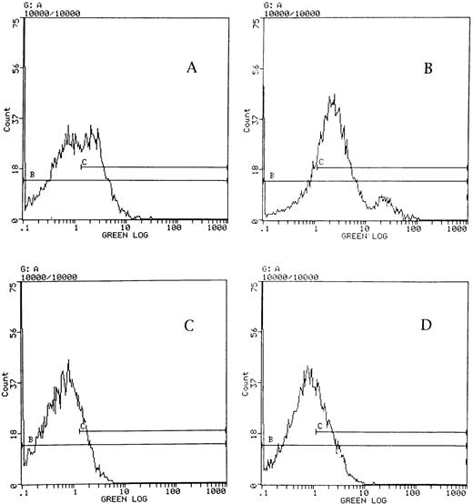
To detect platelet factor XI on the surface of normal platelets, unactivated platelets and platelets activated by various concentrations of thrombin, thrombin receptor peptide, ADP, or collagen were incubated with affinity-purified goat anti-factor XI antibody or with preimmune goat IgG as a control. After incubation with FITC-F(ab′)2 fragment rabbit antigoat IgG antibody, flow cytometry was performed. Figure 1shows representative fluorescence histograms of affinity-purified anti-factor XI antibody binding to unactivated platelets (Fig 1A) and to platelets activated by 10 μmol/L thrombin receptor peptide (Fig1B) and of preimmune goat IgG nonspecifically bound to unactivated (Fig1C) and activated platelets (Fig 1D). Shown in Table 1 are the numerical data obtained for analysis of this representative example. The log mean fluorescence intensity of the sample incubated with affinity-purified anti-factor XI antibody minus that of the sample incubated with preimmune goat IgG is the net log mean fluorescence intensity associated with anti-factor XI antibody bound to platelet factor XI on platelets. The total platelet factor XI exposure (100%) is defined as the net log mean fluorescence intensity of platelets activated by 10 μmol/L thrombin receptor peptide. As shown in Table 1, these values for one representative normal sample are 1.64 fluorescence intensity units for activated platelets (100%) and 0.42 fluorescence units (25.6%) for unactivated platelets. It is clear from Fig 1A that unactivated platelets represent a bimodal distribution of platelets with 43% of the platelets present in the C gate (ie, positive for binding factor XI antibody) showing a log mean fluorescence intensity of approximately 2.8 fluorescence intensity units, whereas the remainder represent a population of platelets that are negative for factor-XI antibody binding, displaying a log mean fluorescence intensity of approximately 0.5 fluorescence intensity units. A small proportion of activated platelets (<10%) showed high levels of factor XI expression (log mean fluorescence intensity, ∼25 units) in this experiment, but this was not a consistent finding.
Flow cytometric assay for the detection of platelet factor XI on the surface of normal platelets. The experiment was performed as described in the Materials and Methods. The Y axis displays the number of platelets at any specific fluorescence intensity noted on the X axis as a log scale. The C gate was set at the edge of the background of platelet fluorescence intensity without adding any primary antibody or preimmune goat IgG. A representative result is shown, in which affinity-purified anti-factor XI antibody was bound to unactivated platelets (A) and to platelets activated by 10 μmol/L thrombin receptor peptide (B). Results in (C) depict preimmune goat IgG nonspecifically bound to unactivated and (D) to activated platelets. The mean fluorescence intensity in (A) minus that in (C) or (B) minus (D), respectively, represent the net mean fluorescence intensity due to anti-factor XI antibody binding to platelet factor XI.
Flow cytometric assay for the detection of platelet factor XI on the surface of normal platelets. The experiment was performed as described in the Materials and Methods. The Y axis displays the number of platelets at any specific fluorescence intensity noted on the X axis as a log scale. The C gate was set at the edge of the background of platelet fluorescence intensity without adding any primary antibody or preimmune goat IgG. A representative result is shown, in which affinity-purified anti-factor XI antibody was bound to unactivated platelets (A) and to platelets activated by 10 μmol/L thrombin receptor peptide (B). Results in (C) depict preimmune goat IgG nonspecifically bound to unactivated and (D) to activated platelets. The mean fluorescence intensity in (A) minus that in (C) or (B) minus (D), respectively, represent the net mean fluorescence intensity due to anti-factor XI antibody binding to platelet factor XI.
Detection of Platelet Factor XI by Flow Cytometry in a Normal Platelet Suspension
| . | Log Mean Fluorescence Intensity* . | |||
|---|---|---|---|---|
| IgG . | Anti- Factor XI-Ab . | Difference (Ab-IgG) . | % . | |
| Unactivated platelets | 0.54 | 0.96 | 0.42 | 25.6 |
| Activated platelets | 0.74 | 2.38 | 1.64 | 100.0 |
| . | Log Mean Fluorescence Intensity* . | |||
|---|---|---|---|---|
| IgG . | Anti- Factor XI-Ab . | Difference (Ab-IgG) . | % . | |
| Unactivated platelets | 0.54 | 0.96 | 0.42 | 25.6 |
| Activated platelets | 0.74 | 2.38 | 1.64 | 100.0 |
*The numbers shown are fluorescence intensity units.
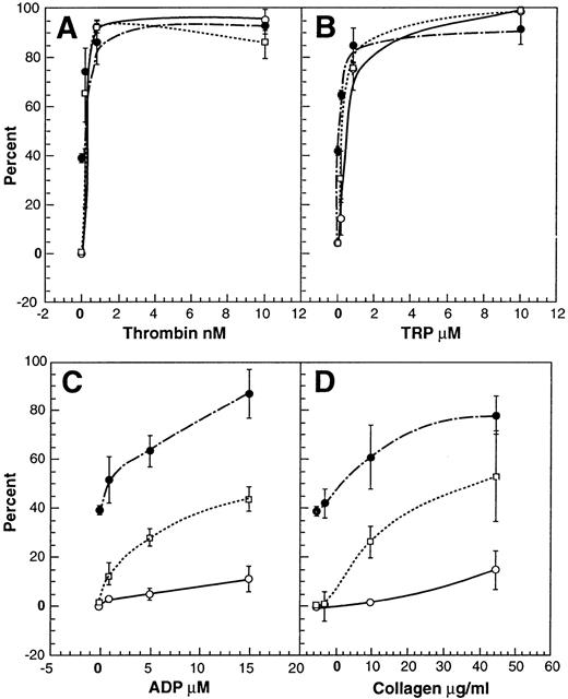
Unactivated platelets expressed a mean of 39.07% (1.91% SEM; n = 4; Fig 2) of total platelet factor XI. The exposure of platelet factor XI on platelets was increased with increasing concentrations of thrombin, thrombin receptor peptide, ADP, and collagen (Fig 2). Concentrations of 0.8 nmol/L (∼0.1 U/mL) thrombin, 0.8 μmol/L thrombin receptor peptide, 15 μmol/L ADP, or 50 μg/mL of collagen stimulated exposure of platelet factor XI almost as great as that of 10 μmol/L thrombin receptor peptide. These results indicate that platelet factor XI is expressed constitutively on unactivated platelets and that the expression of platelet factor XI on activated platelets is increased by various platelet agonists in a concentration-dependent manner.
Comparison of the exposure of platelet factor XI, GPIIb/IIIa, and P-selectin on platelets. One hundred percent binding of anti-factor XI antibody, FITC-PAC1, or FITC-S12 to platelets was defined as the mean fluorescence intensity of platelets activated by 10 μmol/L thrombin receptor peptide (SFLLRN amide) affinity-purified factor XI antibody and incubated with PAC1 antibody as described in the Materials and Methods. The results shown are the means (±SEM) of data obtained with platelets from four normal donors probed for platelet factor XI (•), GPIIb/IIIa (□), or P-selectin (○) .
Comparison of the exposure of platelet factor XI, GPIIb/IIIa, and P-selectin on platelets. One hundred percent binding of anti-factor XI antibody, FITC-PAC1, or FITC-S12 to platelets was defined as the mean fluorescence intensity of platelets activated by 10 μmol/L thrombin receptor peptide (SFLLRN amide) affinity-purified factor XI antibody and incubated with PAC1 antibody as described in the Materials and Methods. The results shown are the means (±SEM) of data obtained with platelets from four normal donors probed for platelet factor XI (•), GPIIb/IIIa (□), or P-selectin (○) .
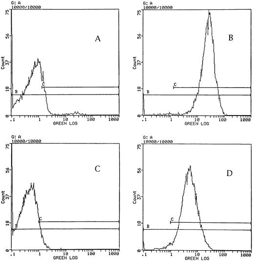
To determine the subcellular localization of platelet factor XI in platelets, two activation-dependent monoclonal antibodies, PAC1 and S12, were used in parallel with anti-factor XI antibody to examine both unactivated platelets and platelets activated by various agonists. Figure 3 shows the binding of PAC1 (Fig 3A and B) and S12 (Fig 3C and D) to unactivated platelets (Fig 3A and C) and to platelets activated by 10 μmol/L thrombin receptor peptide (Fig 3B and D). Whereas 39% of total platelet factor XI was expressed on unactivated platelets, no detectable PAC1 or S12 binding to unactivated platelets was observed (Fig 2), confirming that neither the GPIIb/IIIa epitope for PAC1 nor the P-selectin epitope for S12 is exposed on the surface of unactivated platelets. With increasing concentrations of thrombin, thrombin receptor peptide, ADP, and collagen, close correlations were found between the exposure of platelet factor XI and the expression of GPIIb/IIIa (fibrinogen binding site) indicated by binding of PAC1 (Fig 2). In contrast very low levels of exposure of P-selectin were observed in unstirred platelet suspensions incubated with ADP and collagen (Fig 2C and D), in agreement with similar observations of Shattil et al15 and with the conclusion that “unstirred platelets stimulated with weak agonists undergo minimal secretion yet are capable of expressing most of their fibrinogen receptors.”15,20 Under these conditions, concentrations of ADP and collagen shown to be optimal for the exposure of platelet factor XI caused no significant exposure of S12 binding sites (Fig 2), thereby indicating that the enhanced exposure of platelet factor XI is independent of α-granule secretion.
Flow cytometric assay for the detection of GPIIb/IIIa or P-selectin in activated platelets using PAC1 and S12 antibodies. Two FITC-labeled activation-dependent monoclonal antibodies, PAC1 (A and B) or S12 (C and D), were incubated with unactivated platelets (A and C) or to platelets activated by 10 μmol/L thrombin receptor peptide (B and C), and flow cytometry was performed as described in the Materials and Methods. The Y axis displays the number of platelets at any specific fluorescence intensity noted on the X axis as a log scale. The C gate was set at the edge of the background of platelet fluorescence intensity without adding any antibody. The maximal binding of PAC1 or S12 to platelets was defined as the mean fluorescence intensity of platelets activated by 10 μmol/L thrombin receptor peptide and incubated with FITC-PAC1 (80 μg/mL) or FITC-S12 (16 μg/mL). The mean fluorescence intensity obtained in the absence of antibody was similar to that shown in (A) and (C) and was subtracted from that obtained in (B) and (D) to obtain the net mean fluorescence intensity due to PAC1 binding to GPIIb/IIIa or S12 binding to P-selectin on activated platelets.
Flow cytometric assay for the detection of GPIIb/IIIa or P-selectin in activated platelets using PAC1 and S12 antibodies. Two FITC-labeled activation-dependent monoclonal antibodies, PAC1 (A and B) or S12 (C and D), were incubated with unactivated platelets (A and C) or to platelets activated by 10 μmol/L thrombin receptor peptide (B and C), and flow cytometry was performed as described in the Materials and Methods. The Y axis displays the number of platelets at any specific fluorescence intensity noted on the X axis as a log scale. The C gate was set at the edge of the background of platelet fluorescence intensity without adding any antibody. The maximal binding of PAC1 or S12 to platelets was defined as the mean fluorescence intensity of platelets activated by 10 μmol/L thrombin receptor peptide and incubated with FITC-PAC1 (80 μg/mL) or FITC-S12 (16 μg/mL). The mean fluorescence intensity obtained in the absence of antibody was similar to that shown in (A) and (C) and was subtracted from that obtained in (B) and (D) to obtain the net mean fluorescence intensity due to PAC1 binding to GPIIb/IIIa or S12 binding to P-selectin on activated platelets.
Platelet factor XI in platelets from factor XI-deficient donors.
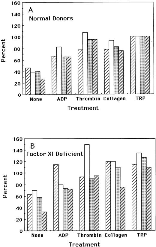
Because previous studies from our laboratory have shown that the platelets from selected patients with severe plasma factor XI deficiency possess normal quantities of platelet factor XI,7 8 we selected four normal donors and four patients with severe plasma factor XI deficiency to study constitutive and agonist-dependent exposure of platelet factor XI by flow cytometry. The results (Fig 4) show normal constitutive expression of platelet factor XI (∼30% to 60% of total) in the plasma factor XI-deficient subjects, compared with the normal subjects (∼30% to 50% of total) and normal responses of the patients' platelets to high concentrations of thrombin (100 nmol/L), thrombin receptor peptide (SFLLRN amide, 10 μmol/L), ADP (100 μmol/L), and collagen (50 μg/mL) in the agonist-dependent exposure of platelet factor XI. Platelets obtained from both normal and factor XI-deficient platelets also had normal responses to all agonists in exposure of GPIIb/IIIa and P-selectin in flow cytometry performed with PAC-1 and S12 antibodies (data not shown).
Effects of platelet agonists on the exposure of platelet factor XI in normal and factor XI-deficient donors. Four normal donors and four patients with plasma factor XI deficiency were tested for the exposure of platelet factor XI on unactivated platelets and on platelets activated by ADP, thrombin, collagen, or TRP as described in the Materials and Methods. The percentage of exposure of platelet factor XI was calculated as described in the text and in the legends to Figs 1 and 3. The total exposure (100%) of platelet factor XI was defined as the net mean fluorescence intensity of normal platelets activated with 10 μmol/L TRP. (A) depicts the mean percentage of platelet factor XI exposure in four normal platelets, whereas (B) depicts results obtained with platelets from four patients with plasma factor XI deficiency.
Effects of platelet agonists on the exposure of platelet factor XI in normal and factor XI-deficient donors. Four normal donors and four patients with plasma factor XI deficiency were tested for the exposure of platelet factor XI on unactivated platelets and on platelets activated by ADP, thrombin, collagen, or TRP as described in the Materials and Methods. The percentage of exposure of platelet factor XI was calculated as described in the text and in the legends to Figs 1 and 3. The total exposure (100%) of platelet factor XI was defined as the net mean fluorescence intensity of normal platelets activated with 10 μmol/L TRP. (A) depicts the mean percentage of platelet factor XI exposure in four normal platelets, whereas (B) depicts results obtained with platelets from four patients with plasma factor XI deficiency.
Functional assay of platelet factor XI.
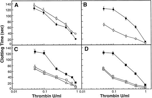
To establish a functional assay for platelet factor XI, normal or factor XI-deficient plasma was titrated with various concentrations of thrombin in the presence of either phospholipids or platelets (Fig 5). In the presence of phospholipids and in the absence of factor XI a prolongation of clotting time at low concentrations of thrombin (0.05 to 0.5 U/mL) is apparent (Fig 5A) compared with results in the presence of factor XI (Fig 5B). This difference is reduced or eliminated at high concentrations of thrombin due to the direct conversion of fibrinogen to fibrin by thrombin. The conclusion that the procoagulant effect of low concentrations of thrombin is factor XI-dependent is confirmed by the observation that the affinity-purified factor XI antibody prolongs the clotting times of normal plasma to levels observed with factor XI-deficient plasma (Fig5A and B). When normal platelets (activated with the thrombin receptor peptide) are substituted for phospholipids in factor XI-deficient plasma (Fig 5C), the clotting times at low concentrations of thrombin were indistinguishable from those obtained in normal plasma (Fig 5D), indicating that the amount of platelet factor XI added with activated platelets at a concentration similar to that in plasma (200,000 platelets/μL) is sufficient to correct the clotting defect of factor XI-deficient plasma observed with low concentrations of thrombin. The conclusion that this correction by platelets of the defect in factor XI-deficient plasma (Fig 5C) is due to platelet factor XI (and not some other coagulant activity of platelets) is confirmed by the observation that the defect is reproduced in the presence of activated platelets by the affinity-purified anti-factor XI antibody (Fig 5C and D). Finally, platelets obtained from the four factor XI-deficient donors were compared with normal platelets and found to give identical results (Fig5C and D), from which it can be concluded that the platelets of these four patients with less than 0.04 U/mL of plasma factor XI have normal amounts of platelet factor XI.
Effects of platelets and factor XI in thrombin-activated coagulation assays. Coagulation assays were performed as described in the Materials and Methods. Either factor XI-deficient plasma (A and C) or normal plasma (B and D) was incubated with phospholipid vesicles (PS:PC 1:3 ratio, 10 μmol/L; A and B) or gel-filtered platelets (200,000 platelets/μL; C and D) activated with the thrombin receptor peptide, SFLLRN-amide (5 μmol/L), and CaCl2 (5 mmol/L). In some assays, affinity-purified goat antihuman factor XI (14.6 μmol/L) was added, followed by thrombin at various concentrations (0.05, 0.1, 0.25, 0.5, and 1.0 U/mL) to initiate clot formation. Data shown are the means (±SEM) for four experiments, each performed in triplicate. In the absence of added thrombin, the clotting times of all samples were greater than 5 minutes. (A) Factor XI-deficient plasma and phospholipid vesicles in the absence (○) or presence (•) of anti-factor XI antibody. (B) Normal plasma and phospholipid vesicles in the absence (○) or presence (•) of anti-factor XI antibody. (C) Factor XI-deficient plasma and normal activated platelets (□) or platelets from factor XI-deficient patients (▵) or normal platelets in the presence of anti-factor antibody (▪). (D) Normal plasma and normal activated platelets (□) or platelets from factor XI-deficient patients (▵) or normal platelets in the presence of anti-factor XI antibody (▪).
Effects of platelets and factor XI in thrombin-activated coagulation assays. Coagulation assays were performed as described in the Materials and Methods. Either factor XI-deficient plasma (A and C) or normal plasma (B and D) was incubated with phospholipid vesicles (PS:PC 1:3 ratio, 10 μmol/L; A and B) or gel-filtered platelets (200,000 platelets/μL; C and D) activated with the thrombin receptor peptide, SFLLRN-amide (5 μmol/L), and CaCl2 (5 mmol/L). In some assays, affinity-purified goat antihuman factor XI (14.6 μmol/L) was added, followed by thrombin at various concentrations (0.05, 0.1, 0.25, 0.5, and 1.0 U/mL) to initiate clot formation. Data shown are the means (±SEM) for four experiments, each performed in triplicate. In the absence of added thrombin, the clotting times of all samples were greater than 5 minutes. (A) Factor XI-deficient plasma and phospholipid vesicles in the absence (○) or presence (•) of anti-factor XI antibody. (B) Normal plasma and phospholipid vesicles in the absence (○) or presence (•) of anti-factor XI antibody. (C) Factor XI-deficient plasma and normal activated platelets (□) or platelets from factor XI-deficient patients (▵) or normal platelets in the presence of anti-factor antibody (▪). (D) Normal plasma and normal activated platelets (□) or platelets from factor XI-deficient patients (▵) or normal platelets in the presence of anti-factor XI antibody (▪).
DISCUSSION
Early evidence for the presence of a contact factor associated with washed platelets21-23 was subsequently reinterpreted as evidence of platelet-associated factor XI activity7,11-13,24-26 and antigen.8,25 Platelet subcellular fractionation studies demonstrated that platelet factor XI is associated with the platelet plasma membrane,7 and three separate groups8-10 have immunoprecipitated and partially purified a protein from platelets with factor XI activity, having an Mr of approximately 220,000 to 240,000 (nonreduced) and Mr of approximately 50,000 to 55,000 (reduced). Despite the consistency of these observations, questions about the existence, biochemical nature, and functional significance of platelet factor XI have remained.6 The possibility that platelet factor XI may have a role in the maintenance of normal hemostasis and may substitute for plasma factor XI has been suggested4,6-11,25,27 on the basis of previous reports of small numbers of selected patients with no detectable plasma factor XI and normal hemostasis with normal amounts of platelet factor XI7,25 and reports of hemostatically abnormal factor XI-deficient patients whose platelets contained no detectable factor XI activity.4 13
Questions about the existence and biochemical nature of platelet factor XI have recently been addressed in our laboratory by the use of the polymerase chain reaction with reverse transcriptase to amplify 12 of 13 exons coding for mature plasma factor XI (ie, exons III-IV and VI-XV, lacking exon V) from platelet mRNA with sequences identical to those encoding plasma factor XI.14 Northern hybridization demonstrated the presence of a factor XI mRNA transcript of approximately 1.9 kb in Meg-01 cells (compared with ∼2.1 kb in liver cells), and in situ amplification and hybridization confirmed the presence of factor XI mRNA lacking exon V in platelets but not other blood cells.28 29 Finally, we have cloned a full-length sequence of platelet factor XI from a cDNA library of a human megakaryoblastic cell line (CHRF-288-11), confirming the conclusion that platelets but not other blood cells contain an alternatively spliced product of the factor XI gene expressed in megakaryocytes that encodes a platelet protein lacking the aminoterminal portion of the Apple 2 domain encoded by exon V (Hsu et al, manuscript submitted).
The purpose of the present studies was to determine the mechanisms and site of exposure of platelet factor XI and to determine whether the expression of functional platelet factor XI can occur in the absence of plasma factor XI. Our results demonstrate that platelet factor XI is constitutively expressed on the surface of unactivated platelets and that, in response to a variety of physiologically relevant platelet agonists (thrombin, thrombin receptor peptide, ADP, and collagen), platelet factor XI is further exposed in an agonist concentration-dependent manner in parallel with the expression of the platelet membrane protein, GPIIb/IIIa, but not in parallel with the α-granule protein, P-selectin. These observations are consistent with our previous demonstration that platelet factor XI is a platelet-plasma membrane-associated protein that is expressed in functional form on the surface of both unactivated and activated platelets.7
To determine whether the platelet factor XI expressed on the platelet surface is functionally active, we have developed a coagulation assay in which normal or factor XI-deficient plasma is titrated with low concentrations of thrombin in the presence or absence of platelets (Fig5). The results demonstrate that activated normal platelets correct the coagulation defect of factor XI-deficient plasma when trace concentrations of thrombin are added to activated platelet factor XI. The concentration of platelets added in this assay is similar to that present in normal blood (∼2 × 10⁸/mL), from which it can be concluded that the amount of platelet factor XI present in normal blood is sufficient to correct the coagulation defect of factor XI-deficient plasma observed with low concentrations of thrombin. This is especially interesting, because it is estimated that, because activated platelets contain approximately 2.5 U of factor XI activity per 1011 platelets,7 the final concentration of factor XI added with 200,000 platelets per milliliter is only 0.5% of the factor XI present in normal plasma. Thus, only a trace of platelet factor XI appears to be required to correct the factor XI-dependent functional defect observed with low concentrations of thrombin (0.05 to 0.5 U/mL).
To address the question of whether the tissue (ie, platelet)-specific expression of platelet factor XI is independent of the expression of factor XI in plasma, we have studied the platelets of patients with severe plasma factor XI deficiency using both flow cytometry and our functional assay that examines the capacity of low concentration of thrombin to activate factor XI in plasma. The results of both assays demonstrate that both platelet factor XI antigen and activity are present in the platelets of four unrelated patients with moderately severe (<0.04 U/mL) plasma factor XI deficiency. We conclude from this observation that platelet factor XI is differentially expressed on platelet membranes in a tissue-specific manner both constitutively and in concentration-dependent fashion by various agonists in the absence of detectable plasma factor XI. This demonstration does not formally address the hypothesis that the presence of platelet factor XI in patients with plasma factor XI deficiency might explain the absence of severe bleeding complications in some factor XI-deficient individuals.7-10 30 A formal test of this hypothesis would require a study in which independent assessments of clinical bleeding histories, platelet factor XI and plasma factor XI determinations, and molecular genetics of factor XI deficiency would be performed in cohorts of patients with normal and abnormal hemostasis.
Our observation that the tissue (ie, platelet)-specific expression of functional platelet factor XI is independent of plasma factor XI expression raises interesting questions about the molecular genetics of factor XI deficiency. A number of DNA mutations responsible for factor XI deficiency have been identified and are especially common among Ashkenazi Jewish families, including one of the most frequent, a mutation (F283L) in which a TTC in exon 9, coding for Phe 283, is mutated to CTC (Leu).31 This missence mutation is located within the Apple 4 domain, which mediates dimer formation of the factor XI molecule.32 The F283L mutant has been constructed by site-directed mutagenesis and demonstrated to cause diminished secretion of factor XI that was attributed to defective intracellular dimerization of the molecule.33 It is therefore postulated33 that the deficiency of plasma factor XI that occurs in patients with the homozygous F283L mutation is due to failure of intracellular dimer formation and the consequent defect of secretion of the protein from liver cells into plasma. The mechanism of synthesis of platelet factor XI is unknown as is the role of dimerization and secretion in the synthesis of the protein. If dimer formation and secretion are not required for synthesis of platelet factor XI, then it is possible that patients with severe plasma factor XI deficiency due to the homozygous F283L mutation could have normal constitutive and agonist-dependent exposure of platelet factor XI. Although all four of the plasma factor XI-deficient patients we have studied are of reported Jewish extraction, the molecular genetics have not yet been determined in any of them.
The other major common mutation producing factor XI deficiency in Jews is a nonsense mutation in exon 5 (E117X) that converts GAA (Glu 117) to TAA (stop), leading to premature polypeptide termination.31However, in platelet factor XI, exon V is spliced out.14,28,29 Therefore, patients with the E117X mutation would be expected to lack plasma factor XI, but to produce platelet factor XI normally, because the stop codon in the spliced out exon V would not be expressed. Therefore, it will be important to define the molecular genetics of plasma factor XI deficiency with normal platelet factor XI and also to determine whether patients exist with deficiencies of both plasma factor XI and platelet factor XI, which have been postulated to cause a more severe hemostatic deficiency state than plasma factor XI deficiency with normal platelet factor XI.4,6-8,11,24 26
ACKNOWLEDGMENT
The authors are grateful to Patricia Pileggi for her assistance in manuscript preparation.
Submitted October 6, 1997; accepted January 12, 1998.
Supported by research grants from the National Institutes of Health (Grants No. HL55407, HL46213, and HL56153) to P.N.W.
Address reprint requests to Peter N. Walsh, MD, PhD, Sol Sherry Thrombosis Research Center, Temple University School of Medicine, 3400 N Broad St, Philadelphia, PA 19140; e-mail:pnw@astro.ocis.temple.edu.
The publication costs of this article were defrayed in part by page charge payment. This article must therefore be hereby marked “advertisement” in accordance with 18 U.S.C. section 1734 solely to indicate this fact.
REFERENCES
Author notes
This paper is lovingly dedicated to the memory of Chang-jun Hu, MD, whose untimely death on March 2, 1998 has deeply saddened his family, friends, and colleagues.

This feature is available to Subscribers Only
Sign In or Create an Account Close Modal