Abstract
T-cell negative selection, a process by which intrathymic immunological tolerance is induced, involves the apoptosis-mediated clonal deletion of potentially autoreactive T cells. Although different experimental approaches suggest that this process is triggered as the result of activation-mediated cell death, the signal transduction pathways underlying this process is not fully understood. In the present report we have used an in vitro system to analyze the cell activation and proliferation requirements for the deletion of viral superantigen (SAg)-reactive Vβ8.1 T-cell receptor (TCR) transgenic (TG) thymocytes. Our results indicate that in vitro negative selection of viral SAg-reactive CD4+ CD8+thymocytes is dependent on thymocyte activation but does not require the proliferation of the negatively signaled thymocytes.
TCR ENGAGEMENT by either a conventional antigen or a superantigen (SAg) provokes a T-cell response that could have opposite effects on the responding T cell depending on its stage of differentiation.1 Mature peripheral T cells undergo a cell activation-mediated proliferative response upon T-cell receptor (TCR) engagement, which is followed by an apoptotic phase involved in the regulation of T-cell homeostasis. However, the ultimate consequence of this response may differ depending on the nature of the antigen: whereas the T-cell response to a conventional antigen generally leads to the generation of an antigen-specific highly responsive memory T-cell pool, upon SAg recognition the initial proliferation response is followed by a massive deletion of the responding T-cell clones.2,3 Immature thymocytes, on the other hand, will be eliminated in the thymus upon recognition of either a specific antigen or a SAg encoded by mouse mammary tumor viruses (MMTV), by a process termed negative selection, whereby intrathymic T-cell tolerance is generated through apoptosis-mediated clonal deletion of potentially autoreactive T cells.4
Although the data generated over the last few years have greatly improved our knowledge of thymocyte negative selection, the molecular mechanisms governing this process are not fully understood. Molecules that play an essential role in signal transduction during T-cell peripheral activation or positive selection such as p56lck, p21ras, and MAPK do not appear to participate in negative selection.5-7 Interestingly, the analysis of negative selection in ovalbumin-specific TCR transgenic (TG) mice crossed with ZAP-70–deficient mice8 has shown the participation of this protein tyrosine kinase which has a crucial role in T-cell activation. In addition, in vitro induction of thymocyte apoptosis through the CD3/TCR complex has been reported to involve a series of signaling events that include activation of protein tyrosine kinases, hydrolysis of phosphatidylinositol, elevation of cytoplasmic-free Ca2+, and activation of protein kinase C (PKC).4 These data suggest that deletion of autoreactive thymocytes is the result of activation-mediated cell death. However, the signaling mechanisms underlying the induction of thymic deletion remain largely unknown. In pioneer studies, treatment with cyclosporin A (CsA) in vivo was shown to interfere with this process.9,10 In addition, it has been recently shown that cell proliferation is required for in vivo bacterial SAg-induced peripheral T-cell apoptosis.11
To get new insights into SAg-mediated negative selection, we have recently analyzed the capacity of different antigen-presenting cells (APCs) to induce the deletion of viral SAg-reactive double-positive (DP) thymocytes in an in vitro system based on the coculture of APCs expressing the Mtv-7 MMTV-encoded SAg withMtv-7–reactive Vβ8.1 TCR TG thymocytes.12 13 In the present report we have used this experimental model to study the cell activation and proliferation dependence of negative selection induced by MMTV-encoded SAgs. Our results indicate that in vitro negative selection of viral SAg-reactive DP thymocytes is dependent on thymocyte activation but does not require the proliferation of the negatively signaled thymocytes.
MATERIALS AND METHODS
Animals.
BALB/c mice were purchased from IFFA Credo (L'Arbresle, France).Mtv-7+ (Mls-1a) congenic BALB/c (BALB.D2) (H-2d, Mtv-7+) mice were bred from breeding pairs originally obtained from Dr H. Festenstein (London Hospital Medical College, London, UK). TCR Vβ8.1 and TCR Vβ8.2 TG mice were obtained from H.P. Pircher (Department of Experimental Pathology, University Hospital, Zürich, Switzerland) and H. Bluethman (Hoffmann-La Roche, Basel, Switzerland), respectively. Both TCR TG mice were crossed on a BALB/c background. In all experiments 4- to 6-week-old female mice were used.
Antibodies.
The following antibodies were used: KT3-1.1, anti-CD3; 172.4, H129.19, and CT-CD4, anti-CD4; CT-CD8a, 53-6.7, and 31M, anti-CD8; AT83, anti-Thy-1.2; H1-2F3, anti-CD69; RA3-6B2, anti-B220; M5-114, anti-MHC class II; C1.A3-1, anti-macrophage antigen F4/80; N418, anti-CD11c; B44, anti-bromodeoxyuridine (BrdU).
Isolation of splenic B cells.
Splenic B cells were purified by depletion of T cells by complement-mediated cytotoxicity. Splenocytes were incubated with anti–Thy-1 (clone AT83), anti-CD4 (clone 172.4), and CD8 (clone 31M) monoclonal antibodies for 60 minutes at 4°C, followed by 30 minutes at 37°C after the addition of rabbit complement (Saxon Europe, Suffolk, UK) and DNAse I (0.5 mg/mL; Boehringer-Mannheim, Mannheim, Germany) to prevent excessive clumping of dead cells and debris. Dead cells were removed by centrifugation on Ficoll-Paque (Pharmacia, Uppsala, Sweden). Analysis of B220, CD4, and CD8 expression confirmed that the B-cell preparation obtained had a purity greater than 95% (data not shown). Contaminating dendritic cells and macrophages represent less than 2% as assessed by the expression of CD11c and F4/80 (data not shown).
In vitro deletion assay.
In vitro deletion assays were performed as described previously.13 In brief, thymocytes (2 × 10⁵/well) from either TCR Vβ8.1 (Mtv-7 SAg-reactive) or TCR Vβ8.2 (nonreactive) TG mice were cultured with splenic B cells (105/well), from either BALB/c (H-2d, Mtv-7−) or BALB.D2 (H-2d, Mtv-7+), in RPMI 1640 medium (Sigma, St Louis, MO) supplemented with 10% fetal calf serum (FCS, Sera-Lab, Sussex, UK), 10 mmol/L HEPES (Sigma), and 100 U/mL penicillin-streptomycin (GIBCO, Grand Island, NY), in round-bottom 96-well plates (Falcon, Plymouth, UK).
Inhibitors.
To study the activation/proliferation dependence of the SAg-mediated deletion of DP thymocytes, in vitro deletion cultures were performed in the presence of 6 μmol/L PKC-specific inhibitor bisindolylmaleimide I (BIM) (Calbiochem, San Diego, CA), 80 nmol/L calcineurin inhibitor CsA (Calbiochem), or 120 μmol/L G1 cell-cycle blocker mimosine (Sigma). Due to the autofluorescence of B cells in the FL2 and FL3 channels of the FACScan when cultured in the presence of BIM, analysis of DP thymocytes was performed after removing magnetically the B cells using anti-mouse Ig-coated magnetic beads (Dynabeads; Dynal, Oslo, Norway). Therefore, to allow the comparison of the results in the different experimental conditions, B cells were also removed in the cultures with CsA or mimosine.
Flow cytometry.
Purity of the B-cell preparation was assessed after double staining with fluorescein isothiocyanate (FITC)-conjugated anti-B220 (clone RA3-6B2; Caltag, San Francisco, CA) and phycoerythrin (PE)-conjugated anti-CD4 and anti-CD8 (clone H129.19, Boehringer-Mannheim; and clone CT-CD8a, Caltag). Deletion of DP thymocytes was determined after double-staining with PE-conjugated anti-CD4 and FITC-conjugated anti-CD8 (clone 53-6.7). Expression of the activation marker CD69 by thymocytes was analyzed after triple staining with tricolor (TC)-conjugated anti-CD4 (clone CT-CD4; Caltag), FITC-conjugated anti-CD8, and biotin-conjugated anti-CD69 (clone H1-2F3) followed by streptavidin-PE (Caltag). FACS data presented correspond to the cells within the viable cell gate, defined on the basis of the light forward scatter (FSC) versus light side scatter (SSC) profiles as previously described.13 Blast formation was estimated by mean of the FSC profile. All the staining steps were performed at 0 to 4°C in phosphate-buffered saline (PBS) containing 5 mmol/L EDTA and 2% FCS. Analysis was performed on a FACScan flow cytometer (Becton Dickinson & Co, Mountain View, CA) at the Flow Cytometry Laboratories of the Faculty of Biology and the Fundación Jiménez Dı́az (Madrid, Spain) and the Ludwig Institute (Lausanne, Switzerland), using Lysys II and PC-Lysys softwares (Becton Dickinson).
Terminal deoxynucleotidyl transferase (TdT)-mediated dUTP nick end-labeling (TUNEL) staining.
At the indicated times, the cells were stained with TC-conjugated anti-CD4 and PE-conjugated anti-CD8, washed in PBS, and incubated with 0.15 mol/L NaCl and 95% ethanol for 30 minutes at 4°C, and then fixed in 1% paraformaldehyde in PBS containing 0.05% Tween-20, for 30 minutes at room temperature followed by 30 minutes at 4°C. The cells were subsequently washed once in PBS, once in TdT buffer (200 mmol/L potassium cacodilate, 25 mmol/L cobalt chloride, 125 mmol/L Tris-HCl, 250 μg/mL BSA, pH 6.6 at 25°C), and incubated in TdT buffer supplemented with 0.7 μmol/L FITC-conjugated dUTP and 25 U/mL TdT (both from Boehringer-Mannheim) for 30 minutes at 37°C.
Bromodeoxyuridine (BrdU) detection.
For this assay, in vitro deletion cultures were performed in RPMI 1640 medium supplemented with 10% FCS, 100 U/mL penicillin-streptomycin, and 10 mmol/L HEPES, and containing 50 μmol/L 2-mercaptoethanol (GIBCO), and 6.5 μmol/L BrdU (Sigma). Deoxycytidine 9 μmol/L (Sigma) and thymidine 8 μmol/L were added to the culture medium to neutralize BrdU toxicity in culture14 and to improve BrdU incorporation by thymocytes, respectively. After 24, 36, and 60 hours in culture, the cells were stained with TC-conjugated anti-CD4 and PE-conjugated anti-CD8, washed in PBS, and incubated with 0.15 mol/L NaCl and 95% ethanol for 30 minutes at 4°C, and subsequently fixed in 1% paraformaldehyde in PBS containing 0.01% Tween-20, for 30 minutes at room temperature followed by 30 minutes at 4°C. The cells were then washed in DNAse buffer (0.15 mol/L NaCl, 4.2 mmol/L MgCl2, 10 mmol/L HCl, pH 8.0) and incubated for 1 hour at 37°C in the same buffer containing 50 U/mL DNAse I (Boehringer-Mannheim). After washing in PBS, cells were incubated in PBS containing 0.5% Tween-20, 5% FCS, and FITC-conjugated anti-BrdU (Becton Dickinson) for 45 minutes at room temperature.
Quantitation method.
The absolute number of DP and single-positive (SP) thymocytes has been obtained by multiplying the total number of cells per well by the percentage of viable cells, and by the percentage of each thymocyte subset calculated within the total viable cell population. To allow the direct comparison of the results obtained in different experimental conditions, the percentages of DP and SP shown in Fig1 correspond to corrected values, which have been calculated considering DP + CD4+SP + CD8+SP = 100%, ie, excluding B cells and double-negative thymocytes. Percent deletion of DP thymocytes was calculated using the formula:
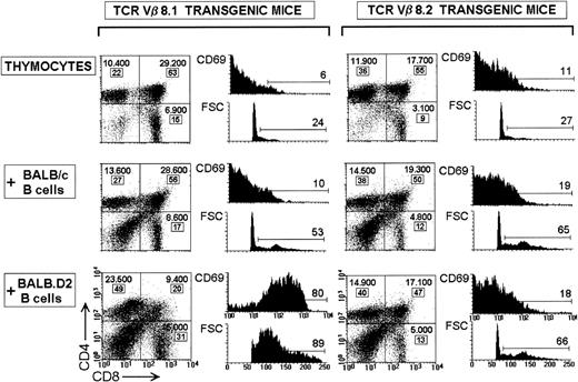
Specificity of the in vitro deletion of TCR Vβ8.1 TG thymocytes. Dot plots show the CD4 versus CD8 profiles (CD4-TC/CD8-FITC staining) of Mtv-7–reactive TCR Vβ8.1 or nonreactive TCR Vβ8.2 thymocytes cultured for 60 hours alone or with B cells fromMtv-7+ BALB.D2 or Mtv-7−BALB/c mice. Percent deletion values of Vβ8.1 DP thymocytes were 2% and 68% when cultured with BALB/c and BALB.D2 B cells, respectively. For Vβ8.2 DP thymocytes percent deletion value was 0% when either BALB/c or BALB.D2 B cells were used. The absolute cell number per well and the percentage (in boxes) are indicated for each thymocyte subset. The percentages correspond to corrected values, which have been calculated considering DP + CD4+SP + CD8+SP = 100%. Histograms represent the CD69 expression and FSC profile of DP thymocytes for each culture condition. The percentage of CD69+ (FL2 > 20) and blast (FSC > 80) DP thymocytes are shown.
Specificity of the in vitro deletion of TCR Vβ8.1 TG thymocytes. Dot plots show the CD4 versus CD8 profiles (CD4-TC/CD8-FITC staining) of Mtv-7–reactive TCR Vβ8.1 or nonreactive TCR Vβ8.2 thymocytes cultured for 60 hours alone or with B cells fromMtv-7+ BALB.D2 or Mtv-7−BALB/c mice. Percent deletion values of Vβ8.1 DP thymocytes were 2% and 68% when cultured with BALB/c and BALB.D2 B cells, respectively. For Vβ8.2 DP thymocytes percent deletion value was 0% when either BALB/c or BALB.D2 B cells were used. The absolute cell number per well and the percentage (in boxes) are indicated for each thymocyte subset. The percentages correspond to corrected values, which have been calculated considering DP + CD4+SP + CD8+SP = 100%. Histograms represent the CD69 expression and FSC profile of DP thymocytes for each culture condition. The percentage of CD69+ (FL2 > 20) and blast (FSC > 80) DP thymocytes are shown.
RESULTS AND DISCUSSION
Specificity of the in vitro deletion of TCR Vβ8.1 TG thymocytes by Mtv-7+ B cells.
The experimental model of in vitro deletion used in this study is based on the coculture of splenic B cells expressing the Mtv-7 SAg, obtained from BALB.D2 mice, with Mtv-7–reactive TCR Vβ8.1 TG thymocytes. Details concerning the potential of splenic B cells to induce the in vitro deletion of SAg-reactive thymocytes in a similar culture system, the kinetics of the process, as well as the optimal APC:thymocyte ratio have been considered in a recent report.13 The specificity of the deletion system is illustrated in Fig 1, which shows the CD4 versus CD8 profile ofMtv-7–reactive TCR Vβ8.1 or control nonreactive TCR Vβ8.2 thymocytes, cultured for 60 hours with Mtv-7+ orMtv-7− splenic B cells, obtained from BALB.D2 and control BALB/c mice, respectively. For each thymocyte subpopulation, the absolute cell number per well and the percentage are shown.
Culture of TCR Vβ8.1 TCR TG thymocytes with BALB.D2 B cells induced a strong deletion of DP thymocytes (68% in the experiment shown in Fig1), as indicated by the decrease in their percentage (20% v63%) as well as in their absolute number (9.400 v 29.200), confirming our previous results using a similar experimental method.13 In the in vitro deletion assay described in the present report, thymocyte viability in culture has been improved by adding 10 mmol/L HEPES to the culture medium, and by using an FCS batch more adequate for these cultures. Percent deletion has been calculated following the formula described in Materials and Methods on the basis of the absolute DP cell number, according to Vasquez et al,15 instead of using the parameter “percentage of DP in the culture” used by Pircher et al,16 because a decrease in the percentage of DP thymocytes in this culture system may reflect not only a deletion process, but also the proliferation of SP thymocytes. In this sense, as previously described,13deletion of SAg-reactive DP thymocytes is paralleled by cell activation, as assessed by the expression of CD69 (80% CD69+ DP cells) and the FSC profile (89% DP with FSC > 80), and by an increase in the absolute number of SP thymocytes per well, particularly of the CD4+ SP subset (23.500 v 10.400). Upregulation of CD69 has also been reported during the in vitro deletion of TCR TG DP thymocytes specific for the lymphocytic choriomeningitis virus,17 or the 2 oxoglutarate dehydrogenase (2C TCR TG mice).18
In contrast, when splenic B cells from Mtv-7−BALB/c mice were used as APCs, no significative deletion (2% deletion) or activation (10% CD69+ DP cells) of TCR Vβ8.1 DP thymocytes were observed. Similar results were obtained when control nonreactive TCR Vβ8.2 thymocytes were cultured with eitherMtv-7+ or Mtv-7− B cells from BALB.D2 or BALB/c mice, respectively. These results indicate that deletion of TCR Vβ8.1 TG DP thymocytes induced by BALB.D2 B cells isMtv-7 SAg specific.
Analysis of cell activation, cell proliferation, and apoptosis during in vitro SAg-mediated deletion of TCR Vβ8.1 DP thymocytes.
Figure 2 compares the phenotype of TCR Vβ8.1 DP thymocytes cultured for 36 or 60 hours alone or with BALB.D2 B cells. A considerable decrease in the absolute cell number per well of DP thymocytes was already observed after 36 hours (77% deletion). DP deletion was accompanied by cell activation, as assessed by CD69 expression and the FSC profile. Analysis of DNA fragmentation by the TUNEL method indicated that around 40% (37% in the experiment shown in Fig 2) of DP thymocytes were apoptotic. However, no BrdU incorporation was detected, indicating that after 36 hours DP thymocytes undergoing in vitro SAg-mediated negative selection did not enter into cell cycle.
Phenotype of TCR Vβ8.1 DP TG thymocytes during in vitroMtv-7 SAg-mediated negative selection. Dot plots show the CD4 versus CD8 profiles (CD4-PE/CD8-FITC staining) of TCR Vβ8.1 TG thymocytes cultured for 36 or 60 hours alone or with BALB.D2 splenic B cells. The absolute cell number of DP and SP thymocytes is indicated. Circles indicate the DP CD4+CD8intpopulation. Histograms represent the FSC, CD69 expression, TUNEL staining, and BrdU incorporation of DP thymocytes for each culture condition. Staining details are given in Materials and Methods. The percentage of CD69+ (FL2 > 20), blast (FSC > 70), TUNEL+ (FL1 > 300), and BrdU+(FL1 > 130) DP thymocytes are indicated. The experiment is representative of four experiments with similar results. (Note that the CD4-TC/CD8-FITC staining used in Fig 1 dot plots does not allow a clear definition of the CD4+CD8int DP population.)
Phenotype of TCR Vβ8.1 DP TG thymocytes during in vitroMtv-7 SAg-mediated negative selection. Dot plots show the CD4 versus CD8 profiles (CD4-PE/CD8-FITC staining) of TCR Vβ8.1 TG thymocytes cultured for 36 or 60 hours alone or with BALB.D2 splenic B cells. The absolute cell number of DP and SP thymocytes is indicated. Circles indicate the DP CD4+CD8intpopulation. Histograms represent the FSC, CD69 expression, TUNEL staining, and BrdU incorporation of DP thymocytes for each culture condition. Staining details are given in Materials and Methods. The percentage of CD69+ (FL2 > 20), blast (FSC > 70), TUNEL+ (FL1 > 300), and BrdU+(FL1 > 130) DP thymocytes are indicated. The experiment is representative of four experiments with similar results. (Note that the CD4-TC/CD8-FITC staining used in Fig 1 dot plots does not allow a clear definition of the CD4+CD8int DP population.)
DP thymocyte deletion was almost complete when TCR Vβ8.1 thymocytes were cultured for 60 hours with BALB.D2 B cells. After this period in culture most DP thymocytes were activated blasts, and approximately 50% of them were apoptotic, as shown by the TUNEL method. Interestingly, around 40% (35% in the experiment shown) DP thymocytes had entered into cell cycle as assessed by BrdU incorporation.
TCR Vβ8.1 DP thymocytes were not activated and did not incorporate BrdU when cultured alone for 36 or 60 hours; a proportion of them, most likely corresponding to apoptotic nonpositively selected thymocytes, were positive for TUNEL staining, in agreement with the findings of Surh and Sprent,19 who suggested that the high proportion of apoptotic cortical thymocytes detected in situ with the TUNEL method is primarily a reflection of lack of positive selection.
Cell activation and cell proliferation requirements of SAg-mediated deletion of TCR Vβ8.1 DP thymocytes.
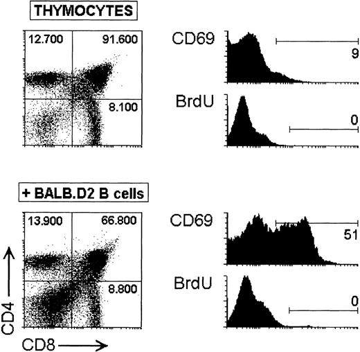
The data shown in Fig 2 indicate that most of the DP thymocytes deleted after 60 hours in culture with BALB.D2 B cells were activated, as more than 75% of DP remaining at 36 hours were eliminated at 60 hours, and up to 70% of DP expressed the CD69 activation molecule at 36 hours. This suggests that cell activation may be required for SAg-mediated DP deletion to occur. However, deletion of DP thymocytes does not appear to be paralleled by cell proliferation, because at 36 hours a strong deletion occurred but no cycling cells were observed, as demonstrated by the absence of BrdU+ cells (Fig 2). To exclude the possibility that DP thymocytes already deleted at 36 hours might have entered into cell cycle during the early phase of our in vitro assay, BrdU incorporation was analyzed after 24 hours in culture. As shown in Fig 3, only 27% deletion of DP thymocytes was observed after 24 hours, and no BrdU incorporation was detected at this timepoint when TCR Vβ8.1 thymocytes were cultured alone or with BALB.D2 B cells, indicating that DP deletion observed at 36 hours was not preceded by entry into cell cycle. Interestingly, around 50% of DP thymocytes were CD69+ after 24 hours in culture with BALB.D2 B cells. Proliferating DP thymocytes detected by BrdU incorporation after 60 hours in culture probably representMtv-7 SAg-stimulated postpositively selected DP thymocytes, which have been shown to be less sensitive to negative selection than mature SP thymocytes.20-22 This is supported by the finding that most DP thymocytes remaining after 60 hours have a CD4+CD8int phenotype (see Figs 2 and4), corresponding to postselected DP.23 Furthermore, most BrdU+ DP cells are located within the CD4+CD8int population (data not shown). In addition, up to 60% CD4+ SP thymocytes were CD69+ BrdU+ cells (data not shown). These data indicate that TCR stimulation by viral SAg may result in deletion or proliferation depending on the thymocyte differentiation stage.
Analysis of cell activation and BrdU incorporation during the early phase of in vitro SAg-mediated deletion of Vβ8.1 DP TG thymocytes. Dot plots show the CD4 versus CD8 profiles of TCR Vβ8.1 TG thymocytes cultured for 24 hours alone or with BALB.D2 splenic B cells. The absolute cell number of DP and SP thymocytes is indicated. Histograms represent the CD69 expression and BrdU incorporation of DP thymocytes. The percentage of CD69+ (FL2 > 20) and BrdU+ (FL1 > 80) DP thymocytes are indicated. The experiment is representative of three experiments with similar results.
Analysis of cell activation and BrdU incorporation during the early phase of in vitro SAg-mediated deletion of Vβ8.1 DP TG thymocytes. Dot plots show the CD4 versus CD8 profiles of TCR Vβ8.1 TG thymocytes cultured for 24 hours alone or with BALB.D2 splenic B cells. The absolute cell number of DP and SP thymocytes is indicated. Histograms represent the CD69 expression and BrdU incorporation of DP thymocytes. The percentage of CD69+ (FL2 > 20) and BrdU+ (FL1 > 80) DP thymocytes are indicated. The experiment is representative of three experiments with similar results.
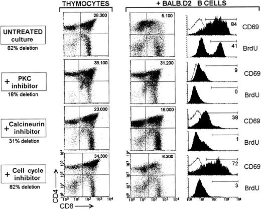
Cell activation and cell proliferation requirements of SAg-mediated deletion of TCR Vβ8.1 DP TG thymocytes. TCR Vβ8.1 thymocytes were cultured for 60 hours alone or with BALB.D2 splenic B cells in control conditions (untreated), or in the presence of 6 μmol/L BIM (PKC inhibitor), 80 nmol/L CsA (calcineurin inhibitor), or 120 μmol/L mimosine (cell-cycle blocker). Dot plots show the CD4 versus CD8 profile (CD4-PE/CD8-FITC staining) after removing magnetically the B cells using anti-mouse Ig-coated magnetic beads (see Materials and Methods for details). The absolute cell number of DP thymocytes is indicated. The statistics corresponding to four similar experiments are given in Table 1. Histograms represent the CD69 expression and BrdU incorporation of DP thymocytes for each culture condition. The percentage of CD69+ (FL2 > 20) and BrdU+ (FL1 > 80) DP thymocytes are indicated.
Cell activation and cell proliferation requirements of SAg-mediated deletion of TCR Vβ8.1 DP TG thymocytes. TCR Vβ8.1 thymocytes were cultured for 60 hours alone or with BALB.D2 splenic B cells in control conditions (untreated), or in the presence of 6 μmol/L BIM (PKC inhibitor), 80 nmol/L CsA (calcineurin inhibitor), or 120 μmol/L mimosine (cell-cycle blocker). Dot plots show the CD4 versus CD8 profile (CD4-PE/CD8-FITC staining) after removing magnetically the B cells using anti-mouse Ig-coated magnetic beads (see Materials and Methods for details). The absolute cell number of DP thymocytes is indicated. The statistics corresponding to four similar experiments are given in Table 1. Histograms represent the CD69 expression and BrdU incorporation of DP thymocytes for each culture condition. The percentage of CD69+ (FL2 > 20) and BrdU+ (FL1 > 80) DP thymocytes are indicated.
To investigate whether DP thymocyte deletion depends on cell activation, but does not require cell proliferation, TCR Vβ8.1 TCR TG thymocytes were cultured for 60 hours with BALB.D2 B cells in the presence of inhibitors of cell activation and/or cell proliferation. BIM and CsA were used as inhibitors of the signaling molecules PKC and calcineurin, respectively, known to play an essential role in T-cell activation. The G1 cell-cycle blocker mimosine was used to inhibit cell proliferation. Each inhibitor was used at a concentration chosen to get the optimum effect with a minimum toxicity, after testing a broad range of concentrations. The results obtained are shown in Fig 4 and Table 1. When cultured in the presence of BALB.D2 B cells and 6 μmol/L of the PKC-specific inhibitor BIM, DP thymocytes were not activated, did not enter into cell cycle, and SAg-mediated DP deletion was almost completely inhibited. Inhibition of PKC activation by staurosporine, calphostin, or H-7 has also been reported to block the in vitro deletion of DP thymocytes from cytochrome c–specific TCR TG mice,15although it has been shown that these inhibitors are poorly selective whereas BIM displays a high degree of selectivity.24Therefore, our results obtained with BIM establish more convincingly the requirement for PKC in the signal transduction pathway underlying SAg-mediated T-cell negative selection.
Effect of Inhibitors of Cell Activation and/or Cell Proliferation on the In Vitro Deletion of TCR Vβ8.1 TG DP Thymocytes
| . | Thymocytes . | +BALB.D2 B Cells . | % Deletion . | n . |
|---|---|---|---|---|
| Untreated culture | 31.300 ± 4.700 | 7.700 ± 2.500 | 74 ± 9 | 5 |
| + PKC inhibitor | 38.600 ± 6.200 | 33.900 ± 7.500 | 14 ± 10 | 4 |
| + Calcineurin inhibitor | 24.100 ± 3.000 | 14.600 ± 3.200 | 40 ± 8 | 4 |
| + Cell-cycle inhibitor | 34.300 ± 3.800 | 8.900 ± 3.000 | 73 ± 10 | 4 |
Data are indicated as the mean ± SD of the absolute number of DP thymocytes per well obtained after culture of TCR Vβ8.1 TG thymocytes alone or with BALB.D2 B cells for 60 hours, in the absence or presence of the indicated inhibitors. A representative experiment is illustrated in Fig 4.
Treatment with the calcineurin inhibitor CsA at 80 nmol/L induced a partial inhibition of DP thymocyte activation after culture with BALB.D2 B cells, and no BrdU incorporation was detected. Percent deletion values were around 40%, indicating a partial inhibition of DP thymocyte deletion. In contrast with the results obtained with BIM, an almost complete inhibition of DP activation and deletion was not reached with CsA, even when used at very high and, therefore, toxic concentrations. These data suggest a direct correlation between the level of DP activation and the degree of DP deletion. In support of this, lower BIM and CsA concentrations induced a lower inhibition of both DP activation and deletion (data not shown). Data dealing with the participation of the calcineurin-mediated signaling pathway in negative selection are controversial. In vivo and in vitro experiments using the calcineurin inhibitors FK50625 and CsA,21respectively, in TCR TG thymocyte deletion assays, argue against calcineurin requirement. However, other in vivo and in vitro experimental evidences have firmly shown the involvement of this calcium/calmodulin-dependent phosphatase in negative selection induced by SAgs,9,10 and by antigenic peptides derived from cytochrome c and the male H-Y antigen, in TCR TG mice.26,27Interestingly, in agreement with our data, in these reports it was shown that after blockage of calcineurin by CsA, negative selection was partially inhibited or delayed. In addition, experiments of TCR TG thymocyte deletion mediated by an antigenic peptide or a peptide analogue have shown that CsA did not interfere with thymocyte deletion in vitro when the antigenic peptide was used at high concentration, but partially inhibited deletion when the peptide analogue or lower concentrations of the antigenic peptide were used.27 These data support the hypothesis that requirement for the calcineurin-mediated signal transduction depends on the intensity of the TCR-coupled signaling triggered by the deleting ligand and may provide an explanation for the reports by Wang et al25 and Vasquez et al21 in which no inhibition of negative selection by CsA or FK506 was observed. Therefore, in contrast to signaling through PKC which appears to have a crucial function in negative selection, calcineurin-mediated signaling may be overcome when the deleting antigen determines a very high-affinity TCR-peptide interaction, but may be required when this interaction has a lower affinity, or when the deleting antigen is present at low concentration. Similar observations have been reported with regard to costimulatory signal requirements during T-cell negative selection.28
BrdU incorporation but not activation of DP thymocytes was inhibited after culture with BALB.D2 B cells in the presence of the cell-cycle blocker mimosine. Interestingly, in accordance with our previous hypothesis, no inhibition of DP deletion was obtained under these culture conditions, demonstrating that in vitro deletion of SAg-reactive TCR Vβ8.1 TG thymocytes does not depends on cell proliferation. This is apparently in contrast with recent data establishing a correlation between proliferation and cell death upon stimulation of peripheral T cell by bacterial SAgs.11However, our results are in agreement with Boehme and Lenardo,29 who reported that progression through the cell cycle was not required for TCR-mediated apoptosis of activated peripheral T cells. Interestingly, these investigators have shown that although peripheral T cells blocked in S phase after treatment with aphidicolin were susceptible to TCR-induced apoptosis, cells blocked in G1 phase by treatment with mimosine had a diminished susceptibility to TCR-induced apoptosis. These data suggest that in vitro SAg-mediated deletion of TCR Vβ8.1 TG thymocytes is less sensitive to cell-cycle blockade in G1 than TCR-induced apoptosis of peripheral T cells.
In conclusion, the data shown in the present report indicate that viral SAg-mediated T-cell negative selection requires cell activation but does not depend on cell proliferation. Finally, the differential inhibition effect on SAg-mediated DP deletion, obtained by blocking PKC or calcineurin, support the hypothesis that synergy between different TCR-mediated signaling pathways may be required depending on the intensity of TCR-antigen interaction mediating the negative selection.
ACKNOWLEDGMENT
We thank Drs Hanspeter Pircher and Horst Bluethmann for the TCR TG mice, and Pilar Martı́n for help in FACS experiments. We are grateful to the Flow Cytometry facility of the Fundación Jiménez Dı́az for making it possible to perform FACS analysis after hours.
Supported in part by grants from the Dirección General de Investigación Cientı́fica y técnica, Ministerio de Educación y Ciencia, Spain (to C.A.) and from the Human Frontiers of Science Program (to H.R.M.). T.R. was supported by a Centennial Fellowship from the Medical Research Council of Canada.
Address reprint requests to Carlos Ardavı́n, PhD, Department of Cell Biology, Faculty of Biology, Complutense University, 28040 Madrid, Spain.
The publication costs of this article were defrayed in part by page charge payment. This article must therefore be hereby marked "advertisement" is accordance with 18 U.S.C. section 1734 solely to indicate this fact.


This feature is available to Subscribers Only
Sign In or Create an Account Close Modal