Abstract
Mutations of the receptor tyrosine kinase c-kit or its ligand stem cell factor (SCF), which is encoded as a soluble and membrane-associated protein by the Steel gene in mice, lead to deficiencies of germ cells, melanocytes, and hematopoiesis, including the erythroid lineage. In the present study, we have used genetic methods to study the role of membrane or soluble presentation of SCF in hematopoiesis. Bone marrow–derived stromal cells expressing only a membrane-restricted (MR) isoform of SCF induced an elevated and sustained tyrosine phosphorylation of both c-kit and erythropoietin receptor (EPO-R) and significantly greater proliferation of an erythrocytic progenitor cell line compared with stromal cells expressing soluble SCF. Transgene expression of MR-SCF inSteel-dickie (Sld) mutants resulted in a significant improvement in the production of red blood cells, bone marrow hypoplasia, and runting. In contrast, overexpression of the full-length soluble form of SCF transgene had no effect on either red blood cell production or runting but corrected the myeloid progenitor cell deficiency seen in these mutants. These data provide the first evidence of differential functions of SCF isoforms in vivo and suggest an abnormal signaling mechanism as the cause of the severe anemia seen in mutants of the Sl gene.
THE PHOSPHORYLATION of proteins on specific tyrosine residues is critical for regulating cell proliferation and differentiation in eukaryotes.1 2 The activation of protein tyrosine kinases (PTKs), resulting in the tyrosine phosphorylation of downstream signaling proteins, is an important event in many signal transduction cascades critical for growth and development. The level of phosphorylation at specific tyrosine residues and the amplification of downstream signals initiated by PTKs to a large extent determine the above cellular outcomes.
The c-kit receptor tyrosine kinase (RTK) encoded by the murineDominant white spotting (W) locus and its ligand stem cell factor (SCF), encoded by the Steel (Sl) locus, define a signaling pathway essential for murine hematopoiesis.3,4 The c-kit RTK is a member of the platelet-derived growth factor (PDGF) receptor family.5,6The interaction of RTKs that belong to the PDGF receptor family and their cognate ligands induce receptor dimerization or oligomerization followed by activation of intrinsic tyrosine kinase and receptor transphosphorylation.7 Many lines of evidence suggest that bivalent binding of ligands is key for inducing receptor dimerization and subsequently receptor activation.8 Although SCF is dimeric in solution and has been shown to induce the dimerization of c-kit,9,10 low levels (3 ng/mL) exist in serum,11 and based on the dimerization Ka, it has been predicted that greater than 90% of the circulating SCF would exist in monomeric form.11 Recent studies using radiolabeled SCF added to serum have shown that 49% to 72% of the circulating SCF exists in monomeric form.11 Further, a detailed examination of dimerization defective variants of SCF showed substantially reduced mitogenic activity, whereas the activity of disulfide-linked SCF dimer was 10-fold higher than that of wild-type (wt) SCF. These results suggest a correlation between dimerization affinity and biological activity. Based on these findings, one would predict that SCF and other cytokines or growth factors that are expressed as membrane-associated (MA) proteins may be more efficient at forming dimers because of an enhanced probability of monomers to encounter each other in the plasma membrane lipid bilayer as compared serum. This may subsequently result in a significantly greater mitogenic activity of MA protein in comparison with its soluble counterpart.
Many cytokines and/or growth factors exist as both MA and soluble isoforms. These proteins may participate in a novel mode of intercellular communication restricted to adjacent cells, termed juxtacrine stimulation.12 Examples of proteins that exist as both soluble and MA forms include transforming growth factor-α (TGF-α),12 tumor necrosis factor (TNF),13 and colony stimulating factor-1 (CSF-1).14,15 Two major isoforms of SCF also exist in both mice and humans as a result of mRNA splicing. A glycoprotein of 248 (SCF248) amino acids (aa) is rapidly cleaved to release a biologically active soluble (S) protein of 164 aa. In contrast, a glycoprotein of 220 aa (SCF220), which lacks the proteolytic cleavage site encoded by differentially spliced exon 6 sequences, remains predominantly MA.16-18 This isoform can also be slowly released from the cell surface through the use of an alternative proteolytic cleavage site in exon 7. Our laboratory has shown that site-directed mutagenesis of the SCF cDNA to ablate both proteolytic cleavage sites results in the generation of a membrane-restricted (MR) and biologically active form of SCF (SCFX9/D3).19 In spite of significant molecular and biochemical characterization of SCF, little is known regarding the physiological role(s) of these isoforms.Steel-dickie (Sld), a viable Slmutant, is the result of an intragenic 4-kb genomic deletion that removes the exons encoding the transmembrane and cytoplasmic domains ofSld-protein and impairs the ability of the protein to anchor in the plasma membrane.20 Thus,Sld mice appear to be capable of producing biologically active soluble SCF, although this truncated protein lacks both exon 7– and exon 8–encoded membrane-proximal aa and therefore differs from the wt isoform of soluble SCF.21 The severe hematologic deficiencies in compound heterozygousSl/Sld and homozygousSld/Sld mice have suggested that the MA isoform of SCF is critical for normal mouse development. Indeed, the severe nature of the erythroid deficiency in Sl/Sldmice in spite of the presence of truncated soluble protein has continued to be an enigmatic feature of the Sl/Sldphenotype years after the cloning of the Sl gene.
The most overt hematopoietic phenotypes of W and Slmutant mice are severe macrocytic anemia and mast cell deficiency. In addition, W and Sl mutants have defects at the level of hematopoietic stem and progenitor cells.3,4,22 In vitro culture assays have shown that soluble SCF synergizes with a number of lineage-restricted hematopoietic growth factors, including erythropoietin (EPO), granulocyte macrophage-colony stimulating factor (GM-CSF), and interleukin-7 (IL-7).23,24 In addition, our laboratory has previously shown differences in the proliferation/survival of human hematopoietic progenitor cells when exposed in vitro to the MA verses soluble isoforms of human SCF.25 The wide range of cellular responses observed following c-kit receptor activation has made it difficult to dissect the molecular events that provide biological specificity to the c-kit/SCF signaling pathway. Recently, the phenotype of mice deficient in EPO suggest that the survival and proliferation of late erythroid progenitors depends on EPO.26Sl/Sld mutants also show a profound deficiency in the erythroid lineage, despite of the presence of high levels of circulating EPO.4 These data suggest that the normal proliferation and/or differentiation of erythroid progenitors to produce mature red cells requires both a functional SCF and EPO signaling pathway. However, to date the isoform of SCF responsible physiologically for the proliferation and/or differentiation of erythroid progenitors to mature red blood cells remains unknown.
In the studies presented here, we have elected to use the MR form of SCF to more clearly elucidate the role of membrane presentation in vitro and in vivo because the wt form of MA SCF (SCF220), at least in vitro, is slowly secreted from the cell surface.18 19 We show that stromal cells expressing only the MR form of SCF induce a more sustained and elevated tyrosine phosphorylation of the EPO receptor (EPO-R) in erythroid cells and stimulate a significantly greater proliferation of an erythrocytic cell line as compared with soluble SCF. In vivo, usingSl/Sld mutant and transgenic mice that overexpress either the soluble or the MR form of SCF, we show a significant increase in the production of red cells, correction of runting, and bone marrow hypocellularity in Sl/Sld mice that express MR SCF as compared with mice that overexpress soluble SCF only. In contrast, overexpression of soluble SCF inSl/Sld mice resulted in complete correction of myeloid progenitor cell deficiency. These in vitro and in vivo observations suggest distinct biological role(s) for SCF isoforms.
MATERIALS AND METHODS
Cell lines.
The murine growth factor–dependent cell line, HCD57, was a gift from Dr H. Lodish (MIT, Boston, MA). This is an EPO-dependent cell line that responds to murine soluble SCF. The biological characteristics of this cell line and culture conditions have been previously described.27,28 HCD57 cells were maintained in Iscove's modified Dulbecco's medium (IMDM) supplemented with 20% fetal calf serum (FCS) and 1 μ/mL of recombinant human (rh) EPO (Amgen, Thousand Oaks, CA). For growth and survival assays, HCD57 cells were factor-starved by washing three times with IMDM and culturing for 18 hours without EPO. Subsequently, starved cells were plated at 1 × 10⁴ cells per mL in medium without growth factor (control), supplemented with rhEPO (1 μ/mL), or cocultured on mitomycin C–treated Sl/Sl4 stromal cells expressing either isoform of SCF in the presence or absence of 1 μ/mL of rhEPO. The culture plates were gently centrifuged for 1 minute at 100gto ensure direct cell-cell contact and further cocultured for 48 hours at 37°C. Viable cells were counted after 48 hours. Sl/Sl4-SCF transfectants were prepared by treating with mitomycin C (5 μg/mL; Sigma, St Louis, MO) for 2 hours at 37°C, washed three times with phosphate-buffered saline (PBS), treated with trypsin, and plated to confluency (1 × 10⁶/well) on 6-well gelatin-coated tissue culture plates (Falcon, Lincoln Park, NJ), and cultured overnight.
Flow cytometric analysis.
To determine the cell surface expression of SCF on Sl/Sl4stromal cells transfected with cDNAs encoding either the soluble or the MR form of SCF, flow cytometric analysis with a rat anti-murine SCF monoclonal antibody (MoAb) was performed. Briefly, 1 × 10⁶ stromal cells were stained separately with either 1 μg of primary rat anti-mouse SCF MoAb (Genzyme, Cambridge, MA) or a rat IgG2a isotype (PharMingen, San Diego, CA) control antibody for 30 minutes at 4°C. Afterward, the cells were washed twice with PBS/0.1% bovine serum albumin (BSA) and subsequently stained with 1 μg of secondary fluorescein-isothiocyanate (FITC)–conjugated goat (F(ab′)2 anti-rat IgG (GIBCO-BRL, Gaithersburg, MD) under identical conditions and analyzed by fluorescence-activated cell sorter (FACS).
Immunoprecipitation and Western blot analysis.
Immunoprecipitation (IP) and Western blot (WB) analyses were performed as previously described.29 Briefly, Sl/Sl4cells expressing either isoform of SCF were treated as described above. These cells were washed and plated on 6-well gelatin-coated plates (1 × 10⁶/well) and cultured for 36 to 48 hours. Factor-starved HCD57 cells (10 × 106/well) were loaded as described above and further cocultured for various periods at 37°C. Thereafter, the supernatants were removed, and cells were lysed in lysis buffer (10 mmol/L K2HPO4, 1 mmol/L EDTA, 5 mmol/L EGTA, 10 mmol/L MgCl2, 1 mmol/L Na2VO4, 50 mmol/L beta-glycerol-phosphate, 10 μg/mL leupeptin, 1 μg/mL pepstatin, and 10 μg/mL aprotinin) at 4°C for 30 minutes. Cell lysates were clarified by centrifuging for 30 minutes at 10,000g at 4°C. IPs were performed by incubating equivalent amounts of cell lysates with either an anti–EPO-R antibody (5 μL per 10 × 106 cells; kindly provided by Dr G. Krystal) or with an anti–c-kit antibody (2 μLs per 10 × 106 cells; PharMingen) for 3 hours at 4°C. Protein A- or protein G-Sepharose beads (Pierce, Rockford, IL) were used to collect the antigen-antibody complexes. The IPs were separated by sodium dodecyl sulfate-polyacrylamide gel electrophoresis (SDS-PAGE), and proteins were electrophoretically transferred onto Immobilon-P membranes (Millipore, Bedford, MA). After blocking residual binding sites on the transfer membrane by incubating the membrane with 2% BSA/TBST (50 mmol/L Tris-HCl, pH 7.4, 150 mmol/L NaCl, 0.05% Tween-20), for 12 hours at 37°C, Western blot analysis using an anti-phosphotyrosine antibody (1:2,000 dilution; Transduction Laboratories, Lexington, KY) and the enhanced chemiluminescence detection system (Amersham, Arlington Heights, IL) were performed according to manufacturer's instructions.
Generation of transgenic mice.
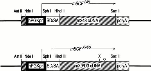
Experiments involving mice described here were reviewed and approved by Animal Use Committee of Indiana University School of Medicine. C3H/HeJ mice were obtained from Jackson Laboratories (Bar Harbor, ME). Transgenic mice were generated by microinjecting into the pronuclei of fertilized C3H/HeJ eggs a 1.3-kb Ndel/Kpnl fragment comprising either the hPGK-mSCF248 or the hPGK-mSCFX9/D3 minigene (Fig 1). Microinjected eggs were transferred to the oviducts of pseudo-pregnant outbred Swiss-Webster females. Offspring were tested for the presence of transgene by analyzing tail DNA. Briefly, tail DNA from mice was digested overnight in digestion buffer (100 mmol/L NaCl, 10 mmol/L Tris base pH 8, 25 mmol/L EDTA pH 8, 1% SDS, and 150 μg/mL proteinase K) at 50°C and extracted the next day with phenol and chloroform. The high molecular weight DNA was subsequently digested with EcoRl, electrophoresed, transferred to filters, and probed using a full-length32P-labeled cDNA murine SCF probe. To examine the in vivo role of the two isoforms of SCF on various lineages in naturally occurring Sl mutants (ie, Sl/Sld), we first crossed transgenic mice overexpressing either the soluble or the MR form of SCF to WC/ReJ-Sl/+ mice to obtainSl/+ mice that overexpress either isoform of SCF. These mice were identified based on their phenotype (ie, forehead blaze and diluted belly) and Southern blot analysis. Transgene positiveSl/+ male mice were further crossed to C57Bl6-Sld/+ (Jackson Laboratory) mice to obtain Sl/Sld transgene positive or negative mice.
SCF transgene constructs. Transgene expression of the mSCF248 and mSCFX9/D3 cDNAs in founder mice was achieved using a minigene cassette consisting of mSCF248 or mSCFX9/D3 cDNA expressed from the human PGK promoter. The PGK promoter, consisting of 514 bp 5′-flanking sequence from the X-linked human phosphoglycerate kinase-1 gene,54 up to but excluding the translational start codon, was subcloned into pGEM-7 as an Aatll/Sph l fragment. The mSCF248 or mSCFX9/D3 cDNA in addition to a splice donor/acceptor and poly A site was subcloned from the expression plasmid V19.8mSCF248 or V19.8mSCFX9/D3 36 as aXho l/Kpn l fragment 3′ to the PGK promoter in the pGEM-7 plasmid vector to generate the final transgene plasmid. hPGKpr: human phosphoglycerate kinase promoter: SD/SA: splice donor/splice acceptor sequences; mSCF248 cDNA: murine SCF cDNA encoding full-length soluble SCF protein: mSCFX9/D3 cDNA: murine SCF cDNA encoding full-length MR19 SCF protein; poly A: polyadenylation sequence. (X) denotes the location of an insertedXho l at the proteolytic cleavage site in exon 6, and (▿) denotes a 12-bp deletion of the secondary proteolytic cleavage site in exon 7.
SCF transgene constructs. Transgene expression of the mSCF248 and mSCFX9/D3 cDNAs in founder mice was achieved using a minigene cassette consisting of mSCF248 or mSCFX9/D3 cDNA expressed from the human PGK promoter. The PGK promoter, consisting of 514 bp 5′-flanking sequence from the X-linked human phosphoglycerate kinase-1 gene,54 up to but excluding the translational start codon, was subcloned into pGEM-7 as an Aatll/Sph l fragment. The mSCF248 or mSCFX9/D3 cDNA in addition to a splice donor/acceptor and poly A site was subcloned from the expression plasmid V19.8mSCF248 or V19.8mSCFX9/D3 36 as aXho l/Kpn l fragment 3′ to the PGK promoter in the pGEM-7 plasmid vector to generate the final transgene plasmid. hPGKpr: human phosphoglycerate kinase promoter: SD/SA: splice donor/splice acceptor sequences; mSCF248 cDNA: murine SCF cDNA encoding full-length soluble SCF protein: mSCFX9/D3 cDNA: murine SCF cDNA encoding full-length MR19 SCF protein; poly A: polyadenylation sequence. (X) denotes the location of an insertedXho l at the proteolytic cleavage site in exon 6, and (▿) denotes a 12-bp deletion of the secondary proteolytic cleavage site in exon 7.
Expression of the transgene.
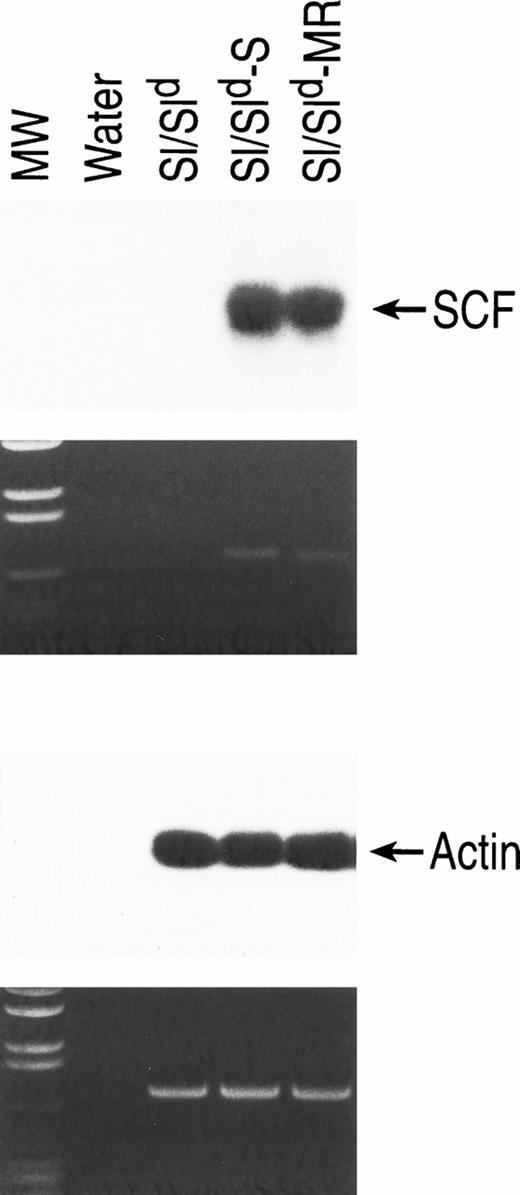
Total cellular RNA was purified from tissues using Tri Reagent (Molecular Research Center, Cincinnati, OH) according to the manufacturer's instructions and used as template. Equal amount of RNA (using actin as an internal control) was used to synthesize single-stranded cDNA by reverse transcriptase (RT) and random hexamers (Perkin Elmer, Branchburg, NJ). cDNA was amplified in a 100 μL reaction mixture by polymerase chain reaction (PCR) usingAmpliTaq DNA polymerase in 35 cycles of 1 minute denaturation at 94°C, 2 minutes of annealing at 55°C, and 3 minutes of synthesis at 72°C using a 5′-GGAGATCTGCGGGAATCC-3′ sense primer and 5′-GGCTGCAGTCCACAATTACACCTCTTG-3′ antisense primer based on published sequence.21 These primers fail to amplify a PCR product from Sl/Sld-encoded mRNA.21 The predicted amplification product from both mSCF248 and mSCFX9/D3 transgene expression is 733 bp. PCR (RT-PCR) products were examined on 1% agarose gel (Fig2). As a control for loading and integrity of total RNA, primers specific for actin (5′-TGGTGGGAATGGGTCAGAAGGACTC-3′ sense primer and 5′-TTGGCATAGAGGTCTTTACGGATGT-3′ antisense primer) were used to amplify cDNA using the described conditions.30 The predicted amplification product of these primers is 732 bp. To further confirm the identity of SCF and actin mRNAs, RT-PCR gels were transfered to nylon membranes (MSI, Westboro, MA), and reverse transcribed mRNAs were detected by hybridization using a32P random-primed full-length mSCF and β-actin cDNA. Hybridizations were performed using ExpressHyb hybridization solution (Clontech Laboratories, Palo Alto, CA) according to manufacturer's instructions.
Analysis of transgene expression in bone marrow–derived stromal cells from Sl/Sld,Sl/Sld-S, andSl/Sld-MR mice. Total cellular RNA was extracted as described in Materials and Methods. SCF-specific primers (upper panel) were used that recognize only the full-length transcript of SCF. As a loading control actin specific primers (lower panel) were used on the same RNA sample as described in Materials and Methods. RT-PCR products were examined on 1% ethidium bromide containing agarose gel and subsequently probed with SCF and actin specific probes. Upper arrow, SCF-specific primers (733 bp); lower panel, actin-specific primers (732 bp). Molecular weight (MW) marker is shown on the left.
Analysis of transgene expression in bone marrow–derived stromal cells from Sl/Sld,Sl/Sld-S, andSl/Sld-MR mice. Total cellular RNA was extracted as described in Materials and Methods. SCF-specific primers (upper panel) were used that recognize only the full-length transcript of SCF. As a loading control actin specific primers (lower panel) were used on the same RNA sample as described in Materials and Methods. RT-PCR products were examined on 1% ethidium bromide containing agarose gel and subsequently probed with SCF and actin specific probes. Upper arrow, SCF-specific primers (733 bp); lower panel, actin-specific primers (732 bp). Molecular weight (MW) marker is shown on the left.
Peripheral blood analysis.
Total peripheral RBC and WBC counts were analyzed on tail vein bleeds with a hemocytometer and Coulter Model ZM electronic particle counter (Coulter Electronics, Hialeah, FL). For WBC counts, RBCs were lysed using Zapoglobin (Coulter Electronics) according to manufacturer's recommendations. Peripheral blood hematocrits were performed by spinning capillary tubes for 5 minutes in a model MB microcapillary centrifuge (IEC, Boston, MA).
Hematopoietic progenitor cell assays.
BM was obtained as described previously,31 and cellularity was determined using a Coulter Model ZM as described. Bone marrow cells to be evaluated for committed progenitor colony formation were plated in IMDM (GIBCO) with 0.9% methylcellulose (Fluka, Hauppage, NY), 30% FCS (GIBCO), 2 U/mL human EPO (Amgen), 100 ng/mL recombinant rat SCF (Amgen), 10 U/mL murine IL-3 (Genzyme), 0.1 mmol/L hemin (Eastman Kodak, Rochester, NY), 2 × 10⁻³ mol/L L-glutamine (GIBCO), and 1 × 10⁻⁵ mol/L beta-mercaptoethanol (Sigma). Cultures were incubated at 37°C in a humidified environment with 5% O2 and 5% CO2 and were scored after 7 to 10 days of incubation as CFU-MIX, CFU-GM, or BFU-E. Primitive colony forming cells with high proliferative potential (HPP-CFC) were also assayed because these progenitors have been shown to be responsive to added recombinant SCF in multiple studies.32-34Double-layer HPP-CFC agar cultures were prepared as described.35 The bottom agar (1%) layer contained 100 ng/mL rat SCF (Amgen), 200 U/mL murine IL-3 (Genzyme), 500 U/mL murine IL-1 α (Genzyme), and 1,600 U/mL murine macrophage colony-stimulating factor (Genetics Institute, Cambridge, MA) BM cells (50,000/mL) were plated in the top agar (0.6%) and incubated at 37°C in a humidified environment at 5% O2, 10% CO2, and 85% N2. HPP-CFC colonies were scored as dense, macroscopic colonies measuring greater than 0.5 mm after 14 days of incubation. LPP-CFC were scored as colonies measuring less than 0.5 mm in diameter and containing greater than 50 cells.
RESULTS
MR isoform of SCF enhances the growth and survival of a factor-dependent erythrocytic progenitor cell line.
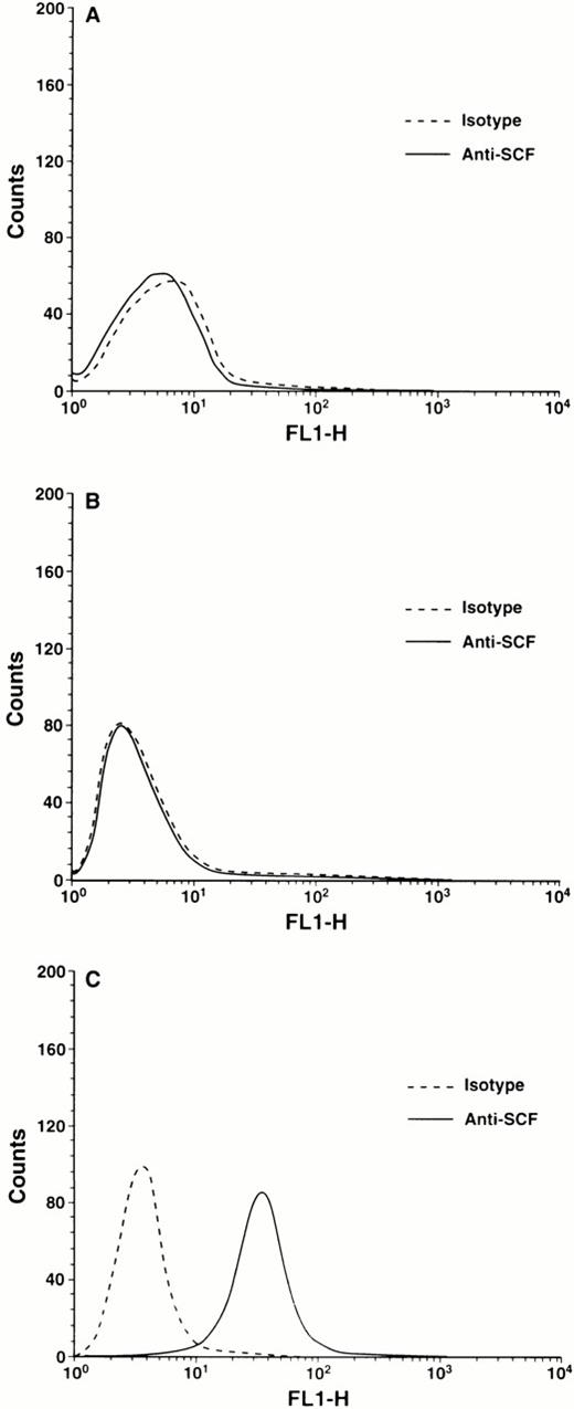
Previously it has been shown that c-kit and EPO-R physically associate in an EPO-dependent and SCF-responsive erythrocytic progenitor cell line, HCD57.27 In the present study we have used this cell line as an in vitro model system to delineate the role of MR and soluble isoforms of SCF in erythropoiesis. We have examined the growth and survival of HCD57 cells and signaling events downstream from c-kit in response to soluble or MR SCF stimulation. A stromal cell line (Sl/Sl4) derived from the fetal liver hematopoietic microenvironment (HM) of animals deficient in SCF as a result of genomic deletion of Sl coding sequences (Sl/Sl homozygotes) and stable transfectants of this line expressing either the MR (Sl/Sl4-MR) or the soluble (Sl/Sl4-S) isoform of SCF were used. These stromal cell lines were treated with mitomycin C to inhibit proliferation, thoroughly washed, then cultured for 24 hours as described in Materials and Methods. Thereafter, factor-starved HCD57 cells were cocultured with subconfluent stromal cell layers for another 48 hours. We have previously shown by immunoprecipitation of soluble and cell-associated SCF that the expression of each isoform is similar in the stromal cell lines used in experiments described here.19Moreover, because of the absence of proteolytic cleavage sites in the cDNA encoding the MR form of SCF, no soluble SCF is detectable in the supernatant of stromal cell cultures.19 However, this isoform was readily detectable on the stromal cell surface by flow cytometry (Fig 3C). In contrast, no detectable expression of SCF was observed on stromal cells that express either the soluble form of SCF (Fig 3B) or that are completely devoid of SCF expression (Fig 3A).
Flow cytometric analysis of the cell surface expression of SCF isoforms on stromal cells. Stromal cells stably transfected with cDNAs encoding either the soluble (Sl/Sl4-S) or the MR (Sl/Sl4-MR) form of SCF were stained for flow cytometric analysis as described in Materials and Methods. (A) Parental Sl/Sl4 cells were stained with either isotype control (dotted line) or rat anti-mouse SCF (solid line) MoAb followed by FITC-conjugated goat F(ab′)2 anti-rat IgG secondary; (B) Sl/Sl4-S cells were stained with either isotype control (dotted line) or rat anti-mouse SCF (solid line) MoAb followed by FITC-conjugated goat F(ab′)2 anti-rat IgG secondary; (C) Sl/Sl4-MR cells were stained with either isotype control (dotted line) or rat anti-mouse SCF (solid line) MoAb followed by FITC-conjugated goat F(ab′)2 anti-rat IgG secondary.
Flow cytometric analysis of the cell surface expression of SCF isoforms on stromal cells. Stromal cells stably transfected with cDNAs encoding either the soluble (Sl/Sl4-S) or the MR (Sl/Sl4-MR) form of SCF were stained for flow cytometric analysis as described in Materials and Methods. (A) Parental Sl/Sl4 cells were stained with either isotype control (dotted line) or rat anti-mouse SCF (solid line) MoAb followed by FITC-conjugated goat F(ab′)2 anti-rat IgG secondary; (B) Sl/Sl4-S cells were stained with either isotype control (dotted line) or rat anti-mouse SCF (solid line) MoAb followed by FITC-conjugated goat F(ab′)2 anti-rat IgG secondary; (C) Sl/Sl4-MR cells were stained with either isotype control (dotted line) or rat anti-mouse SCF (solid line) MoAb followed by FITC-conjugated goat F(ab′)2 anti-rat IgG secondary.
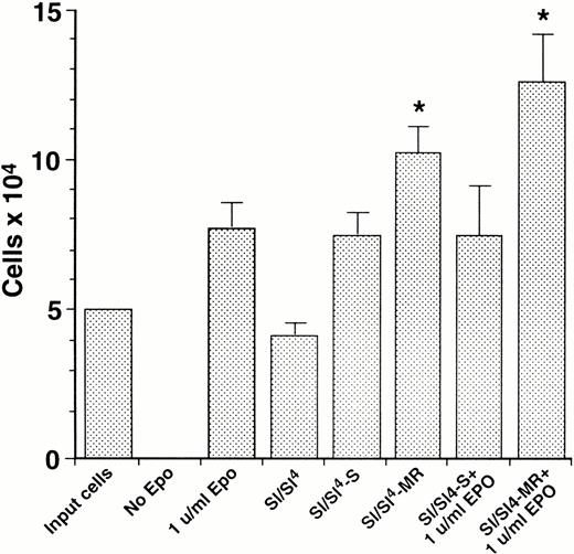
As shown in Fig 4, both Sl/Sl4-S and Sl/Sl4-MR stromal cells support the survival of HCD57 cells in the absence of exogenous EPO. However, Sl/Sl4-MR stromal cells stimulate the proliferation of these cells at significantly higher levels compared with Sl/Sl4-S (Fig 4). Addition of EPO to stromal cell cultures expressing the MR form of SCF showed a significantly greater effect on the growth of HCD57 cells compared with the soluble form of SCF (Fig4).
Survival and proliferation of factor-dependent HCD57 cells in response to in vitro stimulation by SCF isoforms or by a combination of SCF isoforms and rhEPO. HCD57 cells were factor-starved, and stromal cells expressing various forms of SCF were mitomycin C–treated as described in Materials and Methods. HCD57 cells were then plated at 1 × 10⁴ cells per mL on day 1 (Input cells) in medium without growth factor (No Epo), supplemented with rhEPO (1 μ/mL Epo), or cocultured with parental Sl/Sl4 cells (Sl/Sl4) or with stromal cells expressing either the soluble (Sl/Sl4-S) or the MR (Sl/Sl4-MR) form of SCF alone or in combination with rhEPO (Sl/Sl4-S + 1 μ/mL rhEPO or Sl/Sl4-MR + 1 μ/mL rhEPO). Viable cells were counted after 48 hours of culture. Data represent the mean ± SEM (bars) for each group on one of the two representative experiment done in triplicate. (*)P < .05 Sl/Sl4-MR versus Sl/Sl4-S and (*)P < .05 Sl/Sl4-MR + 1 μ/mL rhEPO versus Sl/Sl4-S + 1 μ/mL rhEPO.
Survival and proliferation of factor-dependent HCD57 cells in response to in vitro stimulation by SCF isoforms or by a combination of SCF isoforms and rhEPO. HCD57 cells were factor-starved, and stromal cells expressing various forms of SCF were mitomycin C–treated as described in Materials and Methods. HCD57 cells were then plated at 1 × 10⁴ cells per mL on day 1 (Input cells) in medium without growth factor (No Epo), supplemented with rhEPO (1 μ/mL Epo), or cocultured with parental Sl/Sl4 cells (Sl/Sl4) or with stromal cells expressing either the soluble (Sl/Sl4-S) or the MR (Sl/Sl4-MR) form of SCF alone or in combination with rhEPO (Sl/Sl4-S + 1 μ/mL rhEPO or Sl/Sl4-MR + 1 μ/mL rhEPO). Viable cells were counted after 48 hours of culture. Data represent the mean ± SEM (bars) for each group on one of the two representative experiment done in triplicate. (*)P < .05 Sl/Sl4-MR versus Sl/Sl4-S and (*)P < .05 Sl/Sl4-MR + 1 μ/mL rhEPO versus Sl/Sl4-S + 1 μ/mL rhEPO.
MR SCF induced c-kit activation results in sustained phosphorylation of the EPO-R in HCD57 cells.
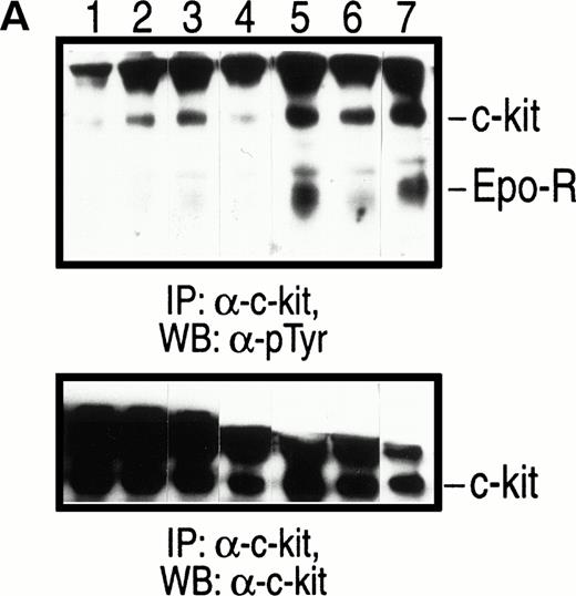
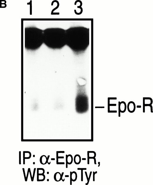
To determine if coculturing HCD57 cells with stromal cells expressing only the MR form of SCF was associated with differences in activation of c-kit and/or downstream signaling from c-kit, mitomycin C–treated confluent stromal cells were further incubated for 24 to 48 hours. Factor-starved HCD57 cells were loaded on these stromal layers and cocultured for various periods of time, then lysed in lysis buffer. Soluble cellular protein derived from the coculture were analyzed by IP using an anti–c-kit antibody followed by Western blot analysis with an anti-phosphotyrosine antibody (Fig 5A). In the presence of SCF ligand expressed in stromal cells, IP with an anti–c-kitantibody resulted in coimmunoprecipitation of c-kit and EPO-R. Consistent with previous observations of this cell line, only the slower-migrating glycosylated form of c-kit was tyrosine phosphorylated upon SCF stimulation (Fig 5A). Despite high level expression of c-kit on HCD57 cells (data not shown), no detectable c-kit tyrosine phosphorylation was observed upon stimulation of HCD57 cells with EPO only (Fig 5B). Tyrosine phosphorylation of both c-kit and EPO-R was induced within 10 minutes of coculture with stroma expressing either form of SCF (Fig 5A, lanes 2 and 3). However, tyrosine phosphorylation of both c-kitand EPO-R was greater in cells cocultured on stroma expressing only the MR form of SCF (Fig 5A, lane 3). This increase was noted as early as 10 minutes after coculture and became more apparent after 60 and 120 minutes. As shown in Fig 5A, lanes 5 and 7, coculture of HCD57 cells with stromal cells expressing only the MR form of SCF resulted in elevated and sustained phosphorylation of both c-kit and EPO-R. In contrast, as noted in Fig 5A, lanes 4 and 6, phosphorylation of EPO-R by stromal cells expressing only the soluble form of SCF was consistently lower than that induced by MR SCF throughout the course of coculture in two separate experiments. An apparent decrease in the phosphorylation of c-kit and EPO-R observed after 60 minutes of coculture with stromal cells expressing the soluble form of SCF (Figure5A, lane 4) was caused by under loading of the protein as seen in Fig5A, bottom panel. As a loading control, Western blot probed with an anti-phosphotyrosine antibody was stripped and reprobed with an anti–c-kit antibody (Fig 5A, bottom panel). In summary, these data suggest that MR form of SCF induces a greater level of phosphorylation of both c-kit and EPO-R in HCD57 cells, which is significantly prolonged compared with soluble SCF.
Coimmunoprecipitation and phosphorylation of c-kit and EPO-R in HCD57 cells. (A) Stimulation with MR and soluble SCF. Factor-starved HCD57 cells were cocultured with mitomycin C–treated stromal cells expressing either the soluble (Sl/Sl4-S) or the MR (Sl/Sl4-MR) form of SCF for various periods of time at 37°C as described in Materials and Methods. Subsequently at various times, cell lysates were collected and subjected to IP with an anti–c-kit antibody and WB analysis with an anti-phosphotyrosine antibody and the enhanced chemiluminescence detection system. Coimmunoprecipitated and tyrosine-phosphorylated Golgi-processed c-kit (slow migrating)27 and EPO-R are indicated. Lane 1 corresponds to parental Sl/Sl4 cells cocultured for 10 minutes; lanes 2, 4, and 6 correspond to Sl/Sl4-S cells cocultured for 10, 60, and 120 minutes, respectively; lanes 3, 5, and 7 correspond to Sl/Sl4-MR cells cocultured for 10, 60, and 120 minutes, respectively. (B) Stimulation with EPO. Factor-starved HCD57 cells were exposed to no growth factor (lane 1) or stimulated with 10 μ/mL rhEPO for 5 minutes (lane 2) and for 10 minutes (lane 3). Cell lysates were subjected to IP using an anti–EPO-R antibody and WB using an anti-phosphotyrosine antibody as described above. Tyrosine-phosphorylated EPO-R is indicated.
Coimmunoprecipitation and phosphorylation of c-kit and EPO-R in HCD57 cells. (A) Stimulation with MR and soluble SCF. Factor-starved HCD57 cells were cocultured with mitomycin C–treated stromal cells expressing either the soluble (Sl/Sl4-S) or the MR (Sl/Sl4-MR) form of SCF for various periods of time at 37°C as described in Materials and Methods. Subsequently at various times, cell lysates were collected and subjected to IP with an anti–c-kit antibody and WB analysis with an anti-phosphotyrosine antibody and the enhanced chemiluminescence detection system. Coimmunoprecipitated and tyrosine-phosphorylated Golgi-processed c-kit (slow migrating)27 and EPO-R are indicated. Lane 1 corresponds to parental Sl/Sl4 cells cocultured for 10 minutes; lanes 2, 4, and 6 correspond to Sl/Sl4-S cells cocultured for 10, 60, and 120 minutes, respectively; lanes 3, 5, and 7 correspond to Sl/Sl4-MR cells cocultured for 10, 60, and 120 minutes, respectively. (B) Stimulation with EPO. Factor-starved HCD57 cells were exposed to no growth factor (lane 1) or stimulated with 10 μ/mL rhEPO for 5 minutes (lane 2) and for 10 minutes (lane 3). Cell lysates were subjected to IP using an anti–EPO-R antibody and WB using an anti-phosphotyrosine antibody as described above. Tyrosine-phosphorylated EPO-R is indicated.
These in vitro observations, along with the fact thatSl/Sld mutants show severe deficiencies in the erythroid lineage, suggest that membrane presentation of SCF may be important in the normal development of these cells in vivo. Therefore, we hypothesized that transgene expression of MR-SCF would have more profound effects than soluble SCF on the development of erythroid lineage in Sl mutants in vivo. We tested this hypothesis using transgenic mice expressing either the soluble or the MR form of SCF on a Sl/Sld genetic background.
Generation of Sl/Sldmice that overexpress soluble SCF or express the MR isoform of SCF.
Transgene positive male founders were obtained following pronuclear injection of the human phosphoglycerate kinase (hPGK)-murine (m) SCF248 and hPGK-mSCFX9/D3 expression plasmids, respectively, (Fig 1) into C3H/HeJ fertilized eggs. The hPGK promoter (pr) was used to express the SCF transgene, because we and others have successfully shown long-term and stable expression of transgenes, including the human SCF cDNA, using this promoter in transduced hematopoietic cells and in transgenic mice.36 37 Selected on the basis of comparable levels of expression of transgenes, two founders, PGK 248-F4 (hereafter referred to as mSCF248) and PGK X9/D3-2 (hereafter referred to as mSCFX9/D3) were bred to C3H/HeJ to establish transgenic lines. As expected the transgene was inherited by 50% of the offspring (data not shown). Founders, as well as their progeny carrying the transgene, appeared normal and fertile with no gross phenotypic abnormalities (data not shown). Very similar levels of expression of both transgenes were observed in stromal cells derived from bone marrow of transgenic mice (Fig 2) and several other primary tissues (data not shown). The steady state level of each transgene mRNA is similar in multiple tissues examined using comparison to actin mRNA derived from the same cells (data not shown). In contrast to transgenic mice and as expected based on the primers used, no SCF transcript was amplified from mRNA derived from bone marrow cells (Fig 2) or primary tissue (data not shown) from Sl/Sld mutant mice.
Compound heterozygous Sl/Sld mutant mice, in which only soluble truncated SCF protein is produced, are viable but show severe hematological abnormalities, including anemia. In an effort to determine the in vivo role of membrane presentation of SCF in hematopoiesis, we performed genetic crosses of transgenic animals expressing the soluble and MR isoforms of SCF intoSld mutants to generateSl/Sld-mSCFX9/D3(Sl/Sld-MR) andSl/Sld-mSCF248(Sl/Sld-S) mice. Briefly, mice carrying either of the transgenes were first bred to Sl/+ mice. Crosses involving these mice produced progeny in the expected ratios, with approximately 25% having the genotype Sl/+and carrying the transgenes (ie,Sl/+-mSCF248 orSl/+-mSCFX9/D3). These mice were identified on the basis of their phenotype (white forehead blaze and a diluted belly) and Southern blot analysis (data not shown).Sl/+-mSCF248 orSl/+-mSCFX9/D3 male mice were chosen for further breedings and were mated toSld/+ female mice. All black-eyed white mice were further characterized along with their wt littermates for the presence or absence of the transgene by Southern blot analysis. As expected, approximately 8% of the progeny were of the genotypeSl/Sld-mSCF248(Sl/Sld-S) orSl/Sld-mSCFX9/D3(Sl/Sld-MR) (data not shown), and these animals and their wt littermates were studied in detail. All mice were killed and analyzed completely at 12 weeks of age.
Expression of MR SCF as a transgene in Sl/Sldmice is critical for erythropoiesis.
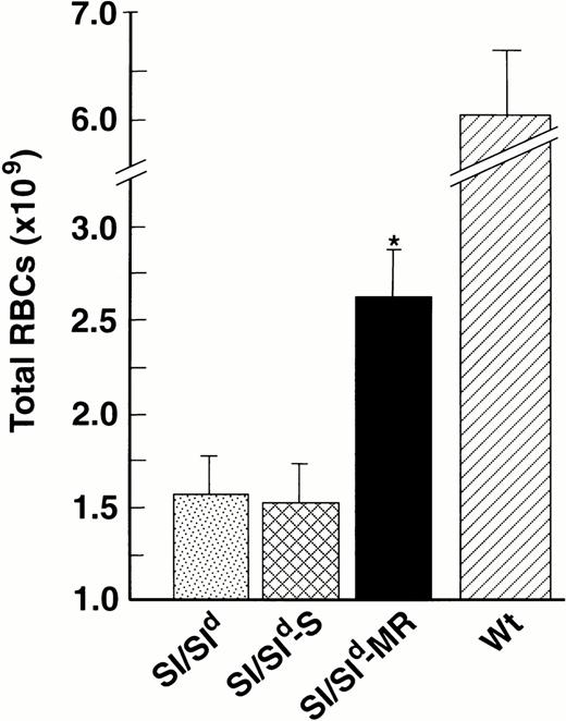
To ascertain if the expression of either transgene in these mice rescued deficiencies in the production of mature red cells, we analyzedSl/Sld, Sl/Sld-MR, andSl/Sld -S mice for total red cell production. Our results show a 40% increase in total red cell production inSl/Sld-MR mice as compared withSl/Sld mice (P = .01) (Fig6). Peripheral red cell counts (Table 1) and hematocrits (data not shown) in Sl/Sld-MR mice were also significantly higher than Sl/Sld orSl/Sld-S mice. In contrast, red cell production was not increased in Sl/Sld-S mice when compared withSl/Sld. As seen in Fig 6 and Table1, in comparison with wt mice the expression of MR SCF transgene in Sld mutants does not completely correct the red cell deficiency, a result which may relate to the level or timing of transgene expression during development.
Expression of MR SCF in Sl/Sld mice improves red cell production. Total red cells were compared betweenSl/Sld (n = 16), Sl/Sld-S (n = 10), Sl/Sld-MR (n = 13), and wt (n = 20) mice. All black-eyed white mice and wt mice at 12 weeks of age were tail bled, and their RBC counts/mL were determined as described in Materials and Methods. Total red cells in each animal were calculated by multiplying the mean blood volume of an adult animal; (3.15 mL/100 g)55 by the total body weight (in grams) and the RBC counts per mL. The results show mean (×109) ± SEM (bars). (∗) P = .01 ,Sl/Sld-MR versus Sl/Sldand Sl/Sld-S.
Expression of MR SCF in Sl/Sld mice improves red cell production. Total red cells were compared betweenSl/Sld (n = 16), Sl/Sld-S (n = 10), Sl/Sld-MR (n = 13), and wt (n = 20) mice. All black-eyed white mice and wt mice at 12 weeks of age were tail bled, and their RBC counts/mL were determined as described in Materials and Methods. Total red cells in each animal were calculated by multiplying the mean blood volume of an adult animal; (3.15 mL/100 g)55 by the total body weight (in grams) and the RBC counts per mL. The results show mean (×109) ± SEM (bars). (∗) P = .01 ,Sl/Sld-MR versus Sl/Sldand Sl/Sld-S.
Comparison of Peripheral Blood Parameters, Bone Marrow Cellularity (BMC), and Multilineage Progenitors Between Transgenic and Nontransgenic Sl/Sld Mice
| Mice . | BMCa . | CFU-MIXa . | RBCs . | WBCs . |
|---|---|---|---|---|
| Sl/Sld | 35.04 ± 1.6 | 5,249 ± 731 | 2,605 ± 252 | 8.25 ± 0.4 |
| Sl/Sld-MR | 45.11 ± 2.8e | 11,162 ± 5,249c | 3,506 ± 277b | 10.6 ± 0.8f |
| Sl/Sld-S | 39.06 ± 2.9 | 11,352 ± 1,713d | 2,649 ± 279 | 10.38 ± 1.1 |
| Wt | 68.22 ± 3.4 | 9,774 ± 998 | 7,949 ± 420 | 10.85 ± 0.9 |
CFU-MIX, white and red cell counts were performed as described in methods. Results are expressed as mean ± SEM.aper 2 hind limbs; bP < .05 versus Sl/Sld; cP < .05 versus Sl/Sld; dP < .01 versus Sl/Sld; P = .01 versus Sl/Sld; fP = .058 versus Sl/Sld. A minimum of 8 mice were analyzed from each group. White and red blood cell counts are expressed as mean × 103/mm3. Bone marrow cellularity (BMC) is expressed as mean × 106 per 2 hind limbs.
Expression of MR SCF as a transgene improves runting associated with Sl/Sldmice.
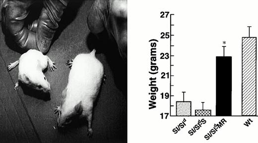
The most obvious phenotypic difference betweenSl/Sld and Sl/Sld transgenic mice, the presence of severe runting, was observed shortly after birth. As seen in Fig 7, left panel, 12-week-oldSl/Sld-MR mice appeared larger and healthier than either Sl/Sld or Sl/Sld-S mice. Quantitatively, the expression of MR SCF as a transgene significantly improved the total body weight of these animals in comparison to nontransgenic mice (P = .01; Fig 7, right panel). In contrast, no increase in body weight was observed inSl/Sld mice that overexpressed the soluble form of SCF as a transgene (Sl/Sld-S).
Expression of MR SCF improves runting associated withSl/Sld mice. Shown in the left panel are pictures of 12-week-old mutant mice that either express the MR form of SCF or overexpress the soluble form. The right panel shows the weights of these mice and their comparison with wt littermates. Weight comparison between Sl/Sld (n = 16),Sl/Sld-S (n = 10),Sl/Sld-MR (n = 14), and wt (n = 20) mice. All black-eyed white mice and wt mice at 12 weeks of age were weighed. The results show mean ± SEM (bars). (*) P = .01,Sl/Sld-MR versus Sl/Sldand Sl/Sld-S.
Expression of MR SCF improves runting associated withSl/Sld mice. Shown in the left panel are pictures of 12-week-old mutant mice that either express the MR form of SCF or overexpress the soluble form. The right panel shows the weights of these mice and their comparison with wt littermates. Weight comparison between Sl/Sld (n = 16),Sl/Sld-S (n = 10),Sl/Sld-MR (n = 14), and wt (n = 20) mice. All black-eyed white mice and wt mice at 12 weeks of age were weighed. The results show mean ± SEM (bars). (*) P = .01,Sl/Sld-MR versus Sl/Sldand Sl/Sld-S.
Overexpression of soluble SCF in Sl/Sldmice increases the frequency of myeloid progenitors in the bone marrow.
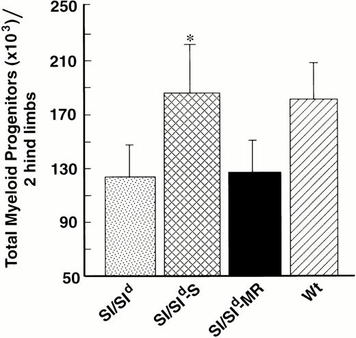
In addition to anemia, Sl/Sld mice show bone marrow hypocellularity and myeloid cell deficiency. To determine if expression of either isoform of SCF as a transgene affected these abnormalities, we compared the bone marrow cellularity of Sl/Sldmice with that of Sl/Sld-S andSl/Sld-MR mice. A significant increase in the BM cellularity was evident in Sl/Sld-MR mice compared with Sl/Sld animals (Table 1). A modest, but nonsignificant increase in the total BM cellularity was observed inSl/Sld mice overexpressing the soluble isoform of SCF. Because marrow cellularity does not necessarily reflect more immature clonogenic compartments, we compared progenitor cell numbers, including high proliferative potential (HPP) and low proliferative potential (LPP) colony-forming cells (CFC), and lineage-restricted progenitors in Sl/Sld,Sl/Sld-S, and Sl/Sld-MR mice. In contrast to the lack of effect on erythropoiesis, transgene overexpression of soluble SCF (mSCF248) inSl/Sld mice completely corrected the deficiency of myeloid progenitors in the bone marrow (Fig8). Total bone marrow myeloid progenitor content (ie, HPP-CFC + LPP-CFC) in Sl/Sld-S was significantly greater than that observed in eitherSl/Sld or Sl/Sld-MR mice (P = .005). The increase in myeloid progenitors inSl/Sld-S mice represented both primitive HPP-CFC (67,601 ± 11,172 v 40,519 ± 4,336,Sl/Sld-S vSl/Sld,respectively) and more mature LPP-CFC (286,034 ± 33,212 v208,598 ± 23,421, Sl/Sld-S vSl/Sld, respectively). This correction in myeloid progenitor content occurred in spite of the BM hypocellularity seen in these animals (Table 1). The expression of either transgene completely corrected the deficiency of multipotential clonogenic cells, colony forming unit-mix (CFU-MIX), which contain cells of both erythroid and myeloid lineages (Table 1). No significant differences were noted in the number of primitive erythroid progenitor blast forming unit-erythroid (BFU-E) between the transgenic mice expressing either isoform of SCF (data not shown). In addition, the peripheral white blood cell counts were improved by expression of either isoform (Table1). This increase in peripheral leukocyte count was mainly caused by an increase in neutrophils (data not shown). Thus, both transgene-expressed isoforms of SCF are biologically active in vivo and can differentially rescue specific hematologic defects inSl/Sld mice.
Overexpression of soluble SCF increases the frequency of myeloid progenitors in the bone marrow. HPP-CFC and LPP-CFC assays were performed on bone marrow cells derived from various transgenic and nontransgenic white mice. Shown are total myeloid progenitors (ie, sum of all HPP and LPPs) from Sl/Sld (n = 22),Sl/Sld-S (n = 13),Sl/Sld-MR (n= 14), and wt (n = 20). HPP and LPP content were determined by multiplying the concentration of HPP-CFC or LPP-CFC by the total bone marrow cellularity. (*) P= .005, Sl/Sld-S versusSl/Sldand Sl/Sld-MR. The results show mean (×103) ± SEM (bars).
Overexpression of soluble SCF increases the frequency of myeloid progenitors in the bone marrow. HPP-CFC and LPP-CFC assays were performed on bone marrow cells derived from various transgenic and nontransgenic white mice. Shown are total myeloid progenitors (ie, sum of all HPP and LPPs) from Sl/Sld (n = 22),Sl/Sld-S (n = 13),Sl/Sld-MR (n= 14), and wt (n = 20). HPP and LPP content were determined by multiplying the concentration of HPP-CFC or LPP-CFC by the total bone marrow cellularity. (*) P= .005, Sl/Sld-S versusSl/Sldand Sl/Sld-MR. The results show mean (×103) ± SEM (bars).
DISCUSSION
The phosphorylation of RTKs on tyrosine by their cognate ligands is central to the regulation of the pathways of signal transduction that control cellular proliferation and differentiation.1 2 The net levels of protein phosphorylation depend on the strength of the receptor-ligand interaction. In the present study we have examined in vitro and in vivo the interaction between the soluble and MR isoforms of SCF and the RTK, c-kit. We show that MR SCF plays an important role in the maturation of erythroid progenitors to mature red cells. In contrast, soluble SCF may be more important for the normal development of myeloid lineage. These results show that expression of different isoforms of SCF may result in different cellular outcomes both in vitro and in vivo depending on the hematopoietic cell lineages in which ligand/receptor interaction occurs.
A large number of studies have shown that recombinant soluble SCF (rSCF) in combination with other cytokines significantly enhances the growth of clonogenic cells in vitro.38-43 Soluble SCF plus EPO primarily increase the number and size of BFU-Es, whereas SCF plus granulocyte colony-stimulating factor result in enhanced neutrophil colonies.23,24 Soluble SCF has also been shown to synergize with other cytokines, such as IL-7, in the development of lymphoid lineage. Mutations in the RTK c-kit and the common cytokine gamma chain reduce cellularity, but are permissive to thymocyte development.44,45 However, mice that lack both c-kit and the gamma chain show complete abrogation of thymocyte development.46 EPO and EPO-R knock out mice have been reported and show that neither EPO nor the EPO-R is required for erythroid lineage commitment.26 However, EPO and EPO-R are crucial in vivo for the proliferation and survival of later erythroid progenitors.26 Despite increased circulating levels of EPO in Sl/Sld mice, these animals still show deficiencies in the erythroid lineage, suggesting that EPO by itself is not sufficient to induce normal proliferation and/or differentiation of committed progenitors to mature red cells inSl/Sld mutants.4,22 Recent observations showing the activation of one receptor by a second receptor of a different cytokine family in response to ligation provides a potential explanation for the diversity of effects seen with different growth factors, including soluble rSCF. In this regard, it has also been shown that soluble SCF can activate the IL-3 receptor in a myeloid cell line in an IL-3– independent fashion.47 More recently, it has been shown that very high concentrations of soluble rSCF (1,000 ng/mL) can lead to transient phosphorylation of the EPO-R and stimulate proliferation of an erythrocytic progenitor cell line in an EPO-independent fashion.27 Although useful, these in vitro and in vivo studies, using high concentrations of soluble rSCF, do not completely define the role of the cytokine in normal hematopoietic microenvironment. In addition, because naturally occurring murineSl mutants are deficient in SCF/c-kit interactions throughout development, the function of the differentially spliced isoforms of SCF in vivo in definitive hematopoiesis is not well understood.
Many proteins exist as both MA and soluble isoforms, and considerable interest has been generated by the recent finding that certain growth factors, classically thought of as autocrine factors, not only exist as MA proteins but also are active as such.12 These factors may participate in a novel mode of intercellular communication restricted to adjacent cells, termed juxtacrine stimulation.12 For instance, cleavage of pro–TGF-α does not result in the generation of an active form of the factor from a precursor, but rather results in the conversion of one active form (MA) to another (soluble).12 For TNF it has been shown that TNF receptor (TNF-R) can be strongly stimulated by MA form of TNF compared with soluble TNF. Moreover, upon activation of the TNF-R by MA TNF, a phenotypic switch of the cellular response pattern to TNF can be observed. In this case, cells fully resistant to the cytotoxic action of soluble TNF become susceptible and are killed upon contact with membrane TNF.13
Previously, we have shown that primary human progenitor cells and hematopoietic cell lines proliferate and differentiate in distinct patterns when exposed to stromal cells expressing either MA or soluble SCF and that these differences are correlated with distinct patterns of c-kit activation.25,48 We have shown that the activation of c-kit tyrosine kinase persists longer when stimulated with MA SCF compared with soluble SCF, and that the length of activation of c-kit could be shortened upon adding soluble SCF to cultures producing MA SCF.48 These data suggest that differences in the kinetics of c-kit activation are dependent on the isoform of SCF presented. However, in vitro stromal cultures favor myeloid over erythroid differentiation, making the interpretation of these experiments with respect to the function of SCF in erythroid cell development difficult. This is a major shortcoming, because anemia is the most prominent hematopoietic phenotypic abnormality seen inSl and W mutants in vivo. To further study the function of each SCF isoform in vitro and in vivo, we have combined in vitro cellular and biochemical studies with an in vivo genetic approach. Our data in vitro using an erythrocytic progenitor cell line shows that MR isoform of SCF induces an elevated and sustained tyrosine phosphorylation not only of c-kit but also of EPO-R and significantly greater proliferation of this cell line compared to soluble SCF. Membrane presentation of SCF in vivo also resulted in the production of significantly more red blood cells. In contrast, expression of either SCF isoform appeared to be sufficient for the generation of multilineage progenitors, CFU-MIX, and for early erythroid BFU-E. Finally, soluble SCF appeared to be a potent growth factor in the development of myeloid progenitors, such as HPP-CFC and LPP-CFC. This finding is not surprising, because at least one group of investigators used HPP-CFC colony stimulation in the cloning of soluble SCF.38 Of relevance to these observations, the differential findings of erythroid and myeloid lineage cells, the similar effects on CFU-MIX, and the similarities in expression of the transgenes would argue against the level of expression of the two transgenes per se being responsible for the observed phenotypic differences.
Previously it has been shown that stimulation of cells expressing c-kit with soluble SCF induces rapid downmodulation of cell surface c-kit expression and degradation of metabolically radiolabeled c-kit protein.49,50 Degradation after internalization of the ligand-receptor complex has been reported in many other receptor systems including the PDGF and the CSF-1 receptors, which are structurally related to c-kit and may be a mechanism of modulation of signaling pathways and cellular responses.50,51 Therefore, we hypothesize that the regulation of EPO-R activation, at least in part, may be caused by the length of time activated c-kit remains membrane-associated in erythroid progenitors in vivo. Alternatively, MR SCF may be more efficient than soluble SCF in forming dimers and, subsequently, activating EPO-R. Recent reports have indicated that EPO activates EPO-R by inducing dimerization. However, c-kit activates the EPO-R probably through phosphorylation of tyrosine residues in the cytosolic domain rather than EPO-R dimerization.52Therefore, during erythroid differentiation, activation of the EPO-R may take place by two different mechanisms, which may lead to different intracellular signals essential for survival, proliferation, and terminal differentiation of committed erythroid progenitors to produce mature red cells. At least one signal emanating from c-kit must use the EPO-R as a downstream signal-transduction protein, because EPO-R−/− fetal liver cells after transduction and expression of exogenous EPO-R require SCF in vitro to further differentiate.52 Thus, our in vitro and in vivo data would suggest that the terminal differentiation and/or proliferation of late erythroid progenitors to mature red cells requires MA SCF/c-kit–mediated activation, presumably because of phosphorylation of the EPO-R both by EPO-mediated and c-kit–mediated ligation. In this regard, we have observed a significant increase in the production of late erythroid progenitors (CFU-Es) in Sl/Sld mice expressing the MR isoform of SCF (R.K. and D.A.W., unpublished observation, August 1997).
Despite the significant improvement in various hematopoietic lineages in Sl/Sld mice expressing the two isoforms of SCF, neither transgene resulted in the rescue of germ cells, mast cells, and melanocytes in these mutants. Characterization of the SCF promoter and subsequent use of this promoter to generate transgenic mice in the future should help to ascertain the in vivo role of SCF isoforms in other lineages. SCF has been shown to be expressed at high levels during embryogenesis in cells associated with both the migratory pathways of melanoblasts and germ cells.53 In this regard, either the dysregulation or level of expression of SCF via the PGK promoter may be the reason for the lack of affects on other lineages as observed in this study. Indirect evidence to support this explanation comes from our previous report of mild melanocyte deficiencies caused by interference of endogenous SCF/c-kit interactions in vivo by PGK-expressed human SCF during development.36,37 On the other hand, once present in the dermis, melanocytes can respond to SCF transgene expression because pigmentation is enhanced when expression of mSCFX9/D3 is directed by an epidermal-specific promoter in wt transgenic mice.56 In this regard, we have occasionally seen mild and scattered pigmentation inSl/Sld mice overexpressing soluble SCF (R.K. and D.A.W., unpublished observation, January 1997).
In summary, we have shown that both soluble and MR SCF can enhance the production of multilineage progenitors. Soluble SCF plays a significant role in the production of myeloid progenitors, however, MA SCF may be more important for the normal development of the erythroid lineage. SCF is a unique example of modulation of target cell responses based on the provision of alternatively spliced forms acting through a common receptor, c-kit.
ACKNOWLEDGMENT
We thank the members of our laboratory and Drs M. Dinauer, W. Clapp, and M. Yoder of the Herman B Wells Center for Pediatric Research for critical reading of the manuscript. We thank Drs Frank Martin and Keith Langley for originally furnishing mSCF cDNAs and Dr Ian McNeice for supplying rrSCF. We also thank Dr H. Lodish for providing HCD57 cell line and Dr G. Krystal for supplying anti–EPO-R antibody.
Supported by R01 DK48605.
Address reprint requests to David A. Williams, Howard Hughes Medical Institute, Herman B Wells Center for Pediatric Research, James Whitcomb Riley Hospital for Children, Cancer Research Building, 1044 W Walnut St, Room 406C, Indianapolis, IN 46202.
The publication costs of this article were defrayed in part by page charge payment. This article must therefore be hereby marked “advertisement” in accordance with 18 U.S.C. section 1734 solely to indicate this fact.

This feature is available to Subscribers Only
Sign In or Create an Account Close Modal