Abstract
Glycoprotein (GP)Ib-IX-V is one of the major transmembrane complexes present on the platelet surface. Its extracellular domain binds von Willebrand factor (vWF) and thrombin, while its intracellular domain associates tightly with the cytoskeleton through the actin-binding protein (ABP)-280, also known as filamin. In the present study, a full-length cDNA coding for a human ABP homologue has been cloned and sequenced. This protein was identified by the yeast two-hybrid screening procedure via its interaction with the intracellular domain of GPIb. Initially, a 1.3-kb partial cDNA was isolated from a megakaryocyte-like cell line (K562) cDNA library followed by a full-length cDNA of 9.4 kb that was identified in a human placenta library. The full-length cDNA encoded a protein of 2,578 amino acids with a calculated molecular weight of 276 kD (ABP-276). The amino terminal 248 amino acids contained an apparent actin binding domain followed by 24 tandem repeats each containing about 96 amino acids. The amino acid sequence of the protein shared a high degree of homology with human endothelial ABP-280 (70% identity) and chicken filamin (83% identity). However, the 32 amino acid Hinge I region in ABP-280 that contains a calpain cleavage site conferring flexibility on the molecule, was absent in the homologue. An isoform containing a 24 amino acid insertion with a unique sequence at the missing Hinge I region was also identified (ABP-278). This isoform resulted from alternative RNA splicing. ABP-276 and/or ABP-278 were present in all tissues examined, but the relative amount varied in that some tissue contained both forms, while other tissue contained predominately one or the other.
© 1998 by The American Society of Hematology.
THE GLYCOPROTEIN (GP)Ib-IX-V complex is the major transmembrane receptor for von Willebrand factor (vWF) on the platelet surface and is present at about 25,000 copies per platelet.1 It contains four type I single-pass transmembrane proteins including GPIbα, GPIbβ, GPIX, and GPV associated in a molar ratio of 2:2:2:1.2 GPIbα and GPIbβ are linked by a disulfide bond,3 while GPIX is linked to the complex by noncovalent bonds.4 GPV is weakly associated with two GPIb-IX molecules in the complex.5 The N-terminal extracellular domain of GPIbα binds vWF and also contains a high-affinity binding site for thrombin,6,7 while the extracellular domain of GPV contains cleavage sites for both thrombin and calpain.5
The GPIb-IX-V complex is involved in mediating several activities important for normal platelet function, including the initial adhesion of platelets to the subendothelium after vascular damage, the activation of platelets by very low concentrations of thrombin, and the regulation of actin polymerization and subsequent platelet shape changes.8 The binding of vWF to the GPIb-IX-V complex initiates these specific intracellular signaling pathways that lead to platelet activation, secretion, and aggregation.9 The intracellular domain of GPIbα associates with the platelet cytoskeleton by a direct interaction with actin-binding protein (ABP, also known as filamin).10,11 This association affects the shape of resting platelets and subsequently allows them to spread after activation.12 The GPIb-IX-V complex may also be a high-affinity thrombin receptor on the platelet surface that is coupled to platelet activation through the protein 14-3-3 ζ,13which has been shown to associate with the GPIb-IX-V complex via the intracellular domain of the GPIbα subunit. The 14-3-3 ζ is known to activate the Raf-1 signaling pathway.14 An increase in the cyclic adenosine monophosphate (cAMP) level in platelets is accompanied by phosphorylation of serine 166 in the intracellular domain of GPIbβ by a cAMP-dependent protein kinase,15mediating the inhibition of actin polymerization.16
ABP-280 is a dimeric protein that self-associates in nonmuscle cells and defines the three-dimensional organization of actin filaments in the submembraneous cortex. The N-terminal of each subunit of ABP-280 contains an actin binding domain, followed by a semiflexible rod-like domain consisting of 24 tandem repeats, each approximately 96 amino acids in length.17 Each repeat sequence is predicted to have six to eight short β-sheets, and these repeats interact intramolecularly to form a rigid rod-like structure. The repeats are interrupted by two insertions located between repeats 15 and 16 and between repeats 23 and 24. These insertions, which are called the Hinge I and Hinge II regions respectively, coincide with swivel regions in the molecule and are believed to confer flexibility to the otherwise rigid rod-like structure. Repeat 24 contains the self-association site,17,18 while repeats 17-19 contain the binding site for the intracellular domain of the GPIbα.19 The gene for ABP-280 has been mapped to Xq28.20 The partial sequence of a homologue of ABP-280, expressed exclusively in skeletal muscle and heart, was 72% identical to ABP-280 over two regions of the molecule.20 The gene for this homologue was mapped to chromosome 7.20 Several membrane proteins, including the acetycholine receptor, the immunoglobulin receptor FcγIR, the CD18 subunit of the β2 integrin, and the thyroid stimulating hormone receptor, have been shown to colocalize and interact with ABP-280.21-24
By using the yeast two-hybrid system, a partial cDNA coding for a protein that interacted with the intracellular domain of GPIbα has been isolated from a megakaryocyte-like cell line K562. Subsequently, a full-length cDNA for this protein was isolated and characterized from a human placenta library. These results show that the encoded protein is a homologue of the two other human ABPs previously described.17 20
MATERIALS AND METHODS
Two-hybrid screening.
The sequence encoding the intracellular domain of GPIbα (His515-Leu610) was amplified by polymerase chain reaction (PCR) from a human erythroleukemia cell (HEL) cDNA library.25 The PCR product was cloned directly into the yeast two-hybrid vector PAS2-1 (Clontech, Palo Alto, CA), creating a fusion of the GAL4 DNA binding domain with the intracellular domain of GPIbα. This fusion bait construct (GPIbα/PAS2-1) was transformed into the yeast strain CG-1945, and the resulting Trp+ strain was used subsequently to screen a K562 cDNA library (Clontech), in which cDNAs were cloned into the PACT2 (Clontech) vector. This generated a hybrid protein with the GAL4 activation domain. Interaction of the cDNA hybrid protein with the GPIbα-GAL4 DNA binding fusion protein led to reconstitution of a functional GAL4 transcription activator and expression of the reporter His3 gene. For further confirmation, transformants with the His+ phenotype were tested for expression of a second reporter gene lacZ using a filter assay for β-galactosidase activity. All yeast manipulations, as well as β-galactosidase enzyme activity assays, were performed as recommended by the supplier (Clontech).
Nucleotide sequencing.
cDNA inserts were sequenced by the dideoxy chain termination method26 using the Sequenase Kit from US Biochemical, Cleveland, OH. All sequences reported were determined on both strands.
Cloning of full-length cDNA.
A subdivided human placenta cDNA library (ZymoGenetics Inc, Seattle, WA) was screened by a combination of PCR and colony hybridization using the partial cDNA sequence as a probe. Briefly, human placenta cDNA was synthesized and cloned into a plasmid vector pZP9 (ZymoGenetics Inc). A total of 500 miniprep DNAs (HPA001-500) were prepared from 500 plates, each containing about 10,000 colonies. The miniprep DNAs were combined into 50 DNA mixtures (HPB01-50) and subsequently into five DNA mixtures (HPC1-5). For library screening, PCR amplification was first performed with a pair of primers at the 5′ end of the partial cDNA sequence, using DNA mixtures HPC1-5 as templates. The positive PCR signal was further traced by screening the corresponding constituent DNA mixtures in the HPB01-50 collection. Eventually, single miniprep DNA in the HPA001-500 collection containing the positive clone was identified. One microgram of DNA from this miniprep was transformed into E. coli strain INVα’F (Invitrogen, Carlsbad, CA), plated, and screened with a radioactively-labeled DNA probe using conventional colony hybridization method.
Analysis of alternative RNA splicing.
To study the presence or absence of a 24 amino acid Hinge I region in ABP-278 or ABP-276, fragments of genomic sequence encompassing the Hinge I region were amplified by PCR with primers P3, P4, P5, and P6, using human genomic DNA (GIBCO/BRL, Gaithersburg, MD) as a template. The primers P3 and P4 were located at the flanking regions of Hinge I and were specific for the ABP homologue, but not ABP-280. The PCR products were cloned into the PCR cloning vector (Invitrogen) and sequenced. Two internal PCR primers (P5 and P6) were also used to determine the size of the introns.
To analyze the tissue distribution of ABP-276 and ABP-278, cDNAs from various tissues (Clontech) were used as templates for PCR amplification with primers P3 and P4. The glyceraldehyde-3-phosphate dehydrogenase (G3PDH) sequence, amplified with primers P7 and P8 and ABP-280 with primers 9 and 10 were used as positive controls for reverse transcriptase (RT)-PCR and a rough estimate of cDNA template.
Oligonucleotides.
Oligonucleotides and their sequences were as follows: P1, GAAATGCCCTTTGACCCCTCTAAAG; P2, CTGGTCCAAAGACTTTGATCCTGCTG; P3, GTGCGCTTCGGTGGTGTTGATA; P4, CACAATCTCAGGTGTGGCTGT; P5, TGAAGGTCGGAGTCAACGGATTTGGT; P6, CATGTGGGCCATGAGGTCCACCAC; P7, TGAAGGTCGGAGTCAACGGATTTGGT; P8, CATGTGGGCCATGAGGTCCACCAC; P9, GGCAAAGTGACGTGCACCGTGTGC; P10, CTGTGATCTCGCCCTTCTTGATGGTG.
RESULTS
Cloning of an ABP homologue of ABP-280.
The yeast two-hybrid system, developed originally by Fields et al,27 is a genetic assay to detect specific protein-protein interactions in vivo and to isolate novel genes encoding proteins that associate with a known protein of interest. To identify the protein(s) interacting with the intracellular domain of platelet GPIbα, nucleotides coding for His515-Leu610 were cloned into a vector to form a fusion protein with the GAL4 DNA binding domain (GPIbα/PAS2-1). The human erythroleukemia cell line K562, which exhibits megakaryocytic characteristics, has been shown to express several subunits of platelet surface glycoproteins including GPIbα, GPIIb, and GPIIIa.28 Thus, it seemed likely that the same cell line would express protein(s) that interact with the intracellular domains of these platelet receptors. Accordingly, a K562 cDNA library, consisting of about one million clones, was screened by the yeast two-hybrid method. Four colonies showed histidine prototrophy and one of the four had β-galactosidase reporter activity. The recovered plasmid from this single isolate conferred His+ prototrophy and expression of β-galactosidase only in the presence of GPIbα/PAS2-1. Characterization and sequencing of the insert (K1.3, Fig 1) showed a cDNA fragment of 1.3 kb in length encoding a peptide that was 74% identical to the human ABP-280 isolated from endothelial cells.17
cDNA clones that span the full-length actin-binding protein homologue sequence. Clone K1.3 was isolated by the yeast two-hybrid screening, clones 1-4 were obtained by rapid amplification of 5′ cDNA ends (5′ RACE), and clones 5 and 6 were the cDNA clones isolated from a human placenta library. The diagram shows an alignment of the ABP cDNA with characteristic functional domains of the ABP subunit. The asterisk indicates the position of the 24 amino acid Hinge I insertion in ABP-278.
cDNA clones that span the full-length actin-binding protein homologue sequence. Clone K1.3 was isolated by the yeast two-hybrid screening, clones 1-4 were obtained by rapid amplification of 5′ cDNA ends (5′ RACE), and clones 5 and 6 were the cDNA clones isolated from a human placenta library. The diagram shows an alignment of the ABP cDNA with characteristic functional domains of the ABP subunit. The asterisk indicates the position of the 24 amino acid Hinge I insertion in ABP-278.
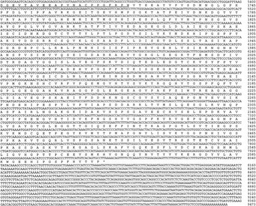
By use of the 5′ rapid amplification of cDNA ends (RACE) technique with several marathon cDNA libraries (Clontech), sequences extending to 3.8 kb were obtained (clones 1-4, Fig 1). Sequences from the immediate 5′ end of clone 4 were then used to screen a subdivided placenta cDNA library by PCR (primers P1 and P2) and colony hybridization. Two positive clones (clones 5 and 6, Fig 1) were identified, isolated, and sequenced. Clone 6 was an apparent full-length cDNA, with an open reading frame encoding a protein of 2,578 amino acids. Clone 5 was a partial cDNA that lacked 2,936 bp at the 5′ end. Clone 5 also differed from clone 6 in having an internal insertion of 72 bp, which coded for 24 additional amino acids. This insertion was also present in the partial cDNA clone 2 and was an alternatively spliced isoform (see below). The composite sequence with the 72-bp insertion is shown in Fig 2, in which the 72-bp insertion is highlighted by a double underline.
The nucleotide and predicted amino acid sequence of ABP-276 and ABP-278. The upstream in-frame stop codon (TAG) is underlined. The 24 amino acid Hinge I insertion in ABP-278 is double-underlined. These data are available from GenBank under the accession number AF043045.
The nucleotide and predicted amino acid sequence of ABP-276 and ABP-278. The upstream in-frame stop codon (TAG) is underlined. The 24 amino acid Hinge I insertion in ABP-278 is double-underlined. These data are available from GenBank under the accession number AF043045.
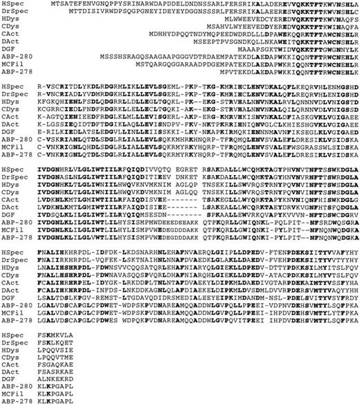
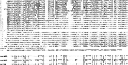
The composite cDNA was 9,473 bp in length and contained an open reading frame of 7,809 nucleotides (nucleotides 166-7974, Fig 2), encoding a protein of 2,602 amino acids with a calculated molecular weight of 278 kD. Clone 6, which did not contain the 72-bp insertion, encoded a protein with a calculated molecular weight of 276 kD. The putative translation initiation site at nucleotide 166 was preceded by an upstream in-frame stop codon, and was flanked by sequences that conformed to the Kozak consensus sequence.29 The 3′ untranslated region (7975-9473) contained a single polyadenylation site. The predicted amino acid sequence was 70% identical to human endothelial ABP-28017 and 83% identical to chicken filamin.30 Accordingly, the encoded protein was a homologue of ABP-280. Clone 6, which coded for the short form, was designated as ABP-276, while the isoform with the 72-bp (24 amino acids) insertion was designated as ABP-278. Both isoforms contained an apparent N-terminal actin-binding domain of 248 amino acids followed by a series of 24 repeats, each about 96 amino acids in length and homologous to those found in human ABP-280. The apparent actin-binding domain also shared substantial similarity with the actin binding domains in chicken filamin,30 and Dictyostelium gelation factor,31 as well as α-actinins,32,33spectrins,34,35 and dystrophins36 37(Fig 3). This domain may be derived from gene duplication and exon shuffling. The actin binding domain in both isoforms was followed by 24 repeats (Fig 4), which interact in a staggered interlocking manner to form a backbone with mechanical resilience. Although endothelial ABP-280 contains two hinge regions, ABP-276, analogous to chicken filamin, lacks a Hinge I region. The 24 amino acid insertion in ABP-278 was located between repeats 15 and 16 and formed an alternative Hinge I sequence. This hinge sequence bore no sequence similarity to the 32 amino acid Hinge I sequence in ABP-280. The Hinge II region, which plays a role in the dimerization of ABP-280, was present in both isoforms ABP-276 and ABP-278, although its sequence was considerably less conserved (43% identity with ABP-280). It is of interest to note that the initial clone K1.3 identified by the yeast two-hybrid technique encoded the region encompassing repeats 20 to 24. This suggests that the binding site on ABP-278 or ABP-276 for GPIbα is located in this region.
Comparison of ABP-278 and ABP-276 amino terminal 248 amino acids with the actin-binding domains of other ABPs. Amino acids conserved in five or more members of the group are highlighted in bold. The group of ABPs includes: human β-spectrin (HSpec),34Drosophilaspectrin (DrSpec),35 human dystrophin (HDys),37 chicken dystrophin (CDys),36 chicken -actinin (CAct),32Dictyostelium -actinin (DAct),33Dictyostelium gelation factor (DGF),31 human ABP-280,17chicken filamin protein (MCFil),47 and human ABP homologues ABP-276 and ABP-278.
Comparison of ABP-278 and ABP-276 amino terminal 248 amino acids with the actin-binding domains of other ABPs. Amino acids conserved in five or more members of the group are highlighted in bold. The group of ABPs includes: human β-spectrin (HSpec),34Drosophilaspectrin (DrSpec),35 human dystrophin (HDys),37 chicken dystrophin (CDys),36 chicken -actinin (CAct),32Dictyostelium -actinin (DAct),33Dictyostelium gelation factor (DGF),31 human ABP-280,17chicken filamin protein (MCFil),47 and human ABP homologues ABP-276 and ABP-278.
ABP-276 and ABP-278 were formed by alternative RNA splicing.
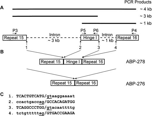
In ABP-280, the 32 amino acid Hinge I region was encoded by a single exon.20 Because the primary structure of ABP-280 and ABP-278 are highly homologous, it seemed likely that the deletion of the Hinge I region in ABP-276 was due to alternative RNA splicing. To investigate the mechanism for the presence or absence of the Hinge I region in the two isoforms, human genomic sequences encompassing the Hinge I region and partial sequences of the flanking repeats, namely repeats 15 and 16, were amplified by PCR and sequenced (Fig5). These data confirmed that the 24 amino acid Hinge I region of ABP-278 was encoded by a single exon of 72 bp. The lengths of intron sequences before and after this exon were about 3 kb and 1 kb, respectively, and were significantly longer than those in ABP-280 (113 bp and 134 bp)38 (Fig 5A). The exon-intron junctions showed the invariant GT-AG dinucleotides (Fig 5C). The genomic structure clearly indicated that alternative RNA splicing lead to the formation of ABP-278 and ABP-276 (Fig 5B).
Alignment of the 24 internal repeats in the ABP-276 and ABP-278 backbone. Alignment of amino acid residues 249-2602 of ABP-278 are shown. The 24 amino acid Hinge I sequence of ABP-278 was inserted between repeats 15 and 16; a 34 amino acid Hinge II sequence of both ABP-276 and ABP-278 was inserted between repeats 23 and 24. A consensus sequence for the repeating unit in ABP-278, derived from residues common to at least 10 of the repeats is listed at the bottom, together with a similar consensus sequence from ABP-28017 and chicken filamin (MCFil).47
Alignment of the 24 internal repeats in the ABP-276 and ABP-278 backbone. Alignment of amino acid residues 249-2602 of ABP-278 are shown. The 24 amino acid Hinge I sequence of ABP-278 was inserted between repeats 15 and 16; a 34 amino acid Hinge II sequence of both ABP-276 and ABP-278 was inserted between repeats 23 and 24. A consensus sequence for the repeating unit in ABP-278, derived from residues common to at least 10 of the repeats is listed at the bottom, together with a similar consensus sequence from ABP-28017 and chicken filamin (MCFil).47
Alternative splicing of the Hinge I region. (A) Schematic representation of the genomic structure encoding the Hinge I region and the PCR products amplified with specific primers P3, P4, P5, and P6. The genomic sequences encoding the Hinge I region and partial sequences of adjacent repeats 15 and 16 are boxed. Intron sequences are represented by dashed lines. The PCR primers specific for ABP-278 (P3, P4, P5, and P6) are labeled. The position of exon-intron junctions are indicated by vertical arrows and numbered. (B) Diagram of the formation of ABP-278 and its isoform ABP-276 by alternative RNA splicing. (C) Partial sequences of exon-intron junctions as numbered in (A). Splice juntions are indicated by slash. The consensus donor (gt) and acceptor (ag) dinucleotides are underlined.
Alternative splicing of the Hinge I region. (A) Schematic representation of the genomic structure encoding the Hinge I region and the PCR products amplified with specific primers P3, P4, P5, and P6. The genomic sequences encoding the Hinge I region and partial sequences of adjacent repeats 15 and 16 are boxed. Intron sequences are represented by dashed lines. The PCR primers specific for ABP-278 (P3, P4, P5, and P6) are labeled. The position of exon-intron junctions are indicated by vertical arrows and numbered. (B) Diagram of the formation of ABP-278 and its isoform ABP-276 by alternative RNA splicing. (C) Partial sequences of exon-intron junctions as numbered in (A). Splice juntions are indicated by slash. The consensus donor (gt) and acceptor (ag) dinucleotides are underlined.
Tissue distribution of ABP-276 and ABP-278.
Distribution of the two isoforms in various human tissues was assessed by RT-PCR with various human cDNA libraries as templates using primers that flank the Hinge I region. With these primers, amplification of ABP-278 produced a PCR product of 349 bp in length, while amplification of ABP-276 produced a product of 277 bp (Fig 6). The PCR products were separated according to size by electrophoresis in a 1.5% agarose gel. As shown in Fig 6, cDNAs from prostate, uterus, lung, liver, thyroid, stomach, lymph node, small intestine, and spleen contained predominantly the ABP-278 isoform, while Daudi cells and spinal cord contained predominantly the ABP-276 isoform. The placenta, bone marrow, brain, umbilical vein endothelial cells (HUVEC), retina, and skeletal muscle contained appreciable or detectable levels of both forms. ABP-280 was present in all tissues and cell types examined (Fig 6) as previously reported.17 An analysis of mRNA from human platelets by RT-PCR indicated the presence of predominantly the ABP-276 isoform, which was about 10-fold less abundant than ABP-280. Little or no ABP-278 isoform was detectable (M. Ling and E. Davie, unpublished observation). Although RT-PCR does not provide a quantitative measurement of mRNA levels, it clearly showed that this homologue to ABP-280 was expressed in all tissues and the predominant isoform varied in each tissue.
Tissue distribution of ABP-276 and ABP-278 by RT-PCR. PCR products, amplified with primers P3 and P4 using various marathon cDNAs (Clontech) as templates, were fractionated on 1.5% agarose gel. The upper band (349 bp) represents the PCR products of ABP-278, while the lower band (277 bp) represents the PCR products of ABP-276. The difference in size is due to the presence and absence of the 72 bp exon sequence encoding the 24 amino acid Hinge I region. The PCR amplification of ABP-280 (P9 and P10) and G3PDH (P7 and P8) sequences were used as controls.
Tissue distribution of ABP-276 and ABP-278 by RT-PCR. PCR products, amplified with primers P3 and P4 using various marathon cDNAs (Clontech) as templates, were fractionated on 1.5% agarose gel. The upper band (349 bp) represents the PCR products of ABP-278, while the lower band (277 bp) represents the PCR products of ABP-276. The difference in size is due to the presence and absence of the 72 bp exon sequence encoding the 24 amino acid Hinge I region. The PCR amplification of ABP-280 (P9 and P10) and G3PDH (P7 and P8) sequences were used as controls.
DISCUSSION
Human ABP is a ubiquitous dimeric phosphoprotein that binds to and promotes orthogonal branching of actin filaments. By associating with the intracellular domain of the GPIbα subunit of the GPIb-IX-V complex, it links the cytoskeleton to the platelet membrane. Several studies showed that ABPs consist of a family of closely related homologues and isoforms. Thus, the gene for ABP-280, located on the X chromosome, is expressed and differentially spliced to give rise to an isoform with an eight amino acid deletion in repeat 15.20In the same study, a partial cDNA for an ABP homologue, highly expressed in skeletal muscle and heart, was also identified. This homologue lacked a Hinge I region and its gene was mapped to chromosome 7. The homologue identified in this study was different in sequence from both of these two ABPs and represents a third member of this gene family. In a preliminary report, Takafuta et al,39 using a similar approach, isolated and cloned a cDNA coding for a human ABP homologue from human placenta. This homologue, designated as hFH-1, appears to be identical to the ABP-278 isoform described in the present study. Chicken filamin, which showed the highest percent identity with ABP-276, appeared to be the equivalent orthologue in chicken. A homologue of chicken filamin, located predominantly at sites of focal adhesion and in the ends of stress fibers, also has been identified.40 However, no sequence information has been presented for this homologue. These results show that several different homologues and isoforms of ABP exist and suggest that they may have specialized functions within different cells.
Variations in the function of ABP isoforms may be attributed in part to variations in the Hinge I region. The Hinge I region of ABP-280, which consisted of 32 amino acids, was postulated to increase the flexibility of ABP and facilitate cross-linking of actin filaments into orthogonal arrays.17 Chicken gizzard filamin, which lacked a Hinge I region, promoted cross-linking of actin filaments into parallel bundles.41 The presence of a 24 amino acid Hinge I region in ABP-278 with no apparent sequence similarity to ABP-280 suggested that it may have a cytoplasmic localization that was different from ABP-280 and may cross-link actin filaments into arrays with different geometric characteristics. Experiments are in progress to determine the intracellular distribution of the various isoforms of ABPs. The Hinge I region in ABP-280 also contained a cleavage site for calpain, a calcium-dependent protease derived from activated platelets. Phosphorylation of the platelet ABP in situ by cAMP-dependent protein kinase protected ABP from proteolysis by calpain, and blocked cytoskeleton reorganization.42 It is unclear if the homologue ABP-276 or ABP-278 contains a comparable calpain cleavage site(s) and phosphorylation site and whether phosphorylation modulates their sensitivity to cleavage by calpain.
Several membrane receptors directly associate with the cytoskeleton and mediate important functions such as shape changes, focal adhesion, motility, signaling, and receptor clustering in response to external ligand binding. This association involves components of the submembraneous skeleton, which lines the cytoplasmic face of the plasma membrane, and includes ABPs, short actin filaments, spectrin, α-actinin, as well as myosin and tropomyosin.43,44 The GPIb-IX-V complex associates with the cytoskeleton through binding to ABP-280, and may also interact with other homologues and isoforms of this family, namely ABP-276 and ABP-278, as suggested by this study. ABP has also been shown to colocalize and directly associate with several membrane receptors, including the acetycholine receptors in chicken myoblasts,21 the immunoglobulin receptor FcγIR,22 and the CD18 subunit of the β2-integrin in leukocytes,23 and the thyroid stimulating hormone receptor (TSH receptor) in endocrine cells.24 In the leukocyte, ABP directly associates with the intracellular domain of the Fc receptor FcγIR when the receptor is not occupied. The binding of IgG to the receptor lowers the affinity of ABP for the Fc receptor and initiates immune defense functions and cell surface changes.42 In cultured thyroid follicular cells, the binding of thyroid-stimulating hormone (TSH) to the TSH receptor induces a rapid and striking reduction in intracellular actin microfilament bundles, accompanied by a dramatic change in the shape of the cell from a flattened to a rounded appearance.45 This reorganization of the actin filaments is apparently mediated in part by association of the intracellular domain of the TSH receptor with a truncated form of ABP, which is identical in sequence to the C-terminal region of ABP-276 and ABP-278, except two single base deletions at nt 7455 and 7922. This truncated form of ABP-276, encompassing a part of repeat 23 and the entire repeat 24, contains the dimerization domain and presumably a region that interacts with the TSH receptor. However, without the N-terminal actin binding domain, this truncated form of ABP-276 may compete with the full-length ABP-276 or other members of the ABP family in binding to the TSH receptor and may modulate association of the receptor to the cytoskeleton.
Although it has been shown that ABP-280 can interact with the cytoplasmic tail of GPIbα, a recent study in an ABP-280–deficient melanoma cell line showed that insertion of the GPIb-IX complex into the membrane did not depend on the GPIbα cytoplasmic tail or the GPIbα-binding site in ABP-280.46 These results suggested that direct cross-linking of actin filaments or direct interaction with receptors may not be necessary for ABP-280 to promote receptor insertion into the membrane. The role of ABP-276 and ABP-278 in this process needs to be further defined and characterized.
ABP-276 and ABP-278, identified in this study, and the truncated form of ABP-276, identified in thyroid cells, together with ABP-280 and its differentially spliced isoform, form a family of structurally and functionally related proteins that may play an important role in linking specific receptors to actin filaments and the cytoskeleton in nonmuscle cells such as platelets. The cloning and identification of these homologues and isoforms makes it possible to further characterize the cell-type specific function of each member and the role they play in response to extracellular ligand binding.
ACKNOWLEDGMENT
The authors are grateful to ZymoGenetics Inc for gifts of HEL cell library, human placenta library, and various cDNAs of human tissues. We also thank Wendy Tsien and Jeff Harris for excellent technical assistance.
Supported by Grant No. HL16919 from the National Institutes of Health, Bethesda, MD.
Address reprint requests to Earl W. Davie, PhD, Department of Biochemistry, Box 357350, University of Washington, Seattle, WA 98195.
The publication costs of this article were defrayed in part by page charge payment. This article must therefore be hereby marked "advertisement" is accordance with 18 U.S.C. section 1734 solely to indicate this fact.


This feature is available to Subscribers Only
Sign In or Create an Account Close Modal