We used a mouse transplantation model to address the recent controversy about CD34 expression by hematopoietic stem cells. Cells from Ly-5.1 C57BL/6 mice were used as donor cells and Ly-5.2 mice were the recipients. The test cells were transplanted together with compromised marrow cells of Ly-5.2 mice. First, we confirmed that the majority of the stem cells with long-term engraftment capabilities of normal adult mice are CD34−. We then observed that, after the injection of 150 mg/kg 5-fluorouracil (5-FU), stem cells may be found in both CD34− and CD34+ cell populations. These results indicated that activated stem cells express CD34. We tested this hypothesis also by using in vitro expansion with interleukin-11 and steel factor of lineage−c-kit+ Sca-1+ CD34− bone marrow cells of normal mice. When the cells expanded for 1 week were separated into CD34− and CD34+ cell populations and tested for their engraftment capabilities, only CD34+ cells were capable of 2 to 5 months of engraftment. Finally, we tested reversion of CD34+ stem cells to CD34− state. We transplanted Ly-5.1 CD34+post–5-FU marrow cells into Ly-5.2 primary recipients and, after the marrow achieved steady state, tested the Ly-5.1 cells of the primary recipients for their engraftment capabilities in Ly-5.2 secondary recipients. The majority of the Ly-5.1 stem cells with long-term engraftment capability were in the CD34− cell fraction, indicating the reversion of CD34+ to CD34−stem cells. These observations clearly demonstrated that CD34 expression reflects the activation state of hematopoietic stem cells and that this is reversible.
AFTER THE SUCCESSFUL hematopoietic reconstitution of baboons with selected CD34+ baboon bone marrow cells by Berenson et al,1 CD34 expression became the hallmark of murine and human hematopoietic stem cells. A number of clinical trials of transplantation of CD34+ human bone marrow and peripheral blood stem cells have documented cases of successful long-term engraftment.2-5 However, this dogma was recently challenged on several fronts. First, Osawa et al6 reported that mouse marrow long-term reconstituting cells are in the lineage (Lin)− c-kit+Ly-6A/E (Sca-1)+ CD34−/low cell population rather than CD34+ cells. Subsequently, Morel et al7 confirmed that CD34− cells contain a significant number of stem cells. Independently from these observations, studies performed by Goodell et al8,9 also raised serious questions about CD34 expression by hematopoietic stem cells. They first reported that murine bone marrow stem cells are highly enriched in a cell population defined by Hoechst 33342 dye as side population (SP).8 They subsequently reported that human and rhesus bone marrow SP cells are largely CD34−/low and that, after 5 weeks of suspension culture on bone marrow stromal cells, the rhesus and human CD34− SP cells became CD34+.9In our laboratory, we have shown previously that the CD34+c-kitlow human bone marrow cells can engraft in sheep bone marrow for more than 1.5 years.10 Subsequently, we found that some human long-term engrafting cells reside also in the CD34− population.11
To solve the apparent controversy, we entertained the hypothesis that CD34 expression reflects activation and/or cycling state of stem cells and tested this hypothesis using a murine transplantation model. We used Lin− c-kit+ Sca-1+ bone marrow cells harvested from normal mice and mice that had been injected with 150 mg/kg 5-fluorouracil (5-FU) 2 days before bone marrow harvest. Stem cells in the latter model have been shown to be in active cell division after 5-FU–induced marrow hypoplasia.12
MATERIALS AND METHODS
Monoclonal antibodies (MoAbs) and hybridomas.
Fluorescein isothiocyanate (FITC)-conjugated RAM34 (anti-CD34; rat IgG2a), phycoerythrin (PE)-conjugated D7 (anti–Ly-6A/E [anti–Sca-1]; rat IgG2a), allophycocyanin (APC)-conjugated 2B8 (anti–c-kit; rat IgG2b), biotin-conjugated A20 (anti–Ly-5.1; rat IgG2a), biotin-conjugated 53-2.1 (anti–Thy-1.2; rat IgG2a), biotin-conjugated RA3-6B2 (anti-CD45R/B220; rat IgG2a), biotin-conjugated RB6-8C5 (anti–Gr-1; rat IgG2b), and biotin-conjugated M1/70 (anti-Mac-1; rat IgG2b) were purchased from Pharmingen (San Diego, CA). Some antibodies were also received as either nonlabeled antibodies or hybridomas. Purified antibodies were conjugated to FITC or to biotin in our laboratory. MoAb ACK4 (anti–c-kit; rat IgG2a) was provided by S.I. Nishikawa (Kyoto University, Kyoto, Japan). Hybridoma RB6-8C5 (anti–Gr-1; rat IgG2b) was provided by R.L. Coffman (DNAX, Palo Alto, CA). MoAb TER-119 (anti-erythrocytes; rat IgG2b) was a gift from T. Kina (Kyoto University). Hybridomas 14.8 (anti-B220; rat IgG2b), M1/70.15.11.5 (anti–Mac-1; rat IgG2b), GK1.5 (anti-CD4; rat IgG2b), and 53-6.72 (anti-CD8; rat IgG2a) were purchased from American Type Culture Collection (Rockville, MD). Hybridoma A20 (anti–Ly-5.1; IgG1) was provided by H. Fleming (Emory University, Atlanta, GA).
Cell preparation.
Ten- to 12-week-old male C57BL/6-Ly-5.1 mice (Jackson Laboratories, Bar Harbor, ME) were used as bone marrow donors. Ten- to 14-week-old female C57BL/6-Ly-5.2 mice (Charles River Laboratories, Raleigh, NC) were used as irradiated recipients and as the source of compromised bone marrow cells.13 In some experiments, the donor mice were treated with an intravenous injection of 5-FU at 150 mg/kg body weight 48 hours before death. Bone marrow cells were flushed from femurs and tibiae, pooled, washed twice with phosphate-buffered saline (PBS) containing 0.1% bovine serum albumin (BSA), made into single-cell suspension by repeated pipetting, and filtered through 40-μm nylon mesh. The cells with densities ranging from 1.063 to 1.077 g/mL were collected by gradient separation using Nycodenz (Accurate Chemical and Scientific Corp, Westbury, NY). Cells reacting to anti–Mac-1, anti–Gr-1, anti-B220, anti-CD4, anti-CD8, and TER-119 were removed by using 2 depletions with immunomagnetic beads (Dynabeads M-450 coupled to sheep antirat IgG; DYNAL, Great Neck, NY). For fluorescence-activated cell sorting (FACS), the resulting Lin− cells were stained with FITC-conjugated anti-CD34 (RAM34), PE-conjugated anti–Sca-1, and biotin-conjugated anti–c-kit. Cells were further stained with streptavidin-conjugated APC (Caltag Laboratories, San Francisco, CA). After the addition of propidium iodide (PI) at a concentration of 1 μg/mL, the cells were washed twice, resuspended in PBS containing 0.1% BSA, and kept on ice until cell sorting. In the serial transplantation experiments, cells were stained with FITC-conjugated anti-CD34, PE-conjugated anti–Sca-1, APC-conjugated anti–c-kit, and biotin-conjugated anti–Ly-5.1. Subsequently, the cells were washed twice and stained with streptavidin-conjugated Red 613 (Life Technologies, Gaithersburg, MD). PI staining was omitted, because its emmision wavelength is similar to that of Red 613. Cell sorting was performed on FACS Vantage (Becton Dickinson, San Jose, CA), with appropriate isotype-matched controls. The bone marrow cells were first enriched for c-kit+, Sca-1+ cells and then the cells were sorted again based on CD34 expression.
Suspension culture.
Five thousand Lin− c-kit+Sca-1+ CD34− cells were stained with PKH26 (Sigma Immunochemicals, St Louis, MO)14 15 and incubated in a 25-cm2 culture flask. The culture medium contained α-modification of Eagle’s medium (αMEM; ICN Biomedicals, Aurora, OH), 20% fetal calf serum, 1% deionized fraction V BSA, 1 × 10⁻⁴ mol/L 2-mercaptoethanol, recombinant steel factor (SF; c-kit ligand), and interleukin-11 (IL-11). SF was purchased from R&D Systems (Minneapolis, MN) and used at a concentration of 100 ng/mL. Human IL-11 (a gift from P. Schendel, Genetics Institute, Cambridge, MA) was used at a concentration of 100 ng/mL. After 1 week of incubation, the cells were washed twice and stained with FITC-conjugated anti-CD34 and were sorted into CD34− and CD34+ fractions.
Hematopoietic reconstitution.
Ly-5.2 mice were administrated with a single 850-cGy dose of total body irradiation using 4 × 10⁶ V linear accelerator. The FACS-sorted cells from donor (Ly-5.1) mice or cultured cells were injected into the tail vein of the irradiated Ly-5.2 mice along with 4 × 10⁵ compromised Ly-5.2 marrow cells. Compromised marrow cells had been subjected to 2 previous rounds of transplantation and regeneration.13 Peripheral blood was obtained from the retro-orbital plexus using heparin-coated micropipets (Drummond Scientific Co, Broomall, PA) 2, 5, and/or 8 months after transplantation. Red blood cells were lysed by 0.15 mol/L NH4Cl. The samples were stained with FITC-conjugated anti–Ly-5.1 and analyzed for donor-derived cells on FACS Vantage. Donor cells in T-cell, B-cell, granulocyte, and monocyte/macrophage lineages at 5 months posttransplantation were analyzed by staining with biotin-conjugated anti–Thy-1.2, biotin-conjugated anti-B220, biotin-conjugated anti–Gr-1, and biotin-conjugated anti–Mac-1, followed by staining with streptavidin-conjugated PE.
Retransplantation.
Mice that had been transplanted with Lin−c-kit+ Sca-1+ CD34+ cells of 5-FU–treated mice were killed 3 months after transplantation. Ly-5.1 Lin− c-kit+ Sca-1+CD34− cells and Ly-5.1 Lin−c-kit+ Sca-1+ CD34+ cells were prepared from the marrow of these mice and were injected into secondary Ly-5.2 recipients along with 5 × 10⁴ Ly-5.2 compromised cells. Peripheral blood was analyzed for Ly-5.1 cells 2 and 5 months after the secondary transplantation.
RESULTS
Transplantation of CD34− and CD34+ cells of normal mice.
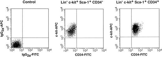
We isolated CD34− and CD34+ cells from the c-kit+ Sca-1+ population of both normal and post–5-FU bone marrow using the FACS regions shown in Fig 1. In Fig 2, we present the result of reanalysis of the sorted CD34+ and CD34−cells of normal mice that showed greater than 99% purity. Because the ratio of the Lin− c-kit+Sca-1+ CD34− (R2) cells to Lin− c-kit+ Sca-1+CD34+ (R3) cells was 1:5, we transplanted 100 CD34− cells or 500 CD34+ cells. The level of engraftment was determined by measuring the percentage of donor (Ly-5.1) peripheral blood nucleated cells at 2 and 5 months after transplantation. The results are presented in Fig 3. The average engraftment levels of the 8 mice transplanted with 100 CD34− cells was 26.5% ± 20.4% at 2 months and 34.7% ± 33.5% at 5 months posttransplantation. Only 1 of 8 mice transplanted with 500 CD34+ (R3) cells engrafted at the levels of 2.8% and 1.8%, respectively, at 2 and 5 months. The remaining 7 mice and the 5 mice receiving only compromised marrow cells showed no Ly-5.1 cells. Similar results were obtained in 2 additional experiments. These results were in agreement with the premise that CD34−cells of normal mice were capable of long-term engraftment.6 7
FACS sorting regions used for preparation of c-kit+ Sca-1+ CD34− and c-kit+ Sca-1+ CD34+ cell populations. Lin+ cells had been removed from mononuclear marrow cells by using immunomagnetic beads before staining for FACS sorting. The Lin− cells were enriched for c-kit+ Sca-1+ cells using the R1 and R4 windows and again sorted on the basis of CD34 expression using R2, R3, R5, and R6 windows.
FACS sorting regions used for preparation of c-kit+ Sca-1+ CD34− and c-kit+ Sca-1+ CD34+ cell populations. Lin+ cells had been removed from mononuclear marrow cells by using immunomagnetic beads before staining for FACS sorting. The Lin− cells were enriched for c-kit+ Sca-1+ cells using the R1 and R4 windows and again sorted on the basis of CD34 expression using R2, R3, R5, and R6 windows.
Results of reanalysis of the cells of normal mice sorted according to R2 and R3 windows of Fig 1.
Results of reanalysis of the cells of normal mice sorted according to R2 and R3 windows of Fig 1.
Percentage of donor nucleated cells in the blood of individual mice transplanted with 100 Lin−c-kit+ Sca-1+ CD34− or 500 Lin− c-kit+ Sca-1+CD34+ marrow cells of normal mice. (○) 2 months posttransplantation; (•) 5 months posttransplantation.
Percentage of donor nucleated cells in the blood of individual mice transplanted with 100 Lin−c-kit+ Sca-1+ CD34− or 500 Lin− c-kit+ Sca-1+CD34+ marrow cells of normal mice. (○) 2 months posttransplantation; (•) 5 months posttransplantation.
Transplantation of CD34− and CD34+cells of 5-FU–treated mice.
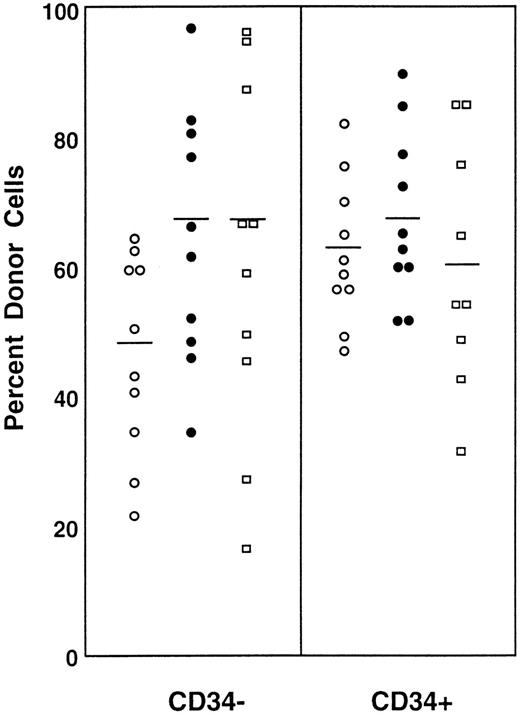
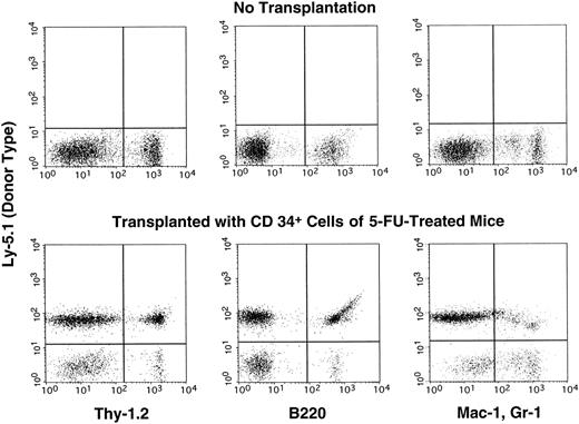
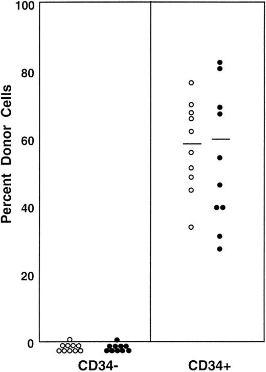
Because the ratio of the CD34− (Fig 1, R5) to CD34+ (Fig 1, R6) cell populations in the bone marrow cells of 5-FU–treated mice was roughly 1:1, we transplanted 100 CD34− or 100 CD34+ cells per mouse for measurement of engraftment capability. In contrast to the cells from normal mice, both CD34− and CD34+ cells engrafted (Fig 4). The average levels of engraftment were 47% ± 16.0% (N = 10) and 62.8% ± 14.6% (N = 10) at 2 months posttransplantation, 64.4% ± 21.9% (N = 10) and 67.8% ± 13.9% (N = 10) at 5 months posttransplantation and 64.3% ± 23.7% (N = 10) and 59.7% ± 23.1% (N = 9) at 8 months posttransplantation for CD34− cells and CD34+ cells, respectively. All mice showed evidence for multilineage engraftment at 8 months posttransplantation. An example of engraftment by CD34+ post–5-FU cells is shown in Fig 5. A similar result was obtained in an additional experiment.
Percentage of donor nucleated cells in the blood of individual mice transplanted with 100 Lin−c-kit+ Sca-1+ CD34− or 100 Lin− c-kit+ Sca-1+CD34+ marrow cells of 5-FU–treated mice. (○) 2 months posttransplantation; (•) 5 months posttransplantation; (□) 8 months posttransplantation.
Percentage of donor nucleated cells in the blood of individual mice transplanted with 100 Lin−c-kit+ Sca-1+ CD34− or 100 Lin− c-kit+ Sca-1+CD34+ marrow cells of 5-FU–treated mice. (○) 2 months posttransplantation; (•) 5 months posttransplantation; (□) 8 months posttransplantation.
An example of multilineage hematopoietic engraftment at 8 months posttransplantation by Lin− c-kit+Sca-1+ CD34+ marrow cells of 5-FU–treated mice.
An example of multilineage hematopoietic engraftment at 8 months posttransplantation by Lin− c-kit+Sca-1+ CD34+ marrow cells of 5-FU–treated mice.
Conversion of CD34− to CD34+ stem cells in culture.
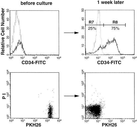
The results listed above strongly suggested that changes in the activation and/or cycling state of the bone marrow are the cause for the phenotypic changes of the stem cells. We wished to further test this premise by using cell culture. Both CD34+ and CD34− populations of Lin−c-kit+ Sca-1+ cells of normal mice were incubated in suspension culture for 1 week in the presence of SF and IL-11. The CD34+ cell populations expanded 4,000-fold. The CD34− cells expanded from 5,000 to 5 × 10⁶ (1,000-fold) and 75% of the cultured cells became CD34+, as shown in Fig 6. Simultaneous staining with PKH26, which binds to membrane lipid, confirmed the divisional history of the cultured cells. All cells that were PKHbright before incubation became PKHdullafter the suspension culture (Fig 6), indicating that all cells divided several times. Because the ratio of CD34− (R7) to CD34+ (R8) cells was 1:3 after incubation, we transplanted either 5 × 10⁴ CD34− cells or 1.5 × 10⁵ CD34+ cells to each recipient. The results of transplantation are shown in Fig7. The levels of engraftment at 2 and 5 months after transplantation of the cultured CD34+ cells were 58.8% ± 13.3% (N = 10) and 60.1% ± 19.2% (N = 10), respectively. In contrast, the levels in the mice transplanted with cultured CD34− cells were 0.4% ± 0.5% (N = 10) at 2 months and 0.3% ± 0.4% (N = 10) at 5 months. These results clearly demonstrated that CD34− stem cells that are stimulated in culture undergo phenotypic changes and express CD34.
Flow cytometric analysis of CD34 expression before and after culture. Sorted Lin− c-kit+Sca-1+ CD34− cells were stained with PKH26 and analyzed before culture (left). After 1 week of incubation with SF and IL-11, cells were analyzed for CD34 expression and PKH26 fluorescence (right). Dotted and solid lines represent cells stained with IgG2a-FITC and CD34-FITC, respectively.
Flow cytometric analysis of CD34 expression before and after culture. Sorted Lin− c-kit+Sca-1+ CD34− cells were stained with PKH26 and analyzed before culture (left). After 1 week of incubation with SF and IL-11, cells were analyzed for CD34 expression and PKH26 fluorescence (right). Dotted and solid lines represent cells stained with IgG2a-FITC and CD34-FITC, respectively.
Percentage of donor nucleated cells in the blood of individual mice transplanted with 5 × 10⁴CD34− cultured cells or 1.5 × 10⁵CD34+ cultured cells. (○) 2 months posttransplantation; (•) 5 months posttransplantation.
Percentage of donor nucleated cells in the blood of individual mice transplanted with 5 × 10⁴CD34− cultured cells or 1.5 × 10⁵CD34+ cultured cells. (○) 2 months posttransplantation; (•) 5 months posttransplantation.
Reversion of CD34+ stem cells to CD34−.
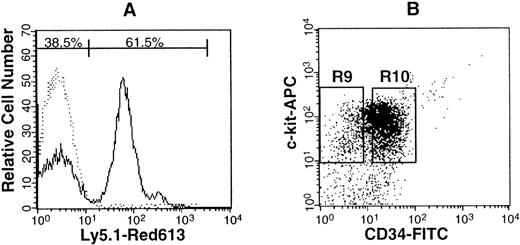
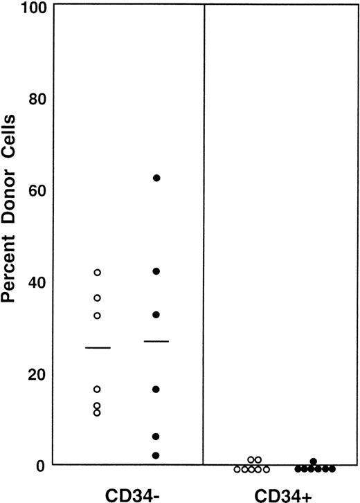
The results in the 2 studies of stem cell activation were concordant and strongly indicated that CD34 expression reflects the activation and/or cycling state of stem cells. In the next experiment, we tested the hypothesis that activated CD34+ stem cells revert to CD34− stem cells when the bone marrow attains a steady state. We transplanted Lin− c-kit+Sca-1+ CD34+ cells from the bone marrow of Ly-5.1 5-FU–treated mice into 10 Ly-5.2 primary recipients. Three months later, 61.5% of the Lin− marrow cells of the primary recipients were of donor-origin (Ly-5.1; Fig 8A). The donor origin Ly-5.1 cells were then isolated from the primary recipients and separated on the basis of CD34 expression (Fig 8B). Each population was transplanted into secondary Ly-5.2 hosts for analysis of engraftment capabilities. Because the ratio of CD34− (R9) to CD34+(R10) cells was 1:5, individual mice received either 1,000 CD34− or 5,000 CD34+ cells. The results are presented in Fig 9. The levels of engraftment at 2 and 5 months posttransplantation by CD34− Ly-5.1 cells were 25.0% ± 13.3% (N = 6) and 27.3% ± 23.2% (N = 6), respectively. Those by CD34+ Ly-5.1 cells at 2 and 5 months posttransplantation were 0.98% ± 0.97% (N = 7) and 0.63% ± 0.65% (N = 7), respectively. These results clearly demonstrated that, when bone marrow recovers from the radiation-induced hypoplasia and reaches steady state, CD34 expression of the cells is downregulated to undetectable levels.
Analysis of Lin− marrow cells from primary recipients transplanted with CD34+ cells. (A) Dotted and solid lines represent cells stained with isotype-matched Ig and Ly-5.1–specific antibody. Sixty-five percent of the Lin−marrow cells of the primary recipients were donor-derived (Ly-5.1) cells. (B) Sorting windows of Ly-5.1+ Lin−c-kit+ Sca-1+ cells. The ratio of the cells in R9 to R10 was 1:5.
Analysis of Lin− marrow cells from primary recipients transplanted with CD34+ cells. (A) Dotted and solid lines represent cells stained with isotype-matched Ig and Ly-5.1–specific antibody. Sixty-five percent of the Lin−marrow cells of the primary recipients were donor-derived (Ly-5.1) cells. (B) Sorting windows of Ly-5.1+ Lin−c-kit+ Sca-1+ cells. The ratio of the cells in R9 to R10 was 1:5.
Percentage of donor (Ly-5.1) cells in the blood of individual secondary recipients. Ly-5.1 Lin−c-kit+ Sca-1+ CD34+post–5-FU marrow cells were transplanted to Ly-5.2 primary recipients. Three months later, 1,000 Ly-5.1 CD34− bone marrow cells or 5,000 Ly-5.1 CD34+ bone marrow cells of the primary recipients were transplanted to individual secondary Ly-5.2 recipients. (○) 2 months posttransplantation; (•) 5 months posttransplantation.
Percentage of donor (Ly-5.1) cells in the blood of individual secondary recipients. Ly-5.1 Lin−c-kit+ Sca-1+ CD34+post–5-FU marrow cells were transplanted to Ly-5.2 primary recipients. Three months later, 1,000 Ly-5.1 CD34− bone marrow cells or 5,000 Ly-5.1 CD34+ bone marrow cells of the primary recipients were transplanted to individual secondary Ly-5.2 recipients. (○) 2 months posttransplantation; (•) 5 months posttransplantation.
DISCUSSION
In this project, we used Lin− c-kit+Sca-1+ cells because these cells in both normal C57BL mice6 and 5-FU–treated C57BL/6 mice12 are highly enriched for stem cells. Injection of a single Lin− c-kit+ Sca-1+CD34− cell from normal mice produced sustained high-level engraftment in an irradiated recipient for 10 months 21% of the time.6 In our laboratory, we repeatedly observed that 100 Lin− c-kit+ Sca-1+ marrow cells from 5-FU–treated mice can provide high levels of long-term engraftment of lethally irradiated mice.15-17
We observed clear-cut association between CD34 expression and the activation state of stem cells with long-term reconstitution capabilities. The majority of stem cells in the bone marrow of normal adult mice were CD34−, as reported by Osawa et al.6 In vivo and in vitro activation of stem cells was associated with expression of CD34, and the activated CD34+stem cells reverted to CD34− state as bone marrow attained stationary phase. Because mouse hematopoiesis has been an excellent model for human hematopoiesis, the observations presented in this report may have important clinical implications. It is possible that, in patients who receive chemoradiotherapy for their malignancies, stem cell CD34 expression may vary depending on the recovery state of the bone marrow. If so, the timing of autologous transplantation using selected CD34+ cells needs to be carefully considered.
Yoder et al18 reported that murine hematopoietic stem cells in the day-9 yolk sac and those in the day-12 to -14 fetal liver are CD34+. Morel et al,7 when studying 4- to 6-week-old mice, reported that both CD34− and CD34+ cell populations contain stem cells. It will be of interest to study whether the developmental changes in the CD34 expression are related to the kinetic state of the stem cells. Although we have shown association between activation state and CD34 expression of stem cells, we did not address the cause-and-result relationship between cell cycling state and CD34 expression of the stem cells. However, Randall and Weissman12 have shown an association between post–5-FU phenotypic changes and the activation/cell cycle state of the stem cells. In this regard, it will be of particular interest to study granulocyte colony-stimulating factor (G-CSF)–mobilized peripheral blood stem cells, because these cells have been shown to reside in G0/G1 state, despite stimulation by G-CSF.19
ACKNOWLEDGMENT
The authors thank Dr Haiqun Zeng for assistance in FACS sorting, Dr Pamela N. Pharr and Anne G. Livingston for assistance in preparation of this manuscript, and the staff of Radiation Oncology Department of the Medical University of South Carolina for assistance in irradiation of mice.
Supported by National Institutes of Health Grants No. RO1-DK32294, RO1-DK/HL 48714, and PO1-CA78582 and by the Office of Research and Development, Medical Research Services, Department of Veterans Affairs.
The publication costs of this article were defrayed in part by page charge payment. This article must therefore be hereby marked “advertisement” in accordance with 18 U.S.C. section 1734 solely to indicate this fact.
REFERENCES
Author notes
Address reprint requests to Makio Ogawa, MD, PhD, Ralph H. Johnson VA Medical Center, 109 Bee St, Charleston, SC 29401-5799; e-mail:ogawam@musc.edu.



This feature is available to Subscribers Only
Sign In or Create an Account Close Modal