It is believed that the 3-dimensional organization of centromeric heterochromatin in interphase may be of functional relevance as an epigenetic mechanism for the regulation of gene expression. Accordingly, a likely possibility is that the centromeres that spatially associate into the heterochromatic structures (chromocenters) observed in the G1 phase of the cell cycle will differ in different cells. We sought to address this issue using, as a model, the chromocenters observed in quiescent normal human hematopoietic cells and primary fibroblasts. To do this, we analyzed the spatial relationships among different human centromeres in 3-D preserved cells using nonisotopic in situ hybridization and confocal microscopy. We showed quantitatively that chromocenters in all cell types do indeed represent nonrandom spatial associations of certain centromeres. Furthermore, the observed patterns of centromere association indicate that the chromocenters in these cell types are made of different combinations of specific centromeres, that hematopoietic cells are strikingly different from fibroblasts as to the composition of their chromocenters and that centromeres in peripheral blood cells appear to aggregate into distinct “myeloid” (present in monocytes and granulocytes) and “lymphoid” (present in lymphocytes) spatial patterns. These findings support the idea that the chromocenters formed in the nucleus of quiescent hematopoietic cells might represent heterochromatic nuclear compartments involved in the regulation of cell-type-specific gene expression, further suggesting that the spatial arrangement of centromeric heterochromatin in interphase is ontogenically determined during hematopoietic differentiation.
Studies on the spatial distribution of centromeric satellite DNA in different cell types and species have shown that centromeres organize into 3-D patterns that appear to be cell-type specific and evolutionarily conserved (reviewed1-9). In mammalian cells, a tendency toward clustering of centromeres around the nucleolus or the nuclear periphery has been consistently observed.3 In addition, it has been shown that centromere positioning changes during the sequential stages of the cell cycle10 and in response to the transcriptional status of the cell (reviewed2). In many cell types, heterochromatic regions (mainly centromeres but also other regions of repetitive DNA) are observed as dark-staining bodies or chromocenters that may fuse, forming aggregate chromocenters.1,3 These structures vary in number and size in different cell types and appear to be restricted to the G1 phase of the cell cycle.11 All these observations led to the proposal that centromeres may behave as structural centers for chromatin organization in interphase, favoring the creation of functional compartments for essential nuclear processes such as gene expression, DNA replication, and cell division.2 Recent observations appear to support this view because it has been shown that repression of gene transcription can be induced by higher-order levels of chromatin organization that spatially juxtapose the euchromatic gene with centromeric heterochromatin.12-14 Accordingly, Brown et al15 observed an inverse correlation of gene activity and association with Ikaros protein, a putative transcriptional regulator that in murine lymphoid cells surrounds constitutive centromeric heterochromatin. These findings, together with earlier observations of position-effect variegation phenomena inDrosophila,16 strongly support the hypothesis that the distribution of heterochromatin in interphase nuclei might indeed be relevant for nuclear metabolism, namely as an epigenetic mechanism of transcriptional regulation.14 If this is so, a likely possibility is that centromeres belonging to specific chromosomes should have specific patterns of spatial association in distinct cell types.
In this study we sought to address this issue using, as a model, the chromocenters formed during the G1 phase of the cell cycle in normal human peripheral blood cells (lymphocytes, monocytes, and granulocytes) and primary human fibroblasts (as representative of a nonhematopoietic cell type). To do this, we analyzed the spatial relationships among different human centromeres in 3-D preserved cells using nonisotopic in situ hybridization and confocal microscopy. We showed that centromeres in all cell types associated with a probability 2 orders of magnitude larger than that expected for a random distribution inside the nucleus and that the chromocenters in hematopoietic cells and fibroblasts were made of different combinations of specific centromeres. The data thus indicates that the spatial arrangements of centromeres in the interphase nucleus give rise to heterochromatic compartments with cell-type-specific composition, a fact likely to have important implications for epigenetic regulation of tissue-specific gene expression.
Materials and methods
Cells
Normal unstimulated human peripheral blood cells from volunteer donors were used. Peripheral blood lymphocytes (PBL) and granulocytes were isolated by centrifugation in a gradient of Percoll (Amersham Pharmacia, Uppsala Sweden) according to the instructions of the manufacturer, harvested by centrifugation at 250g for 5 minutes, applied onto poly-L-lysine-coated 10-mm/10-mm coverslips and immediately fixed and permeabilized in 3.7% paraformaldehyde/0.5% Triton X-100/HPEM (65 mmol/L PIPES, 30 mmol/L HEPES/10 mmol/L EGTA, 2 mmol/L MgCl2, pH 6.9) with gentle shaking for 15 minutes at room temperature. More than 90% of the cells obtained with this procedure were either lymphocytes or granulocytes as assessed by May–Grunwald–Giemsa staining. In experiments aimed at the analysis of cycling cells, PBLs were stimulated by the addition of 0.5% phytohemagglutinin (Biochrome, Berlin, Germany) followed by culture in RPMI-1640 medium (Gibco-BRL, Gaithersburg, MD) with 10% fetal calf serum (Gibco-BRL), 1% penicillin/streptomycin (Gibco-BRL) at 37°C, and 5% CO2 for 72 hours. Cells were then harvested by centrifugation at 250g for 5 minutes, applied to poly-L-lysine-coated coverslips, fixed, and permeabilized as above. To obtain peripheral blood monocytes, mononuclear cells were first isolated by density gradient centrifugation (Ficoll-Paque; Pharmacia, Uppsala, Sweden) followed by incubation in a 25-cm flat culture flask with RPMI-1640 medium (Gibco-BRL) supplemented with 10% fetal calf serum (Gibco-BRL) for 1 hour at 37°C. Then cells in suspension were rejected, and adherent cells were recovered by gentle scraping. More than 90% of the latter were monocytes as assessed by May–Grunwald–Giemsa staining. After isolation, monocytic cells were fixed and permeabilized as above. Experiments of bromodeoxyuridine (BrdU) incorporation17 showed that the 3 isolated hematopoietic cell types were mainly in the G1 phase of the cell cycle (fewer than 5% of the cells were BrdU positive).
Human diploid fibroblasts (Wi-38, ECACC, UK) were seeded at a density of 2 × 10⁴ cells/cm3 onto glass coverslips in minimum essential medium (Gibco-BRL) supplemented with Earle's salts, L-glutamine, 1% nonessential amino acids (Gibco-BRL), and 10% fetal calf serum (Gibco-BRL) at 37%, 5% CO2, and grown until confluence (7 days) in 35-mm Petri dishes. The cells were then fixed and permeabilized as described above. In these growth conditions, fewer than 3% of the cells were found to be in S phase, as assessed by analysis of BrdU incorporation.17
Probes
The presence of chromocenters in all cell types under study was investigated using the pan-centromeric probe p82H, which recognizes alphoid sequences found at the centromeres of all human chromosomes.18 To investigate the association patterns between centromeres belonging to different chromosomes, 3 different centromeres were, whenever possible, simultaneously visualized in each experiment, using specific centromeric probes labeled with different reporter molecules. The probes for centromeres of chromosomes 1, 2, 3, 4, 5/19, 6, 7, 11, 12, 13/21,14/22, 15, 17, 20, and X were plasmid clones kindly provided by Dr M. Rochi (Istituto di Genetica, Bari, Italy) and Dr H. Scherthan (Department of Human Genetics and Human Biology, University of Kaiserslautern, Germany); the probes for centromeres of chromosomes 8, 9, 18, and Y were purchased from ATCC (Rockville, MD), and those for centromeres of chromosomes 7, 9, 10, and 16 were purchased from ONCOR (Gaithersburg, MD). For triple-hybridization experiments, the probes were nick-translated with digoxigenin-dUTP (Boehringer Mannheim, Mannheim, Germany), biotin-dUTP (Boehringer Mannheim), and Cy5-dCTP (Amersham Life Sciences, Arlington Heights, IL) as described.19 The probes were combined with 1 mg/mL hnDNA (Sigma-Aldrich, Madrid, Spain), ethanol precipitated, air dried, and dissolved in hybridization buffer as described.19 The probes from ONCOR were used according to the instructions of the manufacturer.
In situ hybridization
In situ hybridization experiments were performed as described17 with some modifications. Before hybridization, cells were repermeabilized with 0.7% Triton X-100/0.1 N HCl/phosphate-buffered saline (PBS) for 10 minutes on ice with gentle shaking, washed in PBS (3 × for 5 minutes) and 2 × SSC (1 × for 5 minutes), and denatured in 50% formamide/2 × SSC for 20 minutes at 75°C. Probes were denatured for 5 minutes at 75°C and hybridized overnight at 37°C in a moist chamber. Posthybridization washes were conducted in 50% formamide/2 × SSC at 45°C (3 × for 5 minutes). Biotin-labeled probes were detected with Texas-Red avidin (1:200; Vector Laboratories, Burlingame, CA) at 37°C for 30 minutes and washed in 0.05% Tween 20/PBS (3 × for 5 minutes). Digoxigenin-labeled probes were detected with a mouse antidigoxigenin antibody (1:100; Boehringer Mannheim) at 37°C for 30 minutes and washed as described; this was followed by incubation with a FITC-conjugated goat antimouse (1:100; Jackson Immunoresearch Laboratories, West Grove, PA). For the simultaneous visualization of hybridization sites and the nuclear envelope, the samples were incubated immediately after the detection of the hybridization signals with a rabbit anti-laminin-B antibody (kindly provided by Dr S. Georgatos, Heidelberg, Germany) diluted at 1:100 at 37°C for 30 minutes and washed as described, followed by incubation with Texas-Red conjugated goat antirabbit Ig (Jackson Immunoresearch Laboratories) at 37°C for 30 minutes.19
Confocal microscopy and image analysis
The analysis of hybridization signals in 3-D preserved nuclei was performed with the confocal microscope Zeiss LSM-410 (Oberkochen, Germany) as previously described.17 Twenty-five optical sections were obtained for each nucleus from hematopoietic cells (20 sections for fibroblasts) (average increment between sections in the Z axis of 0.25 μm), and the associations between different homologous or heterologous centromeres was determined in each section. To overcome problems with quantitative measures induced by distortion of confocal images, the criterion for centromere association was that of adjacency—partial or total overlapping of the hybridization signals originated by each probe. Because the nucleus of granulocytic cells (mainly neutrophils) is polylobulated, 2 centromeres were considered adjacent only when they were located within the same lobule (the indentations and convolutions of the nuclei were always assessed by the simultaneous labeling of the nuclear lamina) (see above). If cross-hybridization between different probes was observed (this was the case for centromeres 5 and 9, 13 and 21, and 14 and 22), single hybridization experiments were performed and the associations between the 4 signals originated by the individual probe were determined (see “Results”).
Statistical analysis
To investigate the frequency and probability of association of different centromeres in all cell types, we used the following strategy: 100 nuclei from each cell type and for each combination of probes were analyzed. In the analysis of PBL (3100 nuclei), the combinations of probes to be used in the study were chosen so that each individual centromere was represented at least in 2 different combinations in the total data. Twelve triple combinations of probes were used (Table 1). This provided an internal control for the consistency of the data for each donor. Hybridization experiments for centromeres 2 and 3, 2 and 4, 3 and 6, 7 and 8, 7 and 9, 9 and 11, 15 and 16, 15 and 17, and 16 and 17 were further performed in cells from 4 different donors. Probabilities have been calculated within a universe of events in the range 100 to 301. When each of the 2 centromeres from homologous chromosomes in an individual cell were associated with a different centromere, these associations were considered as 2 independent outcomes. To further assess the consistency of the data, a few pairs of centromeres whose frequencies of association (either frequent or rare) could be predicted on the basis of the results obtained in 1 donor were analyzed in the same or different donors. For example, if triple combinations of probes specific for centromeres A, B, C and B, C, D showed that A is associated with B and B is associated with D, then a double hybridization experiment was performed with probes for A and D to see whether the 2 centromeres were also associated, as should have been expected from the previous results. The same reasoning was applied for some low-association expected frequencies (see “Results”). In quiescent monocytes, granulocytes, and fibroblasts, the analysis was specifically focused on selected pairs of centromeres that had been found to be frequently associated (or less frequently associated) in lymphocytes (double-hybridization experiments) (Table 1). To investigate whether the observed results would fit a putative model for a random association of centromeres within the nucleus, the areas of the median optical section of each nucleus and of the hybridization signal were measured. The nuclei of lymphocytes have a mean radius in the order of r0 = 3 μm3, and the fluorescent signals are approximately circular with a radius of r1 = 0.2 μm3. Therefore, if we consider that the PBL nuclei are spherical and have a radius r0 and if the position of centromeres in interphase is random, then the probability of touching or overlapping of 2 signals of the same size is given by P = (2 r1/r0)3 = 0.0024.
Frequencies of association of specific centromeres in quiescent human peripheral blood cells
| Centromeres . | PBL . | n-a . | PBM Centromeres . | PBG a . | a . | |||
|---|---|---|---|---|---|---|---|---|
| A/B . | B/C . | A/C . | A/B/C . | |||||
| 1/2/3 | 0.18 | 0.16 ± 0.01 | 0.23 | 0.01 | 0.56 | 1/4 | 0.38* | 0.47* |
| 2/3/4 | 0.16 ± 0.01 | 0.18 | 0.20 ± 0.04 | 0.01 | 0.61 | 3/4 | 0.24 | 0.24 |
| 3/4/6 | 0.15 | 0.20 | 0.15 ± 0.02 | 0 | 0.55 | 3/6 | 0.23 | 0.30* |
| 6/7/8 | 0.26 | 0.29 ± 0.06 | 0.22 | 0.05 | 0.49 | 4/6 | 0.21 | 0.16 |
| 7/8/9 | 0.29 ± 0.06 | 0.38 | 0.43 ± 0.03 | 0.08 | 0.23 | 7/9 | 0.36 | ND |
| 9/10/11 | 0.24 | 0.42 | 0.35 | 0.05 | 0.34 | 10/11 | 0.26* | 0.24* |
| 10/11/12 | 0.42 | 0.37 | 0.26 | 0.06 | 0.29 | 11/12 | 0.22*,† | 0.52*,† |
| 12/15/16 | 0.24 | 0.46 ± 0.02 | 0.20 | 0.05 | 0.21 | 13/21 | 0.82* | 0.70 |
| 15/16/17 | 0.46 ± 0.02 | 0.25 ± 0.02 | 0.35 ± 0.03 | 0.07 | 0.24 | 14/22 | 0.79 | ND |
| 17/18/20 | 0.34 | 0.41 | 0.21 | 0.07 | 0.22 | 15/16 | 0.44 | 0.34 |
| 20/X/Y | 0.22 | 0.13 | 0.09 | 0.01 | 0.60 | 18/20 | 0.56* | ND |
| Y/1/2 | 0.16 | 0.19 | 0.10 | 0 | 0.57 | |||
| 5/19 | 0.23 | 0.77 | ||||||
| 13/21 | 0.64 | 0.36 | ||||||
| 14/22 | 0.69 | 0.31 | ||||||
| 1/4 | 0.19 | 0.81 | ||||||
| 2/6 | 0.18 | 0.82 | ||||||
| 7/11 | 0.30 | 0.70 | ||||||
PBL, peripheral blood lymphocytes; PBM, peripheral blood monocytes; PBG, peripheral blood granulocytes; A-B-C, triad of centromeres analyzed. A/B, B/C, A/C, A/B/C, respectively, associations between the first and the second centromere of the triad, the second and the third, the first and the third, and all three. Association between homologous centromeres is not indicated (see text). a, associated; n-a, nonassociated; ND, not done.
Comparison with lymphocytes, P < .05.
Comparison between monocytes and granulocytes, P < .05.
Results
Chromocenters in quiescent peripheral blood cells and primary fibroblasts
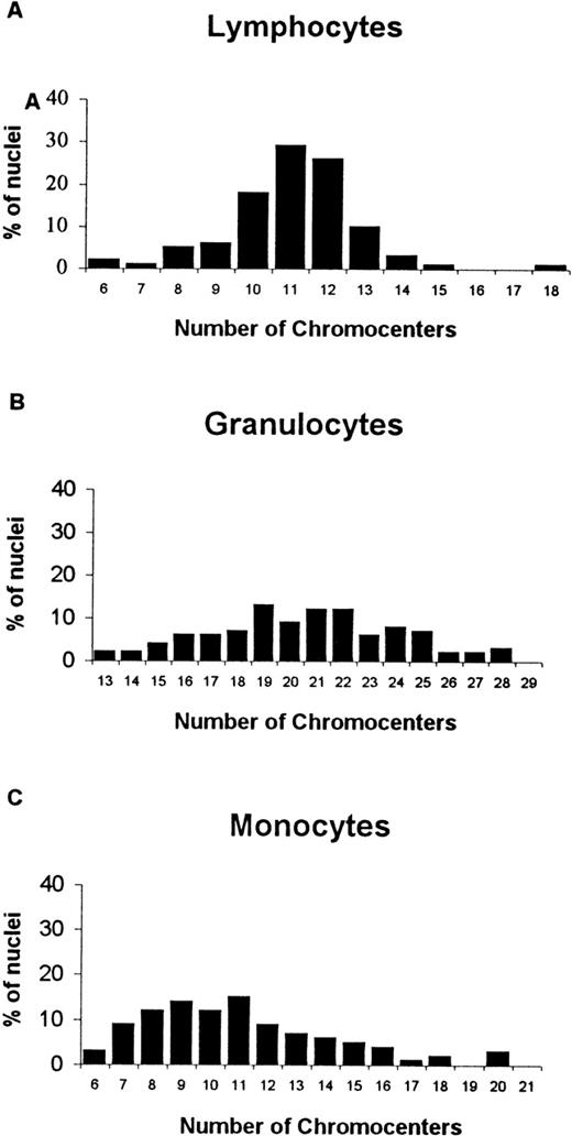
To determine whether centromeres aggregate to chromocenters in quiescent human cells, a pan-centromeric probe was first hybridized to 3-D preserved unstimulated peripheral blood cells and primary fibroblasts in G1. The results in the former showed that instead of the 46 single dot signals that should be expected for normal diploid human cells in G1, the hybridization signals merged into irregular masses of various sizes and shapes, whose number varied from 6 to 18 (mean, 11) per nucleus in lymphocytes, 13 to 28 (mean, 21) in granulocytes, and 6 to 20 (mean, 11) in monocytes and were predominantly associated to the nuclear envelope or the nucleolus (see Figure1 and examples in Figure2A). This differed from what was observed in PBL stimulated with phytohemagglutinin, in which the number of signals was consistently higher (more than 35) and more single-dotted in most of the cells (not shown). As previously reported,11aggregation of centromeres was also observed in quiescent fibroblasts. However, the hybridization patterns differed from those in unstimulated lymphocytes by the higher number of hybridization signals (mean, 25 per nucleus) (Figure 2B). In summary, the data show that centromeres do aggregate to heterochromatic structures in unstimulated peripheral blood lymphocytes, monocytes, and granulocytes, which, in the case of lymphocytes, disperse after stimulation with a mitogen and are larger than those observed in quiescent fibroblasts.
Analysis of chromocenters in quiescent peripheral blood cells using a pancentromeric probe.
Observed distributions of chromocenters per nucleus in lymphocytes (A), granulocytes (B), and monocytes (C) (100 nuclei analyzed for each cell type).
Analysis of chromocenters in quiescent peripheral blood cells using a pancentromeric probe.
Observed distributions of chromocenters per nucleus in lymphocytes (A), granulocytes (B), and monocytes (C) (100 nuclei analyzed for each cell type).
Examples of the analysis of chromocenters in lymphocytes and fibroblasts using confocal microscopy.
(A) Optical series of a lymphocyte nucleus hybridized with the pan-centromeric probe. A reduced number of hybridization signals is observed, corresponding to the coalescence of signals originated by individual centromeres. Note that most of the signals are visible in several sequential optical sections. (B) Analysis of chromocenters in quiescent human fibroblasts. Optical sections obtained in different nuclei. Bars, 10 μm. The nuclear lamina is labeled with an anti-laminin B antibody.
Patterns of centromere associations in chromocenters of peripheral blood cells and fibroblasts
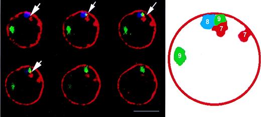
We next sought to investigate whether these structures represented random associations of different centromeres or the preferential clustering of specific centromeres in each cell type. To do this, probes specific for each of the human centromeres were first hybridized to quiescent lymphocytes. As shown in Table 1, different centromeres have different frequencies of association. The highest are between centromeres of the acrocentric chromosomes (pairs 13-21 and 14-22, associated in more than 60% of the cells), which were almost always in proximity to the nucleolus (as visualized by the superimposition of fluorescent and phase-contrast images), followed by centromeres 15 and 16 (46% ± 0.02%), the latter also in proximity to the nucleolus; 10 and 11 (42%); 18 and 20 (41%); 7 and 9 (43% ± 0.03%); 8 and 9 (38%); 15 and 17 (38% ± 0.03%); 11 and 12 (37%); and 17 and 18 (34%) (see example in Figure 3). Associations were consistently observed between heterologous centromeres because those from homologous chromosomes were rarely associated (less than 2% of the cells in each experiment for each pair of homologs). To verify whether the observed associations varied among different donors, hybridization experiments with probes for a few pairs of centromeres that had been found to have higher (centromeres 7 and 8, 7 and 9, 15 and 16, 15 and 17, 16 and 17) or lower (centromeres 2 and 3, 2 and 4, 3 and 6) frequencies of association in 1 donor were subsequently performed in samples derived from 4 other donors. No significant differences were observed among different donors for the more-associated or the less-associated pairs of centromeres (allP > 0.05) (Table 1; cases with mean values ± SD). To further verify the consistency of the data, pairs of centromeres whose patterns of association could be predicted from the analysis using triple combinations of probes were subsequently analyzed in cells from different donors in double-hybridization experiments. More specifically, triple hybridization for centromeres 1-2-3 showed that associations between centromeres 1 and 2 occurred in 18% of the cells, whereas the combination of probes for centromeres 2-3-4 showed a mean association between centromeres 2 and 4 of 20%. Double hybridization with probes for centromeres 1 and 4 was then performed and showed an association of 19% for this pair of centromeres. Similarly, the triple combinations of probes for centromeres 2-3-4 and 3-4-6 showed a frequency of association of 16% and 15% for centromeres 2 and 3 and 3 and 6, respectively. Double hybridization with probes for centromeres 2 and 6 in another donor was 18%. The triple combinations for centromeres 7-8-9 and 9-10-11 showed frequencies of associations of 43% for centromeres 7-9 and of 35% for centromeres 9-11. Centromeres 7 and 11 were found to be associated in 30% of the cells from a different donor (Table 1). Probabilities of association between pairs of centromeres were found to be the same for these double- and triple-hybridization experiments. They had an error of 1% except for centromere pair 7 and 8, in which the error of association was 5%. Taken together, the data show that the association patterns between specific centromeres are maintained in cells derived from different donors.
Optical sections of a lymphocyte nucleus showing the clustering of 3 different centromeres (arrows).
The respective chromosome numbers are indicated on the diagram on the right. Note the proximity of 3 centromeres to the nuclear lamina (see text), which is labeled with an anti-laminin B antibody (red). Bar, 5 μm.
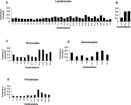
As to the centromeres belonging to the acrocentric chromosomes 13, 14, 21, and 22 and to chromosomes 5 and 19, the analysis was hampered by the cross-hybridization of the respective probes, a fact that makes it impossible to discriminate accurately between heterologous centromeres. To overcome this problem, single-hybridization experiments were performed, which originated 4 single-colored signals per nucleus, and the association of homologous centromeres was assumed to be a rare event for these chromosomes. Accordingly, every coalescence of hybridization signals was considered as corresponding to associations between heterologous centromeres. As expected, centromeres belonging to acrocentric chromosomes were frequently associated in the nuclei of these cells (more than 60% of the cells). This contrasted with the low association between centromeres 5 and 19 (23%) (Table 1). The analysis of probability based on all the data confirmed the nonrandom association of centromeres in the nucleus of quiescent lymphocytes (as assessed by the magnitude of P values, which differed markedly from those predicted for a random association of centromeres in the nucleus) (Figures 4A, 4B) (see “Materials and Methods”).
Probabilities of associations of specific centromeres in quiescent peripheral blood cells and fibroblasts as assessed by hybridization experiments using probes for different centromeres.
(A, B) Lymphocytes. Analysis in triple-hybridization (A) or single-hybridization (B) experiments (see text). (C) Monocytes. (D) Granulocytes. (E) Fibroblasts.
In summary, the chromocenters present in the nuclei of unstimulated PBL can be considered the emerging pattern of the preferential association of specific centromeres. They can be assigned to 2 main groups: 1 containing the centromeres of acrocentric chromosomes (probability of association, 60%), and the other containing the centromeres of the remaining chromosomes (probability of association, 10% to 40%; mean, 20%).
We subsequently asked whether the chromocenters in the other hematopoietic cell types were made of the same clustered centromeres as in quiescent lymphocytes. Thus, the frequencies and probabilities of association between selected pairs of centromeres were determined in monocytes (1100 nuclei) and granulocytes (800 nuclei). The following groups of centromeres were studied (see Table 1): 1 corresponding to those with lower frequencies of association in lymphocytes (centromeres 1 and 4, 3 and 4, 3 and 6, 4 and 6) and the other corresponding to those with higher frequencies of association in lymphocytes (centromeres 7 and 9, 10 and 11, 11 and 12, 13 and 21, 14 and 22, 15 and 16, 18 and 20). The data show that in monocytes and in granulocytes, the highest frequencies of association were again observed for centromeres of acrocentric chromosomes: 13 and 21 (82% in monocytes, 70% in granulocytes) and 14 and 22 (79% in monocytes, not investigated in granulocytes). As to the remaining pairs, the frequencies of association between specific centromeres were shown to differ in the 3 cell types. Thus, when comparing lymphocytes with monocytes, significant differences (P < .05) were observed for 4 of 9 pairs of centromeres from nonacrocentric chromosomes analyzed (44%) (see Table 1). When comparing lymphocytes with granulocytes, significant differences were observed for 4 of 7 pairs from nonacrocentric chromosomes analyzed (57%) (Table 1). In contrast with these results, the comparison between granulocytes and monocytes showed only 1 significant difference for centromeres 11 and 12 (52% vs 22% for granulocytes and monocytes, respectively) (1 of 7 pairs from nonacrocentric chromosomes, 14%) (Table 1). The probability analysis in monocytes and granulocytes is shown in Figures 4C and 4D. In summary, the data show that marked differences in the spatial arrangements of specific centromeres can be observed when comparing PBL with granulocytes and monocytes. This is in contrast with the more similar patterns of centromere association observed in the latter 2 cell types.
Subsequently, the associations between the same pairs of centromeres were analyzed in fibroblasts, as representative of a nonhematopoietic cell type (1300 nuclei analyzed), and were compared with the other cell types. As shown in Table 2, centromeres belonging to acrocentric chromosomes 13 and 21 and 14 and 22 were also the most frequently associated in fibroblasts. As to the remaining pairs analyzed (total, 9), significant differences were observed in 6 pairs when comparing lymphocytes with fibroblasts (67%) (Table 2). When fibroblasts were compared with monocytes, significant differences were found for 8 of 9 pairs of centromeres (89%) (belonging to nonacrocentric chromosomes) and, when compared with granulocytes (7 pairs of nonacrocentric centromeres analyzed), significant differences were observed for 6 pairs (86%) (Table 2). The analysis of probability in fibroblasts is shown in Figure 4E.
Frequencies of association of specific centromeres in quiescent human primary fibroblasts: comparison with hematopoietic cells
| Centromeres . | Fibroblasts a . | PBL a . | PBM a . | PBG a . |
|---|---|---|---|---|
| 1/4 | 0.1 | 0.19 | 0.38* | 0.47* |
| 3/4 | 0.08 | 0.18 | 0.24* | 0.24* |
| 3/6 | 0.08 | 0.15 | 0.23* | 0.30* |
| 4/6 | 0.08 | 0.20* | 0.21* | 0.16* |
| 7/9 | 0.13 | 0.43* | 0.36* | ND |
| 10/11 | 0.12 | 0.42* | 0.26* | 0.24* |
| 11/12 | 0.15 | 0.37* | 0.22 | 0.52* |
| 13/21 | 0.55 | 0.64 | 0.82* | 0.70* |
| 14/22 | 0.35 | 0.69* | 0.79* | ND |
| 15/16 | 0.22 | 0.46* | 0.44* | 0.34 |
| 18/20 | 0.18 | 0.41* | 0.56* | ND |
PBL, peripheral blood lymphocytes; PBM, peripheral blood monocytes; PBG, peripheral blood granulocytes. a, associated; ND, not done.
Comparison with fibroblasts P < .05.
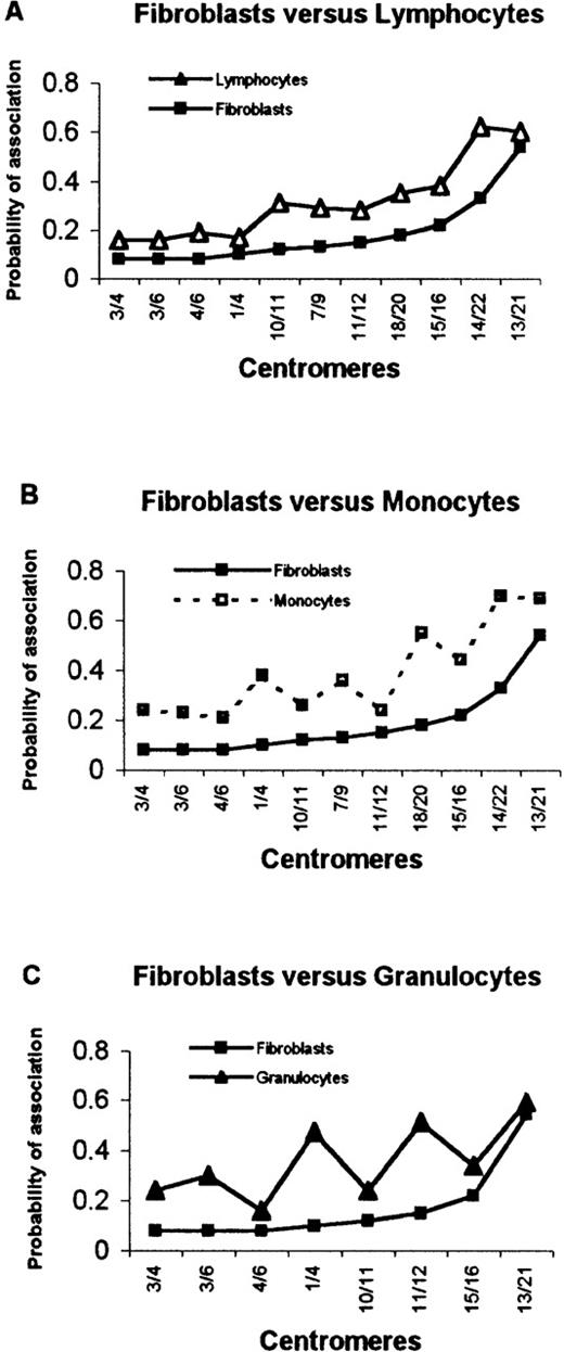
Then the probabilities of association for the same pairs of centromeres were compared between lymphocytes and the other cell types under analysis. To do that, the probability of association for similar centromeric pairs was plotted as a function of the corresponding pairs. As shown in Figures 5A to 5C, there was a marked difference between fibroblasts and each hematopoietic cell type as evidenced by the nonmonotonicity of the correlation curves for lymphocytes (significant differences observed for 6 of the 9 pairs of nonacrocentric centromeres, 67%), monocytes (significant differences observed for 8 of the 9 pairs of nonacrocentric centromeres, 89%), and granulocytes (significant differences observed for 6 of the 7 pairs of nonacrocentric centromeres analyzed, 86%), though a trend to follow the same correlation curve, with a few inflections of the derivative, can be observed in all cell types. As to the comparison between different hematopoietic cells, depicted in Figure 5, lymphocytes differed from monocytes (in 4 of 9 nonacrocentric centromere pairs, 44%) and granulocytes (4 of 7 nonacrocentric pairs analyzed, 57%), a less conspicuous difference than from fibroblasts (Figures 5D,5E). This is in contrast with what was observed for the curves for granulocytes and monocytes, which were similar, except for the comparison of centromeres 11 and 12 (Table 1) (Figure 5F). Therefore, this correlation analysis shows that different cell types have distinct patterns of centromere associations and that within hematopoietic cells, 2 distinct emerging patterns appeared to exist, 1 common to cells of the myeloid lineage (monocytes and granulocytes), the other characteristic of cells of the lymphoid lineage.
Probabilities of centromeric association in hematopoietic cells and fibroblasts as a function of the same pairs of centromeres.
(A to C) Comparison between fibroblasts and hematopoietic cells. In all cases, the order by which centromeric pairs are depicted was chosen so that a monotonous curve was obtained for fibroblasts (increasing order of frequencies). (D to F) Comparison between different hematopoietic cells. Note that the ordering of centromeric pairs was changed so that a monotonous curve was obtained for lymphocytes (D, E) or granulocytes (F). See Tables 1 and 2 for statistically significant differences.
Discussion
In this study we investigated the spatial association of centromeres into chromocenters of quiescent human peripheral blood cells and fibroblasts. The data show that these interphase heterochromatic structures correspond to nonrandom spatial arrangements of specific centromeres in all cell types, that the chromocenters observed in these cells are made of different combinations of centromeres, and that, as to the emerging patterns of centromere associations, fibroblasts are strikingly different from hematopoietic cells, and, within the latter, cells of myeloid origin (monocytes and granulocytes) are similar to each other and different from lymphoid cells.
We started by characterizing the number, size, and location of chromocenters in all cell types. As consistently reported in previous studies and for different mammalian cell types, centromeres are nonrandomly distributed in the nucleoplasm and show a tendency to cluster around the nucleolus; peripherally, they are in proximity to the nuclear envelope (see for example1,20). A detailed analysis of the composition of chromocenters in quiescent PBL was subsequently performed, using sequential combinations of probes specific for each of the human centromeric α-DNA sequences. Two main findings emerged from the study: first, the observed frequencies of association showed that the centromeres close to the nucleolus mostly belonged, as expected, to chromosomes containing nucleolar organizers (NOR). However, the nucleolus-related clusters of centromeres were not exclusive of NOR-containing chromosomes because the centromeres of chromosomes 16 (which does not contain a NOR) were frequently associated with those of chromosome 15, near the nucleolus, a fact that might be related to the presence of large constitutive pericentromeric heterochromatic blocks in chromosome 16.10 Still, it must be noted that the question of whether the observed clustering is just the reflex of some sort of physical attraction driven by the respective heterochromatic regions14,21 or obeys any, as yet unknown, nucleolus-related functional imperative, is open to question. The analysis of fibroblasts, here used as representative of a nonhematopoietic cell type, showed that a similar global topography of centromeres exists for these cells, thus strengthening the idea that physical constraints imposed either by ribosome biogenesis or heterochromatin associations to the nuclear lamina22-24 are responsible for a general, nontissue-specific, meta-organization of these chromosomal domains within the nucleus. But the second and most striking finding of the current study is that the nucleolus-unrelated chromocenters in the interphase nuclei of quiescent cells are also formed by the preferential association of centromeric regions belonging to specific chromosomes and are not the consequence of a random assortment of centromeres. In lymphocytes, centromeres from chromosomes 7 to 20 were the most frequently associated (varying from 38% to 47% of the cells among the different combinations analyzed), whereas centromeres from chromosomes 1 to 9, X and Y, were the least frequently associated (varying from 10% to 26% of the cells). Importantly, the comparison of a few selected pairs of centromeres in lymphocytes, monocytes, granulocytes, and fibroblasts further showed that different centromeres have different probabilities of association in these cell types. Two main findings emerged from this comparative analysis. First, the most conspicuous differences were observed between fibroblasts and hematopoietic cells (irrespective of the cell type). Second, within the latter, 2 distinct patterns of heterochromatic compartments were clearly observed—a myeloid pattern, as evidenced by the overall similar arrangement of centromeres in monocytes and granulocytes, and a lymphoid pattern, present in quiescent lymphocytes.
Therefore, taken together, these observations indicate that the chromocenters in quiescent cells might represent tissue-specific organizations of centromeric heterochromatin. Although the biologic mechanisms underlying such spatial arrangements are totally unknown, the clustering of specific centromeres into chromocenters, as shown in this study, is likely to have important functional implications for the cell. Recent evidence indicates that centromeres and other heterochromatic regions may behave as sequestering compartments for factors regulating gene activity and that these higher-order chromatin structures can sustain a stable and heritable inactivation of transcription (reviewed14,21). This, together with the evidence that genes may shuttle from transcriptionally competent to heterochromatic repressive environments (and vice versa), in apparent relation to their differentiation stage-dependent activity15raises the hypothesis that the repressive environments themselves must be organized in a tissue-specific manner. The data presented in this study indicate that this might well be the case, further suggesting that heterochromatic compartments in the cell nucleus might be ontogenically determined, as evidenced by the differential patterns observed in different hematopoietic cell-lineages. It is conceivable that such arrangements would favor the creation of functional atmospheres in the nucleus with intrinsic coherence, in that they are formed by the aggregation of specific centromeres surrounded by a “cloud” of euchromatic regions belonging to the respective chromosomes. It is, in this respect, noteworthy that an apparent functional clustering of specific chromosomes into novel transcriptional domains has been recently reported in human cells in G1.25 If this is so, a further (and testable) hypothesis is that the specific combination of chromosomes related to different chromocenters must be predominantly enriched in 2 main types of genes, those that are not specifically engaged in the active genetic makeup of that particular tissue (constitutively inactive genes for that cell type) and those with tissue-specific expression but whose activity is differentially regulated during, for example, development or cell ontogeny (akin to the genes investigated by Brown et al15). The former genes would be the nearest neighbors of the repressive compartment, the chromocenter, whereas the latter would be the farthest, but at a distance compatible with their shuttling to and from the chromocenter. It must be noted that this last aspect is of importance because the dynamics of chromosomes or chromosomal subregions in interphase appears to be determined by physical constraints that limit their range of movement within the nucleoplasm (reviewed26). In a scenario such as this, the aggregated centromeres in the chromocenters of the interphase nucleus could then be envisaged as major regulatory compartments for tissue-specific gene expression.
Acknowledgment
The authors thank M. Carmo-Fonseca for critical review of the manuscript.
Supported by a grant from Program PRAXIS XXI. IA was supported by a PRAXIS XXI fellowship.
Reprints:Leonor Parreira, Instituto de Histologia e Embriologia, Faculdade de Medicina de Lisboa, Avenida Prof. Egas Moniz 1699, Lisboa Codex, Portugal; e-mail: hleonor@correio.fm.ul.pt.
The publication costs of this article were defrayed in part by page charge payment. Therefore, and solely to indicate this fact, this article is hereby marked “advertisement” in accordance with 18 U.S.C. section 1734.


This feature is available to Subscribers Only
Sign In or Create an Account Close Modal