Abstract
Jun N-terminal kinase (JNK) and p38, members of the mitogen-activated protein kinase family of serine/threonine kinases, are activated as a result of cellular stress but may also play a role in growth factor-induced proliferation and/or survival or differentiation of many cells. A recent report has implicated JNK and p38 in the induction of apoptosis in the erythropoietin (EPO)-dependent erythroid cell line HCD57 following EPO withdrawal, whereas our previously reported data did not support a role for JNK in growth factor withdrawal-induced apoptosis in HCD57 cells. Therefore, further testing was done to see if JNK was activated in EPO withdrawal-induced apoptosis; the study was extended to p38 and characterized the effect of EPO on JNK and p38 activities. Treatment of HCD57 cells with EPO resulted in a gradual and sustained activation of both JNK and p38 activity; these activities decreased on EPO withdrawal. Transient activation of p42/p44 extracellular signal-related kinases (ERK) was also detected. Inhibition of ERK activity inhibited proliferation in EPO-treated cells but neither induced apoptosis nor activated JNK. Inhibition of p38 activity inhibited proliferation but did not protect HCD57 cells from apoptosis induced by EPO withdrawal. Treatment of HCD57 cells with tumor necrosis factor-alpha induced JNK activation but did not induce apoptosis. These results implicate JNK, p38, and ERK in EPO-induced proliferation and/or survival of erythroid cells but do not support a role for JNK or p38 in apoptosis induced by EPO withdrawal from erythroid cells.
The glycoprotein hormone erythropoietin (EPO) is the primary regulator in the control of erythroid cell maturation. Cells at the proerythroblast stage or colony-forming unit-erythroid stage of differentiation depend on EPO for continued differentiation. Apoptosis or programmed cell death occurs when EPO is withdrawn in vitro.1-4 Recently, a family of serine-threonine protein kinases that is structurally similar yet functionally distinct has been identified. These mitogen-activated protein kinases (MAPKs) fall into 4 distinct groups: the extracellular signal-related kinases (ERKs)5,6 the cJun-amino terminal kinases (JNKs),7 p38 map kinase (p38),8 and Erk5/BMK1.9 Although these kinases all represent the end of pathways involving multiple serine-threonine kinases that are activated in a cascade,10 they exhibit different physiological effects on cell development. The ERK pathway is primarily associated with promoting proliferation, whereas the role of the JNK and p38 pathways is more complex. Apoptosis-inducing agents, such as UV irradiation,11 ion irradiation,12 growth factor deprivation,13,14 and inflammatory cytokines such as tumor necrosis factor α (TNF-α),15-17 induce JNK and p38 activation. However, JNK and p38 are also activated by survival factors, such as EPO, stem cell factor (SCF),18-20interleukin 4 (IL-4),21 thrombopoietin, IL-3, and granulocyte macrophage-colony stimulating factor. There is also evidence that TNF-α treatment may activate JNK to promote proliferation of some hematopoietic cells and tumor cells22-25 and to promote liver regeneration after partial hepatectomy.26 Therefore, it is clear that JNK also plays a role in mitogenic and growth factor signaling.
The end result of activation of signal transduction cascades is often the phosphorylation and activation (or deactivation) of transcription factors. JNK and p38 activation results in the phosphorylation of their substrates activator protein-1 (AP1) and activating transcription factor-2 (ATF-2), respectively.27,28 AP1 comprises members of the Fos and Jun families of proto-oncogene and binds to DNA in a sequence-specific manner to activate or to repress transcription.29 AP1 has long been associated with cell cycle progression, tumor promotion, and proliferation. JNK phosphorylates the N-terminus of the cJun protein and increases its transactivation potential. Of the 3 known Jun family members (cJun,30 JunB,31 and JunD32), cJun is the only member that can serve as an efficient substrate for JNK.33,34 The ATF family of transcription factors includes ATF-1, ATF-2, ATF-3, ATF-4, and the cyclic AMP responsive binding protein (CREB).35 Although ATF-2 is the only member of this family known to be phosphorylated directly by p38, CREB is activated indirectly in response to p38 activation via activation of the ribosomal SK 6 kinases, which then activate CREB.36 37 JNK and p38, therefore, can have multiple effects on transcription factor activation.
Recently, JNK and p38 have been implicated in the regulation of erythroid proliferation and survival. JNK and p38 activation were initially reported to be induced by EPO,20,38 and recent reports39,40 have suggested that p38 and JNK are necessary for the initiation of erythroid differentiation. Our laboratory has previously reported on the role of AP1 and JNK in the regulation of erythroid cell proliferation and apoptosis.41 By using the murine erythroleukemia cell line HCD57, we demonstrated that AP1 DNA-binding activity was induced in either the proliferative and growth factor withdrawal states but that different AP1 factors were involved in the two processes. cJun DNA-binding activity and JNK activity were induced in the presence of EPO, whereas EPO withdrawal resulted in a decrease in JNK activity and an increase in JunB DNA-binding activity. In contrast, a recent report by Shan et al.42 has implicated JNK and p38 in the induction of apoptosis induced by EPO withdrawal in HCD57 cells. Their report suggested a reciprocal relationship between p42/p44 ERK activation and JNK activation similar to that observed in growth factor-deprived PC12 neuronal cells.14 To clarify our position, we present here further studies into the role of JNK, ERK, and p38 in erythroid proliferation and initiation of apoptosis in HCD57 cells. These studies confirm our previous contention that JNK and cJun activities are not associated with the induction of apoptosis in HCD57 cells but are instead associated with EPO-induced proliferation. In addition, p38 activation appears to participate in EPO-dependent proliferation but not in apoptosis induced by EPO withdrawal.
Materials and methods
HCD57 cells
The EPO-responsive erythroid cell line HCD57 was originally established by Drs Hankins, Chin, and Dons in 1987.43 The cell line was established from leukemic cells that arose from a newborn mouse infected with a Friend helper virus and subsequently passed in vivo in mice before being adapted to tissue culture. The HCD57 cells extensively used in this laboratory since 1989 were obtained from Dr Sandy Ruscetti (Frederick, MD) as part of a joint collaboration with Dr Hankins.44 In this paper, these HCD57 cells are referred to as HCD57(R) cells. The HCD57(R) cell line was subcloned from a single cell. HCD57(R) cells are ideally suited to EPO signaling studies because these cells will survive EPO withdrawal for 24 hours without undergoing significant apoptosis. This withdrawal of EPO overnight results in up-regulation of the cell surface receptors for EPO by 10-fold or more over cells maintained in EPO.45 In addition, this prolonged absence from EPO also results in complete quiescence of EPO signaling such that a dramatic burst of signaling is activated on EPO treatment in HCD57 cells.46 47
HCD57 cells also used in this study were obtained from Dr Linda Kelley, who obtained early passage HCD57 cells from Dr Hankins to conduct a collaborative study.48 This cell line is referred to as HCD57(K) in this paper. It is likely that the HCD57(K) cell line is derived from a heterogeneous population of cells transplanted from the leukemic mouse. In contrast to the HCD57(R) cells, HCD57(K) cells undergo extensive apoptosis following EPO withdrawal for 24 hours. Thus, these cells can only be deprived of EPO for a short period (4 hours or less) prior to EPO signaling studies. We will compare HCD57(R) and HCD57(K) cells in some studies to test if these cells differentially use JNK or p38 activity to induce apoptosis following EPO withdrawal.
Materials
Murine TNF-α and inhibitors PD98059, SB203580, and LY294002 were purchased from Calbiochem (La Jolla, CA). Recombinant SCF was purchased from Intergen (Purchase, NY). Phospho-specific antibodies against JNKs (Thr183/Tyr185), ERKs (Thr/Tyr204), AKT (Ser473), and p38 (Thr 180/Tyr182) were obtained from New England Biolabs (Beverly, MA). Rabbit- and goat-polyclonal antibodies recognizing both phosphorylated and nonphosphorylated forms of JNK1 (C-17), JNK2 (FL), ERK1 (C-16), ERK2 (D-2), and p38 (C-20) were obtained from Santa Cruz Biotechnologies (Santa Cruz, CA).
Cell culture
HCD57(R) cells were cultured in Iscove modified Dulbecco medium (IMDM) (Life Technologies Inc, Gaithersburg, MD), 25% fetal calf serum (Hyclone, Logan, UT), and 10 μg/mL gentamicin (Life Technologies Inc) at 37°C in a 5% CO2 environment and maintained in 1 U EPO/mL media. HCD57(K) cells were cultured in the same media with the exception that 30% fetal calf serum was used. For each time point, 2.5 × 10⁶ HCD57 cells were used. For EPO-deprivation studies, the cells were washed 3 times in media and incubated in the absence of EPO for the times indicated in the figure legends. For EPO-induced proliferation studies, HCD57(R) cells were washed 3 times and incubated for 18 hours in the above media minus EPO. The cells were then treated with 10 U EPO/mL for the times indicated in the figure legends. For studies with the MEK inhibitor PD98059 and the p38 inhibitor SB203580, cells were washed 3 times to remove all EPO from the cells and then cultured in media containing EPO, EPO + DMSO, EPO + 50 μmol/L PD98 058, EPO + 20 μmol/L SB203580, or no additional growth factor for the times indicated in the figure legends. Cell viability was determined by counting several hundred cells on a hemocytometer in the presence of 0.2% trypan blue. For TNF-α studies, HCD57(R) cells were washed 3 times to remove all EPO from the cells and then cultured in media containing no EPO for 18 hours. The cells were then treated with various concentrations of TNF-α, 10 SCF/mL or 1 U EPO/mL as indicated in the figure legends.
Western blot analysis
Following treatment of the cells under the different conditions, HCD57(R) or HCD57(K) cells were harvested and lysed immediately in sample buffer (0.05 mol/L Tris, pH = 8, 2% sodium dodecyl sulfate, 0.1% bromophenol blue, 10% glycerol, and 10% β-mercaptoethanol) and sonicated for 10 seconds each to shear the genomic DNA. Equal volumes (40 μL) of sample were electrophoresed on a 10% acrylamide SDS-PAGE gel (for JNK, AKT, and ERK blots) or a 12% acrylamide SDS-PAGE gel (for the p38 blots) and transferred to nitrocellulose. The blots were blocked for 1 hour in TBST buffer (25 mmol/L Tris, pH = 7.8, 125 mmol/L NaCl, and 0.25% Tween-20) containing 5% nonfat milk and then incubated in primary antibody overnight at 4°C in TBST buffer containing 5% bovine serum albumin (BSA). The blots were washed in TBST buffer, and specific reactive proteins were detected by using enhanced chemiluminescence (Amersham Pharmacia Biotech, Piscataway, NJ). The blot was then stripped as previously described46 and re-probed successively with phospho-specific antibodies as indicated in the figures. To ensure equal loading of proteins, the blots were last probed with antibodies that recognize both the phosphorylated and nonphosphorylated forms of JNK-1, ERK1, or p38 (as indicated in the figure legends). Molecular weights are indicated in kilodaltons (kD). For the in vitro kinase assays, total cell extracts were immunoprecipitated as previously described with anti-JNK-1 and subjected to an in vitro kinase assay as previously described41 using 1 μg GST-Jun fusion protein as a substrate (a generous gift from Dr Paul Dent). In the case of the kinase assays in Figures 1 and 3, following electrophoresis of the samples on a 10% acrylamide SDS/PAGE gel, the proteins were transferred to nitrocellulose and exposed to autoradiography 18 hours at −80°C with an intensifying screen to visualize phosphorylated GST-Jun. The blots were then probed with the polyclonal anti-JNK1 antibody to ensure equal loading of proteins.
Detection of apoptosis of HCD57 cells
Apoptosis of HCD57(R) and HCD57(K) cells was detected by using flow cytometry analysis of propidium iodide-stained cells. Following cell treatment, HCD57 cells were fixed in 70% ethanol overnight at 4°C. The cells were then washed in phosphate buffered saline (PBS) and stained overnight in 3 mmol/L NaCitrate, 2 μmol/L propidium iodide, and 50 μg/mL RNAse A at 4°C in the dark. The cells were then collected, washed once in 1X PBS, and analyzed by using the FACScan flow cytometer (Becton Dickinson, Rutherford, NJ). Cells containing sub-G0/G1 DNA indicative of apoptosis were gated and shown as a percentage of the total number of cells.
Results
JNK and p38 activities decrease on EPO withdrawal in HCD57 cells
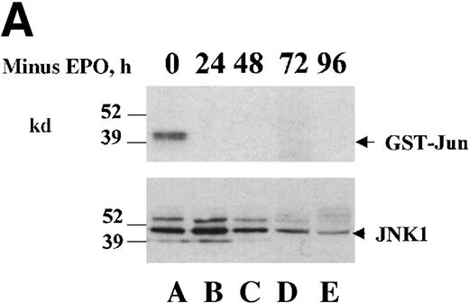
To test if JNK and/or p38 are activated following EPO withdrawal to induce apoptosis, we utilized in vitro assay of JNK activity and activation specific antibodies for JNK and p38. Two variants of the HCD57 cell line were used in the following studies. One cell line, which we have designated HCD57(K), undergoes rapid apoptosis within 24 hours of EPO withdrawal. The other cell line, designated HCD57(R), undergoes apoptosis more slowly and does not exhibit significant apoptosis until 48 to 72 hours following EPO withdrawal. We have previously shown that removing EPO from HCD57(R) cells by using an EPO-neutralizing antibody resulted in decreased JNK activity over a 24-hour period (Figure 5 of reference 41). To ensure that there was no activation of JNK in the period of time from 24 to 96 hours that may correlate with the later activation of apoptosis in this cell line, we repeated our in vitro kinase assay of EPO-deprived HCD57(R) cells over a 96-hour period. Figure 1A shows that JNK activity was observed in the presence of EPO and was no longer detectable 24 hours following EPO withdrawal. JNK activity was also not detected at later time points. To further confirm this observation of a loss of JNK activity in EPO-deprived HCD57(R) cells, whole cell extracts were subjected to Western blot analysis by using an antibody specific to the active form of JNK (phospho-JNK antibody; Figure 1B). JNK1 and JNK2 activation were detected in the presence of EPO but decreased greatly on EPO withdrawal. These results confirmed our earlier observation that JNK activity decreased on EPO withdrawal and did not correlate with the initiation of apoptosis in HCD57(R) cells.
JNK and p38 activities are high in HCD57 cells in the presence of EPO and decrease on EPO withdrawal.
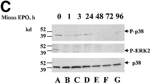
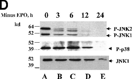
HCD57 cells were deprived EPO for up to 96 hours and samples were taken at 24-hour intervals. (A) Rabbit-anti-JNK-1 immunoprecipitates were subjected to an in vitro kinase assay using γ-32P-ATP and GST-Jun fusion protein as a substrate and transferred to nitrocellulose. Following exposure to show the phosphorylated GST-Jun (upper arrow), the proteins were immunoblotted with goat anti-JNK-1 to visualize JNK proteins (lower arrow). (B) Whole cell lysates were immunoblotted with an anti-phospho-JNK antibody (upper panel). The blot was then stripped and reprobed with anti-JNK1 to ensure equal loading of proteins (arrow, lower panel). (C) Whole cell lysates were immunoblotted with an anti-phospho-p38 antibody (upper panel). The blot was then stripped and reprobed with anti-phospho-ERK antibody (middle panel) to detect phosphorylated ERK (arrow, middle panel) and p38 (lower panel) to ensure equal loading of proteins (arrow, lower panel). (D) HCD57(K) cells were deprived of EPO for the times indicated. Whole cell lysates were immunoblotted with anti-phospho-JNK antibody (top panel). Arrows indicated phosphorylated JNK-1 and -2 proteins. The blot was then stripped and reprobed with anti-phospho-p38 antibody to detect phosphorylated p38 (arrow, middle panel) and anti-JNK1 to ensure equal loading of proteins (arrow, lower panel).
We next investigated the effect of EPO deprivation on p38 and ERK activity in HCD57(R) cells by using an antibody directed to the activated from of p38 (phospho-p38). p38 was active in cells cultured in EPO, and EPO withdrawal induced a rapid decrease in p38 activation 1 hour following EPO withdrawal and was undetectable 48 hours following EPO withdrawal (Figure 2C, lane E). Reprobing this blot with the ERK phospho-specific antibody revealed that ERK activation was also lost within 1 hour of EPO withdrawal (Figure 1C, middle panel, lane B). Therefore, p38 activation did not correlate with the induction of apoptosis on growth factor withdrawal in HCD57(R) cells.
JNK and p38 are activated on EPO addition in HCD57 cells.
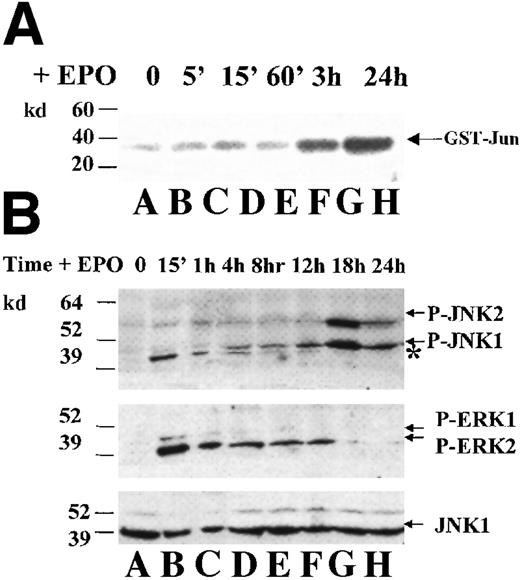
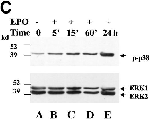
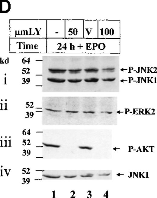
HCD57(R) cells were deprived of EPO for 18 hours and then incubated in EPO for the times indicated. (A) Rabbit-anti-JNK-1 immunoprecipitates were subjected to an in vitro kinase assay using γ-32P-ATP and GST-Jun fusion protein as a substrate. Arrow indicates phosphorylated GST-Jun. (B) Whole cell lysates were immunoblotted with an anti-phospho-JNK antibody (upper panel). Asterisk indicates phosphorylated ERK2 protein that cross-reacts with the phospho-JNK antibody. The blot was then stripped and reprobed with anti-phospho-ERK antibody (middle panel) to detect phosphorylated ERK-1 and -2 (arrows, middle panel) and anti-JNK1 (lower panel) to ensure equal loading of proteins (arrow, lower panel). (C) Whole cell lysates were immunoblotted with an anti-phospho-p38 antibody (upper panel). The blot was then stripped and reprobed with anti-ERK-1 (lower panel) to ensure equal loading of proteins (arrow, lower panel). (D) ERK and JNK are not activated in a PI 3-kinase dependent manner in HCD57 cells. HCD57(R) cells were incubated for 24 hours in the presence of EPO alone (lane 1), EPO + DMSO (lane 3) or EPO + 50 μmol/L or 100 μmol/L LY294002 (lanes 2 and 4). Whole cell lysates were immunoblotted with an anti-phospho-JNK antibody (i). The blot was then stripped and reprobed with anti-phospho-ERK (ii, arrow), anti-phospho-AKT antibody to detect phosphorylated AKT (iii, arrow), and anti-JNK1 to ensure equal loading of proteins (iv, arrow).
The cell line we used for the above studies is slow to undergo apoptosis induced by EPO withdrawal. It is possible, therefore, that subcloning or continuous passage of these cells has resulted in a cell line that has lost JNK- and p38-related apoptosis signals. We, therefore, assessed MAPK activity in the HCD57(K) cells that undergo apoptosis within 24 hours of EPO withdrawal. Figure 1D shows that both JNK and p38 activation were high in HCD57(K) cells and decreased quickly on EPO withdrawal to undetectable levels by 24 hours. The time course of the decrease in activity was different for the 2 kinases, however. JNK phosphorylation decreased within 3 hours of EPO withdrawal (Figure 1D, top panel, lane B), whereas p38 activity did not decrease until 12 hours following EPO removal (Figure 1D, middle panel, lane D). This result does not support a role for JNK or p38 signals that contribute to apoptosis but does suggest that different upstream signals activated by EPO distinctly regulate JNK and p38 activities.
JNK and p38 are activated by EPO in HCD57 cells
It has been previously reported20 39 that JNK was activated in erythroid cells within 15 minutes of EPO addition to erythroid cells. To investigate JNK activity in response to EPO, HCD57(R) cells were washed extensively to remove EPO and cultured in the absence of EPO for 18 hours to up-regulate the EPO receptor and turn off EPO signals. The cells were then stimulated with EPO. When we assessed JNK activity over a 24-hour period following EPO addition, we observed a slow and gradual increase in JNK-1 and JNK-2 activity (Figure 2). We obtained this result by using both conventional in vitro kinase assays (Figure 2A) and the detection of phosphorylated JNK (Figure 2B) by using the phospho-JNK antibody. This activity was maintained for at least 72 hours following EPO addition (data not shown). ERK activation was also detected in HCD57(R) cells, but the time course of activation was quite different from JNK activation; both p42 and p44 were activated to maximum levels within 15 minutes of EPO addition and then decreased to a basal level that is maintained as long as EPO is present. Twenty-four hours following EPO addition, activation of ERK2 was detected in an extended exposure of the Western blot (Figure 2B, center panel, and Figure 2Dii). Similar to the activation of JNK, p38 activity was low in the absence of EPO and increases over a 24-hour period (Figure 2C). Therefore, whereas ERK activation was immediate and transitory, the addition of EPO did not appear to immediately activate JNK and p38 but instead resulted in later activation in HCD57(R) cells.
Some reports49,50 suggest that PI 3-kinase is an upstream regulator of JNK in response to growth factors, such as epidermal growth factor (EGF) and platelet-derived growth factor (PDGF). Our laboratory and others have demonstrated EPO-induced PI 3-kinase activity in erythroid cells.51 52 We, therefore, tested whether EPO-induced JNK and ERK activation was mediated via PI 3-kinase activity in HCD57(R) cells. Treatment of these cells with the PI 3-kinase inhibitor LY294002 did not affect JNK activation (Figure 2Di) or ERK activation (Figure 2Dii), although it abolished EPO-dependent activation of AKT, a kinase activated downstream of PI 3-kinase activity (Figure 2Diii). Therefore, JNK and ERK activation are not mediated through PI 3-kinase activation in HCD57(R) cells.
Inhibition of ERK activity does not induce JNK activity or apoptosis in HCD57 cells
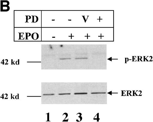
ERK and JNK have been shown to have opposing effects on apoptosis induced by growth factor withdrawal in PC12 cells.14 In this study, both suppression of ERK activity and activation of JNK correlated with the induction of apoptosis. To determine if the inhibition of ERK activity affected JNK activity or induced apoptosis in our system, HCD57(R) cells were treated for 48 hours in EPO in the absence or the presence of 50 μmol/L PD98059, a potent inhibitor of MEK, which activates ERK1 and ERK2. JNK activity was assessed by in vitro kinase activity of JNK1/2 immunoprecipitates. As we observed in the experiments outlined above, JNK activity was undetectable in the absence of EPO but high in the presence of EPO (Figure3A, lanes 1 and 2). JNK activity was unaffected by treatment with the PD98059 (Figure 3A, lane 4). Western blot analysis of samples isolated in parallel to the JNK immunoprecipitates using an ERK phospho-specific antibody revealed that ERK activation was completely suppressed in the PD98059 treated cells, indicating that the inhibitor was functional (Figure 3B, lane 4). By counting the cells with the use of trypan blue exclusion, we determined that the number of cells was dramatically decreased in the PD98059-treated cells (Figure 3C). We then stained these cells with propidium iodide and analyzed the stained cells by using flow cytometry to look for DNA fragmentation indicative of apoptosis. The results of this flow cytometry are shown in Figure 3D. Whereas cells cultured in the absence of EPO showed an increase in the number of apoptotic cells (Figure 3D, top left panel), cells treated with the PD98059 inhibitor in the presence of EPO showed no evidence of DNA fragmentation (Figure3D, lower right panel), even though the cell number was decreased compared with control cells (Figure 3C). It appears, therefore, that inhibition of ERK activity inhibited proliferation of HCD57 cells, but it induced neither JNK phosphorylation nor apoptosis.
Inhibition of ERK activity inhibits proliferation but does not induce apoptosis or JNK activation in HCD57 cells.
HCD57(R) cells were washed and treated with either EPO (EPO, lane 2), EPO + DMSO vehicle (V, lane 3), EPO + 50 μmol/L PD98 059 (PD, lane 4), or no additional growth factor (No EPO, lane 1) for 48 hours. (A) Rabbit-anti-JNK-1 immunoprecipitates were subjected to an in vitro kinase assay using γ-32P-ATP and GST-Jun fusion protein as a substrate and transferred to nitrocellulose. Following exposure to show the phosphorylated GST-Jun (upper arrow), the proteins were immunoblotted with goat-anti-JNK to visualize JNK proteins (lower arrows). (B) Rabbit-anti-ERK immunoprecipitates were immunoblotted with mouse-anti-phospho-ERK antibody to visualize phosphorylated ERK proteins (upper arrow). The blot was then stripped and reprobed with anti-ERK antibody to ensure equal loading of proteins (arrow, lower panel). (C) Proliferative response of HCD57(R) cells to EPO and PD98059 24 hours (columns 1-4) and 48 hours (columns 5-8) following addition of EPO (columns 2-4 and 6-8) or no growth factor (columns 1 and 5) in the presence of PD20859 (columns 4 and 8). Data are indicated as the number of cells as a percentage of the starting number of cells. In this experiment, nonviable cells were 5% or less. (D) Cells incubated for 48 hours with no additional growth factor (upper left panel), EPO alone (upper right panel), EPO + DMSO (lower left panel), or EPO + PD98059 (lower right panel) were stained with propidium iodide as indicated in the “Materials and methods” section and analyzed by using flow cytometry. The number of apoptotic cells is indicated as a percentage of the total number of cells containing sub-G0/G1 DNA and is indicated as M1 on plots.
Inhibition of p38 activity inhibits proliferation but not induction of apoptosis
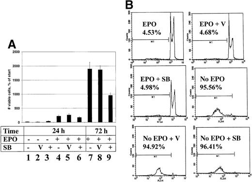
The activation of p38 in HCD57 cells in response to EPO suggests a role for p38 in EPO-induced proliferation or survival. To determine if p38 contributes to either proliferation or survival of these cells, we treated HCD57(K) cells with the specific p38 inhibitor SB20850 and assessed both cell proliferation and apoptosis. We found that treatment of HCD57(K) cells with the SB20850 inhibitor for 72 hours in the presence of EPO suppressed proliferation (Figure4A, 72 hours + SB). Treatment with the inhibitor in the absence of EPO for 24 hours, however, did not suppress apoptosis (Figure 4B, No EPO + SB). This result supports the hypothesis that p38 activity contributes to EPO-induced proliferation and not to the induction of apoptosis.
Inhibition of p38 activity inhibits proliferation but not induction of apoptosis in HCD57(K) cells.
(A) Proliferative response of HCD57 cells to EPO and SB203580. HCD57(K) cells were washed and treated with no additional growth factor (columns 1-3) or EPO (columns 4-9) in the presence of DMSO vehicle (V, columns 2, 5, and 8), or 20 μmol/L SB203580 (SB) (columns 3,6, and 9) for 24 hours (columns 1-6) or 72 hours (columns 7-9). Data are indicated as the number of cells as a percentage of the starting number of cells. In this experiment, nonviable cells were 5% or less. (B) Cells incubated 24 hours in the presence or absence of EPO or inhibitor as indicated were stained with propidium iodide as indicated in “Materials and methods” and analyzed by using flow cytometry. The number of apoptotic cells is indicated as a percentage of the total number of cells containing sub-G0/G1 DNA and is indicated as M1 on plots.
TNF- induces JNK activity but does not induce apoptosis in HCD57 cells
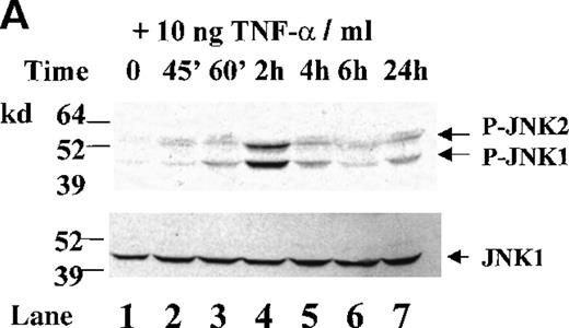
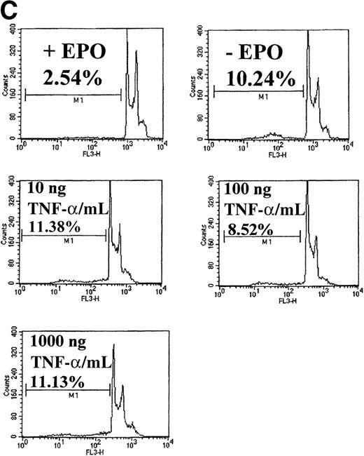
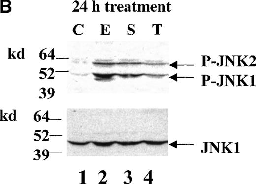
TNF-α has been shown to activate JNK in a number of systems. Depending on the system, this activation may induce proliferation or apoptosis. We were interested in whether TNF-α could activate JNK in HCD57 cells and if so, whether this JNK activation induced apoptosis. We found that HCD57(R) cells treated with exogenous TNF-α in the absence of EPO resulted in JNK activation after 1 hour (Figure5A, lane 3); JNK activation was still detected 24 hours following TNF-α treatment (Figure5A, lane 7, and 5B, lane 4). An assessment of the DNA content of cells incubated in TNF-α for 24 hours, however, revealed that no DNA degradation indicative of apoptosis was detected greater than that induced by the removal of EPO. Therefore, although treatment with TNF-α can clearly induce JNK activation, it does not induce apoptosis in HCD57(R) cells.
TNF- induces JNK activation but does not induce apoptosis in HCD57 cells.
(A) HCD57(R) cells were deprived of EPO for 18 hours and then treated with 10 ng TNF-α/mL for the times indicated. Whole cell lysates were immunoblotted with an anti-phospho-JNK antibody (upper panel, arrows). The blot was then stripped and reprobed with anti-JNK1 to ensure equal loading of proteins (lower panel, arrow). (B) HCD57 cells were deprived of EPO for 18 hours and then treated with no additional growth factor (lane 1), 1 U EPO/mL (lane 2), 10 ng SCF/mL (lane 3), or 10 ng TNF-α/mL (lane 4) for 24 hours. Whole cell lysates were immunoblotted with an anti-phospho-JNK antibody (upper panel, arrows). The blot was then stripped and reprobed with anti-JNK1 to ensure equal loading of proteins (lower panel, arrow). (C) HCD57 cells were deprived of EPO for 18 hours and then treated with 1 U EPO/mL (+EPO); no additional growth factor (No EPO); or 10 ng, 100 ng, or 1000 ng TNF-α/mL for 24 hours. Cells were stained with propidium iodide as indicated in “Materials and methods” and analyzed by using flow cytometry. The number of apoptotic cells is indicated as a percentage of the total number of cells containing sub-G0/G1DNA and is indicated as M1 on plots.
Discussion
It is clear from the literature that AP1 and JNK may exhibit proliferative, pro-survival, or pro-apoptotic effects, depending on the cell type. In neuronal cells for instance, cJun phosphorylation and JNK activation appear to be critical for the induction of apoptosis during both growth factor deprivation- and stress-induced apoptosis.53-55 We have examined the possibility that JNK activation may play a role in the induction of apoptosis induced by EPO deprivation in EPO-dependent erythroid cells. We have previously reported that AP1 DNA binding increased on EPO withdrawal of HCD57 cells; however, we observed that JNK activity was high in the presence of EPO and disappeared on EPO withdrawal41 (and Figure 1 of this report). This result led us to explore the role of the specific AP1 family members in the induction of apoptosis. We determined that JunB was present in the AP1 complex when EPO was withdrawn, whereas cJun was present in the AP1 complex in the presence of EPO but not when EPO was absent. Taken together, these results implicated cJun and JNK in the proliferation and/or survival and JunB in the initiation of apoptosis of HCD57 cells.
A recent report suggesting that JNK triggered apoptosis in HCD57 cells led us to reexamine our previous data and to further explore the role of the map kinases ERK, JNK, and p38 in EPO-induced proliferation. We deprived HCD57 cells of EPO for periods up to 96 hours and assessed JNK, ERK, and p38 activity during the apoptotic process. In agreement with previously published data, JNK activity decreased over 24 hours. Furthermore, JNK activity was undetectable for the rest of the time course, a finding not consistent with a role for JNK in inducing apoptosis. Western blot analysis of phosphorylated JNK also confirmed this result. ERK activation decreased 1 hour after EPO withdrawal, whereas p38 activity decreased more slowly but was undetectable 48 hours following EPO removal. The slower inactivation of p38 relative to JNK suggests that the dephosphorylation of these proteins may occur through different mechanisms. Thus, neither JNK nor p38 seem to trigger apoptosis in these cells. Furthermore, when we treated HCD57 cells with TNF-α, we observed an increase in JNK activity that did not induce apoptosis. Therefore, direct induction of JNK activity failed to initiate apoptosis of HCD57 cells, supporting our contention that JNK activity does not contribute to apoptosis induced by EPO withdrawal.
When HCD57 cells were deprived of EPO for 18 hours and then stimulated with EPO, we observed an increase in ERK, JNK, and p38 activation. The time course of these EPO-dependent activations were very different, however. ERK activation occurred within 15 minutes of EPO addition and decreased to a low but detectable level within 18 hours that remains as long as EPO is present. This result was consistent with previous reports from our laboratory and from others.40,47,56Whereas EPO-dependent JNK and p38 activation were detected within 15 minutes of activation in these previous studies, maximum JNK and p38 activities were reached 18 to 24 hours after EPO treatment (Figure 2). These results differ from earlier reports39,40 on JNK activation in SKT6 erythroid cells that reported an immediate increase in JNK activation necessary for EPO-induced differentiation. The reasons for a difference in the time course of JNK and p38 activation between HCD57and SKT6 are not clear; however, the JNK activity was not tested for longer than 60 minutes after EPO treatment of the SKT6 cells. In the SKT6 cell line, early JNK and p38 activation may be necessary for EPO-induced differentiation. Because HCD57 cells do not differentiate in response to EPO, it is possible that these cells do not have the early signals that activate JNK and differentiation. JNK and ERK activation are not affected by treatment with the PI 3-kinase inhibitor LY294002, which is in contrast to some reports in other cell types.57 58 The upstream activators of JNK in erythroid cells, therefore, remain to be determined.
We tested the hypothesis that ERK activity might suppress JNK by using the MEK inhibitor PD98059. When HCD57 cells are treated with PD98059 in the presence of EPO, ERK activity and proliferation are suppressed (Figure 3B and 3C). JNK activity is high in the presence of EPO and is not enhanced further by the addition of the PD98059. Inhibition of ERK activity did not induce apoptosis in HCD57 cells (Figure 3D). Therefore, suppression of ERK did not induce JNK activation or apoptosis. Because ERK activation decreases as JNK activation increases in EPO-treated HCD57 cells, we cannot rule out the possibility that a decrease in ERK activity may facilitate an increase in JNK activity in this system. However, suppression of ERK activity is insufficient to induce apoptosis in HCD57 cells.
The results of our study stand in sharp contrast to the recent report from Shan et al42 that implicates JNK and p38 in the induction of apoptosis in HCD57 cells. Our previous work on the inhibition of apoptosis by a dominant negative AP1 in HCD57 cells was cited by Shan et al42 as supporting a role of JNK inducing apoptosis; however, they made no mention of our contradictory finding that JNK activity decreased on EPO withdrawal from HCD57 cells. The differences between the work by Shan et al42 and our work presented here are the following: (1) Whereas we report here that both JNK and p38 are stimulated by EPO and that EPO withdrawal results in no stimulation of either JNK or p38, only suppression of activity; Shan et al42 find just the opposite. They reported that in the presence of EPO there was no JNK or p38 activity and that EPO withdrawal stimulated both JNK and p38 activity to high levels in a time course preceding apoptosis. (2) Whereas we saw a complete suppression of ERK-1, -2/MAP kinase activity following EPO withdrawal for only 1 hour, Shan et al42 found ERK still detectable following EPO withdrawal for 24 hours. (3) Whereas we saw a rapid and transient activation of ERK-1/-2 by EPO, Shan et al42 found a gradual increase in ERK-1/-2 phosphorylation over the 24-hour time course. (4) Although we found that inhibition of the MAP kinase cascade with the MEK inhibitor PD98059 effectively blocked ERK activity and slowed the proliferation of cells, this inhibitor did not have any effect on JNK activity nor induced apoptosis. In contrast, Shan et al42 found that treatment of HCD57 cells with PD98059 induced JNK activity, and it was implied that apoptosis was induced by PD98059 treatment of HCD57 cells (but no data were shown). (5) Although we found that inhibition of p38 activity with the p38 SB inhibitor (SB203580) slowed the proliferation of HCD57 cells, we found the treatment with this inhibitor neither caused apoptosis in the presence of EPO nor protected these cells from apoptosis when EPO was withdrawn. In sharp contrast, Shan et al42 reported that inhibition of p38 protected HCD57 cells from apoptosis following EPO withdrawal for 4 days.
What factors might explain such sharply contrasting data detailed above in experiments using the same cell line? The first possibility was that the Shan et al42 study was conducted with HCD57 cells that were different from the HCD57 cells we used; either in passage number, culture conditions, or different origins. We have repeated our study of JNK activity during EPO deprivation, using the exact same cell culture and cell lysis conditions outlined in the Shan et al42paper. We found that JNK and p38 activity were high in the presence of EPO and decreased on EPO withdrawal in a manner similar to that seen with our culture and cell lysis conditions (data not shown).
Another possibility to explain the difference between these studies is that 2 different HCD57 cell lines were used. We are aware of 2 slightly different HCD57 cell lines. As described in detail in “Materials and methods,” the subcloned HCD57(R) cells are somewhat more resistant to apoptosis than the HCD57(K) cell line of the original HCD57 cells. Shan et al42 did not list the origin of their HCD57 cells; however, it is possible that they might have used both populations of HCD57 cells because they reported in their first figure that the HCD57 cells underwent rapid apoptosis (60% apoptotic after 24 hours without EPO) but very slow apoptosis in the last figure (Figure 7 of Shan et al42 shows only 25% apoptosis after 24 hours of EPO withdrawal and 4 days for complete apoptosis of the HCD57 cells). As we show here (Figures 1 and 2), our results with either type of HCD57 cell line were comparable and clearly showed that neither activity of p38 nor JNK increased following EPO withdrawal. We have also obtained HCD57 cells from S. Brandt at Vanderbilt University, who also supplied HCD57 cells to Shan et al,42 and repeated the JNK and p38 activity experiments by depriving this HCD57 cell line of EPO, using the exact cell culture and lysis conditions described by Shan et al42; we confirmed the result we are reporting here: We observed a decrease in JNK and p38 activity on EPO withdrawal (data not shown).
The difference seen between our experiments and the study by Shan et al42 on MAP-kinase/ERK activation are likely because our experiments were performed with HCD57(R) cells deprived of EPO overnight such that the EPO receptors were up-regulated 10-fold45 and the basal signaling of the MAP kinase pathways was completely turned off. In contrast, the Shan et al42 study only deprived cells of EPO for 4 hours such that a weaker response to EPO in ERK activity would be expected.
We do not have a ready explanation of why inhibition of the MAP kinase cascade by the MEK inhibitor PD98059 induced JNK activity in the Shan et al42 study but did not do so in our study. Shan et al42 attributed an increase in JNK activity to treatment with the PD inhibitor and a “decrease in the number of viable cells,” but they did not suggest the mechanism by which this decrease occurs. Although we did see a decrease in the number of cells in the presence of the inhibitor, we observed no increase in apoptosis via flow cytometric analysis of propidium iodide-stained cells (Figure3). It is clear, therefore, that the MEK inhibitor totally blocked the MAP kinase cascade and suppressed proliferation but did not induce apoptosis in HCD57 cells. It is notable that they used a higher concentration of inhibitor well beyond that required to totally inhibit the MAP kinase cascade such that JNK activity be activated by a nonspecific effect of the inhibitor unrelated to MAP-kinase inhibition.
We have no explanation as to why the inhibition of p38 activity seems to protect HCD57 cells from apoptosis following EPO withdrawal in the study by Shan et al42 yet was completely ineffective in protecting HCD57 cells from apoptosis in our study (Figure 4). The fact that treatment of HCD57 cells with SB203580 in this study dramatically slowed the proliferation of the cells serves as a control that the inhibition of p38 affects HCD57 cells. These data are more consistent with previous reports38 39 of p38 playing a role in the EPO signaling pathway rather than a mediator of apoptosis.
JNK is thought to function by phosphorylating the N-terminus of cJun and increasing its transactivation potential. Because the other Jun proteins, JunD and JunB, lack proper docking sites for JNK (in the case of JunD) or phosphorylation sites (in the case of JunB), they are poor substrates for JNK.34 Therefore, one would expect that for JNK to have a pro-apoptotic effect, it would need to have cJun, its substrate, present. The loss of JNK activity concurrent with the loss of cJun-DNA binding and an increase in JunB-DNA binding fits with our model of JunB as the important AP1 family member in the induction of apoptosis in HCD57 cells. Likewise, an increase in JNK activity concurrent with the increase in cJun-DNA binding in the presence of EPO fits with our model of cJun and JNK playing an important role in EPO-induced proliferation. Taken together, the results of this study support our model implicating JNK in EPO-induced proliferation and/or protection from apoptosis in HCD57 cells. We have also extended these studies to show that p38, JNK, and ERK appear to play a role in EPO-induced proliferation but not in the induction of apoptosis observed on EPO withdrawal in HCD57 cells.
Acknowledgments
We thank Dr Amy Lawson and Dr Haifeng Bao for their helpful discussions regarding this manuscript.
Supported by grant R01DK39781 (S.T.S.) from the National Institutes of Health, a grant from the American Heart Association (S.M.J.-H.), and grant IN-105 from the American Cancer Society (J.J.R.).
Reprints:Stephen T. Sawyer, Department of Pharmacology/Toxicology, PO Box 980613, Richmond, VA 23298; e-mail:ssawyer@hsc.vcu.edu.
The publication costs of this article were defrayed in part by page charge payment. Therefore, and solely to indicate this fact, this article is hereby marked “advertisement” in accordance with 18 U.S.C. section 1734.




This feature is available to Subscribers Only
Sign In or Create an Account Close Modal