Abstract
The mi transcription factor (MITF) is a basic-helix-loop-helix-leucine zipper transcription factor that is important for the development of mast cells. Cultured mast cells (CMCs) of mi/mi genotype express abnormal MITF (mi-MITF), but CMCs of tg/tg genotype do not express any MITFs. It was previously reported thatmi/mi CMCs showed more severe abnormalities thantg/tg CMCs, indicating that mi-MITF had inhibitory function. Whereas mi-MITF contains a single amino acid deletion in the basic domain, MITF encoded bymiewallele (ew-MITF) deletes 16 of 21 amino acids of the basic domain. Here the effect of a large deletion of the basic domain was examined. Inmiew/miew CMCs, the expression pattern of genes whose transcription was affected by MITF was comparable to that of tg/tg CMCs rather than to that ofmi/mi CMCs. This suggested that ew-MITF lacked any functions. The part of the basic domain deleted inew-MITF appeared necessary for either transactivation or inhibition of transactivation.
Introduction
The mi locus of mice encodes a member of the basic-helix-loop-helix-leucine zipper protein family of transcription factors (hereafter called MITF).1 MITF plays an important role in the development of mast cells.2-9Mast cells of mi/mi genotype express normal amounts of abnormal MITF (mi-MITF), whereas mast cells oftg/tg genotype do not express any MITFs.10-12Because tg is considered to be a null mutant allele, thetg/tg mice are useful as a standard for evaluating the function of other mutant MITFs.13-15 We previously compared the phenotype of mast cells of mi/mi mice with that of tg/tg mice.13 The transcription of mouse mast cell protease (mMCP)–4, mMCP-5, and mMCP-6 genes in cultured mast cells (CMCs) derived from mi/mi mice is reduced to the level comparable to that of tg/tg CMCs.13 The transcription of c-kit, granzyme B (Gr B), and tryptophan hydroxylase (TPH) genes is significantly reduced in mi/miCMCs, but the reduction of transactivation of these genes is moderate in tg/tg CMCs.13 This shows that themi-MITF possesses an inhibitory effect on the transcription of c-kit, Gr B, and TPH genes.13 Themi-MITF deletes one arginine in the basic domain, whereas MITF encoded by the miew mutant allele (ew-MITF) deletes 16 of 21 amino acids of the basic domain.10 11 In the present study, we compared the mast cell phenotypes of miew/miew mice with those of tg/tg and mi/mi mice to examine the effect of a large deletion of the basic domain.
Study design
Mice and cells
The C57BL/6-tg/tg and C57BL/6-mi/mi mice (hereafter tg/tg and mi/mi mice) were described previously.13 The C57BL/6-miew/+mice16 were sent from the laboratory of M. L. Lamoreux, and C57BL/6-miew/miew mice were raised in our laboratory. C57BL/6-+/+ mice were used as a control. CMCs and P815 cells have been described.13
Northern blot analysis
Total RNA prepared from CMCs was electrophoresed and blotted. The intensity of the signal for each messenger RNA (mRNA) was quantified by a densitometer (Molecular Dynamics, Sunnyvale, CA).
Concentration of serotonin
The concentration of serotonin was measured using high-performance liquid chromatography as previously described.17
Staining of skin mast cells
Pieces of dorsal skin were removed from mice aged 20 days and embedded in paraffin. A section was stained with alcian blue and nuclear fast red, and an adjacent section was stained with berberine sulfate.18
Transient cotransfection assay
Results and discussion
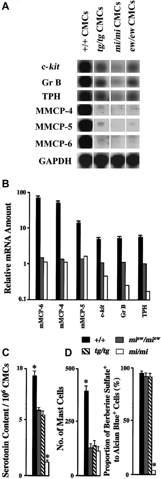
The phenotype of miew/miew mast cells was compared with that of +/+, tg/tg, andmi/mi mast cells. First, CMCs were obtained from spleens of mice of each genotype.13 The expression of genes that were known to be affected by MITF19-23 was compared with Northern blot. The amounts of c-kit, Gr B, and TPH mRNAs inmiew/miew CMCs were comparable to those of tg/tg CMCs, and the amounts were intermediate between those of +/+ and mi/mi CMCs (Figure1A). Although the amounts of mMCP-4, mMCP-5, and mMCP-6 mRNAs were very low in all kinds of CMCs examined here, their amounts in miew/miewCMCs were comparable to those of tg/tg and mi/miCMCs (Figure 1A).
Comparison of abnormalities of CMCs and skin mast cells of tg/tg, mi/mi, andmiew/miew mice.
(A) Northern blot analysis of RNAs obtained from CMCs of +/+,tg/tg, mi/mi, ormiew/miew genotype. The blot was hybridized with 32P-labeled complementary DNA probe of c-kit, Gr B, TPH, mMCP-4, mMCP-5, mMCP-6, or GAPDH as described previously.11 Twenty micrograms of total RNA was loaded in each lane, and each 32P-labeled complementary DNA was reprobed using the same membrane. A representative experiment is shown. (B) The amount of mMCP-6, mMCP-5, mMCP-4, c-kit, Gr B, and TPH mRNAs was quantified by the densitometry, and the ratio to the mRNA amount of tg/tg CMCs, which was defined as relative mRNA amount, was calculated. The bars represent the mean ± SE of 3 independent experiments. In some cases, the SE was too small to be shown by the bars. (C) The serotonin contents per 106 +/+,tg/tg, mi/mi, andmiew/miew CMCs were measured. The bars represent the mean ± SE of 3 experiments. *P < .05 by t test when compared with the value of tg/tg CMCs. (D) Mast cells were counted, and the number was expressed as mast cells per centimeter of skin. Berberine sulfate–positive mast cells were also counted, and the proportion of berberine sulfate–positive cells to alcian blue–positive cells was calculated. The bars represent the mean ± SE of 5 mice. In some cases, the SE was too small to be shown by the bars. *P< .01 by t test when compared with the value oftg/tg mice.
We quantified the amounts of mRNAs using densitometry and calculated the ratio of each mRNA amount of +/+, mi/mi, ormiew/miew CMCs to that oftg/tg CMCs (Figure 1B). We defined the ratio as relative mRNA amount. The relative mRNA amounts of all examined genes were more than 1.0 in +/+ CMCs, indicating that the +-MITF had positive effect on transcription. In mi/mi CMCs, the relative mRNA amounts of mMCP-4, mMCP-5, and mMCP-6 genes were about 1.0, but those of c-kit, Gr B, and TPH genes were less than 1.0. This suggested that the mi-MITF had negative effect on the transcription of c-kit, Gr B, and TPH genes. The relative mRNA amounts of all examined genes were about 1.0 inmiew/miew CMCs, indicating that theew-MITF had neither positive nor negative effects on transcription.
Because TPH is the rate-limiting enzyme of the serotonin synthesis,24 the serotonin contents were compared among various CMCs. The serotonin content ofmiew/miew CMCs was comparable to that of tg/tg CMCs. Both values were significantly smaller than that of +/+ CMCs and significantly greater than that ofmi/mi CMCs (Figure 1C, P < .05 by ttest). Serotonin contents of +/+, tg/tg, mi/mi,and miew/miew CMCs were well correlated with the mRNA expression levels of TPH gene in CMCs of each genotype (+/+ > miew/miew ∼tg/tg > mi/mi).
Second, the number of mast cells and the proportion of berberine sulfate–positive mast cells were compared among skin tissues of +/+,tg/tg, mi/mi, andmiew/miew mice. The number of skin mast cells of miew/miew mice decreased to one third that of the +/+ mice and was comparable to that of tg/tg and mi/mi mice (Figure 1D). Most skin mast cells were berberine sulfate–positive in +/+ mice.25The proportion of berberine sulfate–positive to alcian blue–positive mast cells in the skin of miew/miewmice was comparable to that of +/+ mice and to that of tg/tgmice (Figure 1D). In contrast, the proportion of berberine sulfate–positive mast cells was only 3% in the skin of mi/mimice as reported previously.7 The abnormalities ofmiew/miew skin mast cells were comparable to those of tg/tg skin mast cells rather than to those of mi/mi skin mast cells.
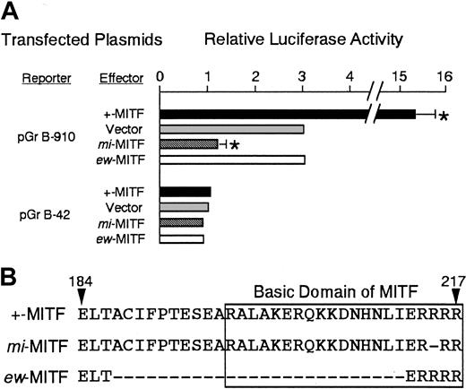
We examined the effect of ew-MITF on the transactivation of Gr B promoter using the transient cotransfection assay. The luciferase construct containing the Gr B promoter was cotransfected into P815 cells with the expression plasmid containing no insert, +-MITF,mi-MITF, or ew-MITF complementary DNA. The coexpression of +-MITF significantly increased the luciferase activity, whereas that of mi-MITF significantly reduced it as reported previously (Figure 2A).13The expression of ew-MITF showed luciferase activity comparable to the value obtained by the expression vector alone (Figure2A). The ew-MITF had no effect on the activity of Gr B promoter. This was consistent with the fact that the abnormalities of CMCs and skin mast cells ofmiew/miew mice were similar to those of tg/tg mice. The part of the basic domain that was deleted in ew-MITF (Figure 2B) appeared necessary for both positive and negative functions.
Function and structure of +-MITF, mi-MITF, and ew-MITF.
(A) The effect of coexpression of +-MITF, mi-MITF, orew-MITF on the luciferase activity under the control of the Gr B promoter. Various reporter and effector constructs were introduced into P815 cells by electroporation. The value of the luciferase activity was divided by the value obtained with the transfection of pGr B-42 alone and was shown as the relative luciferase activity. The bars represent the mean ± SE of 3 independent experiments. In some cases, the SE was too small to be shown by the bars. *P < .01 by t test when compared with the control, in which expression vector containing no insert was cotransfected. (B) Comparison of amino acid sequence among +-MITF, mi-MITF, and ew-MITF. The amino acids are numbered from the initiation codon. Bars in mi-MITF orew-MITF show the deleted amino acids. One of 3 arginines (amino acid 215-217) is deleted in mi-MITF. It is unclear which one has been deleted. Twenty-one amino acids that compose the basic domain are boxed.
Supported by grants from the Ministry of Education, Culture, Sports, Science and Technology and Uehara Memorial Foundation.
The publication costs of this article were defrayed in part by page charge payment. Therefore, and solely to indicate this fact, this article is hereby marked “advertisement” in accordance with 18 U.S.C. section 1734.
References
Author notes
Eiichi Morii, Dept of Pathology, Rm C2, Osaka University Medical School, Yamada-oka 2-2, Suita 565-0871, Japan; e-mail: morii@patho.med.osaka-u.ac.jp.


This feature is available to Subscribers Only
Sign In or Create an Account Close Modal