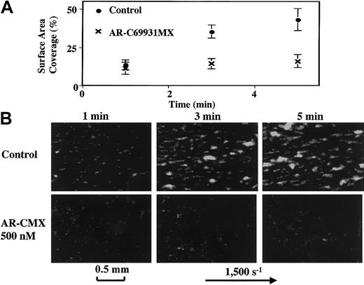
We read with interest the article presented by Dr Turner and colleagues demonstrating the effects of P2Y1 and P2Y12 adenosine 5′-diphosphate (ADP) receptor blockade on platelet aggregation and thrombus formation under flow conditions.1 However, we have serious concerns about their statement, “blockade of P2Y12 alone or blockade of P2Y1 alone did not reduce thrombus formation on VWF-collagen surface,” not only because these results are not in agreement with previously published findings using prodrugs of thienopyridine antiplatelet agents2 that inhibit the P2Y12 receptor, but also because they are not in agreement with our own experimental results (Figure1). We used a similar flow chamber system equipped with an epi-fluorescent videomicroscope3 and perfused whole blood containing platelets rendered fluorescent by the addition of mepacrine on immobilized bovine type I collagen, in a manner similar to that described by Turner et al1 and at a slightly lower shear rate of 1500 s-1. However, our results, shown in Figure 1, clearly demonstrate that AR-C69931MX alone at the dose of 500 nM was sufficient to inhibit platelet thrombus formation on the surface of immobilized collagen. We used the specific and reversible antithrombin agent argatroban instead of heparin as the anticoagulant, to exclude the possible pleiotropic effects of heparin.3,4 Also, we perfused the blood on the collagen surface for 5 minutes instead of for 1 minute. We believe that the second of the aforementioned differences explains the differences in our results, because the inhibitory effects of AR-C69931MX were not clear at 1 minute even in our study (Figure 1). Thus, we believe that the authors should have extended the perfusion period further in their experiments before concluding that “blockade of P2Y12alone” “did not reduce thrombus formation on VWF-collagen.” Indeed, numerous clinical studies have clearly demonstrated the effectiveness of thienopyridine antiplatelet agents that block the P2Y12 receptor in preventing conditions associated with arterial thrombosis, such as acute myocardial infarction,5in which VWF-mediated platelet thrombus formation plays an important role.6 Therefore, we believe that P2Y12inhibition by itself is sufficient to inhibit platelet thrombus formation at sites of endothelial damage exposed to arterial levels of shear stress.
Effects of AR-C69931MX on platelet thrombus formation on the surface of collagen under flow conditions.
Blood containing platelets rendered fluorescent by the addition of mepacrine and anticoagulated with the specific antithrombin agent argatroban was perfused on collagen-coated glass coverslips at a wall shear rate of 1500 s-1 for 5 minutes, either in the absence (Control) or presence of the specific P2Y12 antagonist AR-C69931MX at a final concentration of 500 nM. Platelet thrombi formed on the collagen surface were detected by inverted-stage epi-fluorescent videomicroscopy (DM IRB, 1RB-FLUO, Leica, Germany), as demonstrated in the lower panel of the figure. The microscopic images were digitized online with a photosensitive CCD camera (L-600, Leica, Germany). Surface coverage by the platelets was calculated using National Institutes of Health (NIH) Image software (public domain software by Dr Wayne Rasband, NIH, Bethesda, MD, version 1.62), and the results are shown in the upper panel of the figure. These results are the mean ± SD of the 3 experiments, while the results shown in the lower panel show 1 representative result of the 3 experiments.
Blockade of adenosine diphosphate receptors P2Y12and P2Y1 is required to inhibit platelet aggregation in whole blood under flow
The Letter to the Editor by Goto et al contains some potentially interesting data but also some generally misleading comments.
We found that ARMX alone did not inhibit platelet thrombus formation onto the collagen surface at 3000 s−1 after 1 minute of flow.1-1 Goto et al also show in the upper portion of their Figure 1 that after 1 minute of flow at 1500 s−1 there is no difference in the platelet surface coverage between control samples and ARMX addition. Only at longer periods of flow (3 or 5 minutes) did Goto et al find that ARMX reduced the percent of the collagen surface covered by platelets (by about 25% to 30% at 1500 s−1). They also fail to mention the striking inhibition of platelet deposition seen after 1 minute of perfusion when both P2Y12and P2Y1 inhibitors are present1(figs5-7). No data on dual inhibition are presented in the letter of Goto et al.
Goto et al fail to mention that we reported in our paper that ARMX alone inhibited shear-induced platelet aggregation by more than 50% at both 750 and 1500 s−1 for 30 seconds in the viscometer1(fig1). This inhibition was accentuated by concurrent addition of A3P5P. There are no results presented on bulk shear (viscometry studies) in the letter by Goto et al.
In summary, we believe the limited data presented by Goto et al are consistent with our data1-1 at the same time points of perfusion, even though the shear rates differ by a factor of 2. The relatively small differences at longer time points with just the P2Y12 inhibition may be true, but the magnitude of change in platelet deposition is actually not impressive compared to what is seen with blockade of both P2Y12 and P2Y1receptors concurrently. Data on limited effects of each inhibitor separately in bulk shear stress (viscometry) studies were already presented in our earlier paper.1-1
References
We acknowledge the financial support from a Grant-in-Aid for Scientific Research in Japan 13670744; grant JSPS-RFTF97I00201 from the Japanese Society for the Promotion of Science; and a grant from the Science Frontier Program of MESSC of Japan.


This feature is available to Subscribers Only
Sign In or Create an Account Close Modal