Key Points
Spleen filtration function is altered in infants with SCA as early as 3-6 months of age.
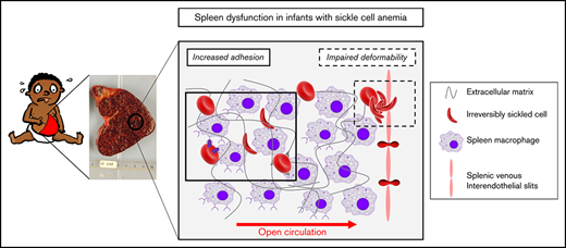
Both impaired deformability and increased adhesion of sickle RBCs play a key role in splenic loss of function.
Abstract
Spleen dysfunction is central to morbidity and mortality in children with sickle cell anemia (SCA). The initiation and determinants of spleen injury, including acute splenic sequestration (ASS) have not been established. We investigated splenic function longitudinally in a cohort of 57 infants with SCA enrolled at 3 to 6 months of age and followed up to 24 months of age and explored the respective contribution of decreased red blood cell (RBC) deformability and increased RBC adhesion on splenic injury, including ASS. Spleen function was evaluated by sequential 99mTc heated RBC spleen scintigraphy and high-throughput quantification of RBCs with Howell-Jolly bodies (HJBs). At 6 and 18 months of age, spleen filtration function was decreased in 32% and 50% of infants, respectively, whereas the median %HJB-RBCs rose significantly (from 0.3% to 0.74%). An excellent correlation was established between %HJB-RBCs and spleen scintigraphy results. RBC adhesion to laminin and endothelial cells increased with time. Adhesion to endothelial cells negatively correlated with splenic function. Irreversibly sickled cells (ISCs), used as a surrogate marker of impaired deformability, were detected at enrollment and increased significantly at 18 months. %ISCs correlated positively with %HJB-RBCs and negatively with splenic uptake, indicating a relationship between their presence in the circulation and spleen dysfunction. In the subgroup of 8 infants who subsequently experienced ASS, %ISCs at enrollment were significantly higher compared with the asymptomatic group, suggesting a major role of impaired deformability in ASS. Higher levels of %HJB-RBCs were observed after the occurrence of ASS, demonstrating its negative impact on splenic function.
Introduction
Splenic dysfunction is central to morbidity and mortality in children with sickle cell anemia (SCA). The spleen is morphologically and functionally normal at birth, but when the rise of the mutated hemoglobin (HbS) allows Hb polymerization to occur, it is inferred that sickling-related injury takes place in the spleen and results in progressive or acute ischemia. The spleen is indeed the first organ to be clinically symptomatic during the course of the disease in early infancy.1,2 Although early therapeutic intervention (hydroxyurea, hematopoietic stem cell therapy, and blood transfusion programs) may allow a variable degree of reversal of spleen loss of function,3-5 ultimately, repeated spleen injury leads to the fibrosis of the organ, a loss also referred to as autosplenectomy.
Altogether, splenic clinical manifestations in children with SCA include acute splenic sequestration (ASS), splenomegaly, chronic sequestration (or hypersplenism), and spleen atrophy. All these complications remain somewhat unpredictable and furthermore may combine with a variable degree of loss of function, with no clear relationship between the size of the spleen and its function. ASS, defined by the sudden enlargement of the spleen with an acute drop in Hb level of >2 g/dL, is a major life-threatening event in infancy, occurring in 10% to 30% of infants with SCA, with 75% of first cases occurring at <2 years of age.6 Little is known about determinants or predictive factors of ASS and their specific impact on spleen function. By contrast, splenic loss of immune and filtering functions, central to pneumococcal defense in childhood, is a well-described phenomenon associated with an increased risk of death in the absence of prophylaxis or treatment.7,8
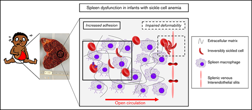
The spleen allows efficient filtering and clearance of senescent or infected red blood cells (RBCs) and bacterial antigens.9 The red pulp of the spleen filters the blood and is composed of cords and venous sinuses. It is known to be the major source of mononuclear phagocytic cells involved in the filtering function of the organ. In the splenic cords, RBCs circulate freely in direct contact with spleen resident cells, macrophages and endothelial cells lining the splenic sinuses,9 which remove senescent or abnormal cells and contribute to the pitting mechanism, a process that clears nuclear remnants or parasites from the RBCs before they recirculate.10,11 This circulation in the red pulp is therefore termed “open” and is also slow, because of a high hematocrit of ∼60%, which favors cell–cell interaction.12 In order to join the venous circulation, RBCs flowing through the red pulp must pass through interendothelial slits (1-3 µm wide) that line the sinuses. Within this ultimate checkpoint, RBCs that are not deformable are trapped. Altogether, if the filtering function of the spleen is altered, the clearance of nuclear remnants fails, and they are found in circulating RBCs as Howell-Jolly bodies (HJB-RBCs).
At least 2 major characteristics of sickle RBCs—namely, impaired deformability and increased adhesion—may play an important role in splenic injury, notably ASS. Impaired deformability of sickled RBCs promotes their becoming trapped during their circulation, particularly upstream of the narrow interendothelial slits.6 Likewise, increased RBC adhesion may contribute to the congestion of the red pulp through prolonged RBC interactions with cellular and matrix components. Such abnormal interactions could involve the erythroid adhesion marker Lu/BCAM (CD239; Lutheran/basal cell-adhesion molecule), whose expression is increased on reticulocytes of infants with SCA as early as 3 to 6 months of age13 and whose ligand is laminin,14 a major component of the spleen’s extracellular matrix.15
Our study’s primary objective was to evaluate the initiation of spleen loss of function in a cohort of SCA infants enrolled very early in life (3-6 months of age) and thereafter longitudinally followed up to 24 months of age. Specifically, we measured splenic function sequentially using flow cytometry–based measurements of HJB-RBCs, coupled with 99mTc heated RBC spleen scintigraphy. Our secondary objective was to explore determinants of spleen injury focusing on impaired deformability and increased adhesion properties of sickle RBCs as potential major contributors. We therefore chose to measure, at enrollment and thereafter longitudinally, the usual SCA biomarkers and, more specifically, irreversibly sickled cells (ISCs) as a surrogate marker of RBC deformability and the expression of the adhesion marker Lu/BCAM and its functional activity, in relation to spleen injury, notably ASS.
Methods
Patients and blood samples
Infants diagnosed with SCA following neonatal screening were enrolled in a multicenter prospective study on prognostic factors in SCA (ClinicalTrials.gov: NCT01207037) from September 2010 through March 2013, described in Brousse et al.16 Inclusion criteria were (1) SS or S-β° sickle genotype, (2) age <6 months, and (3) no prior episode of ASS. At each study visit, complete clinical workup and blood sampling were performed, and relevant medical events were recorded. Patients were followed up with scheduled visits planned at enrollment (3 and/or 6 months), 12, 18, and 24 months. All received standard age-appropriate care for SCA; however, none was treated with hydroxyurea, in accordance with national treatment guidelines during the study period. No child was lost to follow-up. The protocol was approved by the ethics committee (Comité pour la Protection des Personnes Ile de France II) and by the French agency for security of health products (Agence Française de Sécurité Sanitaire des Produits de Santé). ASS was defined by the sudden enlargement of the spleen (>2 cm compared with basal) with a decrease in Hb level (>2 g/dL, compared with the previous measurement) and reticulocytes >100 000/mm3. Asymptomatic patients were defined as patients who did not experience any ASS, vaso-occlusive crisis, transfusion, or hospitalization for sickle cell disease (SCD)–related events during the follow-up period up to 24 months of age.
At each visit, blood analysis for routine and SCA-specific biomarkers was performed as described elsewhere.16 In addition, RBCs reserved in ID-CellStab (Bio-Rad) were cryopreserved in the Centre National de Référence pour les Groupes Sanguins. Blood samples from 7 healthy adult donors obtained from the Etablissement Français du Sang were used as negative controls.
In this cohort of 57 patients, HJB-RBCs and ISCs quantification was performed in 45 patients for whom sufficient sample volumes were available at 2 different time points (3-6 and 18 months) and who had not been transfused in the prior 2 months (supplemental Figure 1). However, 7 of these infants were transfused at some point during the follow-up. Adhesion assays were performed in 15 randomly selected nontransfused patients for whom samples were available at all time points for longitudinal analysis.
Spleen scintigraphy was initially part of the systematic splenic exploration in all infants enrolled in the study but, in response to parental reluctance, an amendment to the protocol was agreed upon, and spleen scintigraphy exploration became optional. Transfusion was not an exclusion criterion. It was performed in 25 patients at the first time point (3-6 months), 17 of whom underwent a sequential exploration at 18 months of age. The subgroup in whom spleen scintigraphy was performed was therefore based on parental acceptance and technical possibility (eg, adequate venous access). Of note, this subgroup did not differ from the rest of the cohort in terms of baseline clinical, biological, or splenic characteristics (data not shown). Among the 25 patients who underwent a spleen scintigraphy, 7 were transfused at some point during the course of the study, 2 after spleen scintigraphy.
Quantification of HJBs
Quantification of %HJB-RBCs was performed by using imaging flow cytometry (IFC), as previously described.17 Briefly, 10 μL of the RBC pellet was suspended in 1 mL of buffer (phosphate buffered saline; 0.5% bovine serum albumin), 80 μL of the suspension was added to 120 μL of buffer, and Hoechst 33342 (Life Technologies) was added at a final concentration of 40 μg/mL and incubated for 5 minutes at room temperature. Importantly, this HJB-RBCs quantification technique can also be applied using a classic flow cytometer. Classic May-Grünwald Giemsa smears were performed in parallel for comparison purposes.
Quantification of ISCs
ISCs, a subpopulation of dense cells with marked decreased deformability, were quantified using an IFC-based method.18 Briefly, 2 µL of packed RBCs were suspended in 200 µL of ID-CellStab (Bio-Rad) and 50 000 events were acquired using ImageStream X Mark II Imaging Flow Cytometer. ISCs were quantified using IDEAS (version 6.2). We optimized an analysis based on morphological and shape features,19 to finely evaluate the percentage of ISCs in blood samples, as previously described.20
Flow cytometry analysis
Expression of CD239/Lu/BCAM was quantified, as previously described.13 Samples were analyzed by flow cytometry with a BD FACScanto II flow cytometer (Becton Dickinson) and FACSDiva software (version 6.1.3).
Adhesion assays
RBC adhesion to immobilized laminin 521 (Biolamina, AB) and transformed human bone marrow endothelial cell (TrHBMEC) monolayers was determined under physiological flow conditions, using Vena8 Endothelial+ biochips (internal channel dimensions: length, 20 mm; width, 0.8 mm; and height, 0.12 mm) and ExiGo nanopumps (Cellix), as described.21-23 TrHBMECs were pretreated with tumor necrosis factor-α (100 U/mL) 24 hours before adhesion experiments. Adhesion experiments were performed at the 4 time points in 15 patients with characteristics comparable to the rest of the cohort.
Phosphorylation assays
Evaluation of the activated state of the Lu/BCAM long isoform Lu was performed through phosphorylation assays. In brief, RBCs were lysed for 45 minutes at 4°C with lysis buffer (Tris, 20 mM; NaCl, 150 mM; EDTA, 5 mM; and Triton X100, 1%). Phosphorylation of Lu/BCAM was performed after immunoprecipitation with a specific anti-Phospho-Lu antibody (Institut National de la Transfusion Sanguine). The quantification of the proteins was achieved using Quantity One Software (Bio-Rad).
Spleen scintigraphy
Spleen/liver scintigraphy, using heat-denatured 99mTc-labeled RBCs, was performed as previously described.24 Spleen scintigraphy with heated autologous red cells measures primarily the filtration capacity of the splenic red pulp because heated RBCs are poorly deformable and therefore get trapped in the splenic microvasculature.25,26 Radionuclide images were taken of the posterior, left lateral, and anterior views of the spleen. Splenic uptake was compared with that of the liver and expressed in a semiquantitative percentage using a computational method: 0% to 25% (lowest splenic uptake), 25% to 50%, 50% to 75%, 75% to 100%, and 100% to 150% (highest splenic uptake).
Splenic volume was calculated by computed tomography and was estimated after 1.5 hours of tomography acquisition.
Statistics
Data were analyzed by 2-tailed Wilcoxon test or paired Wilcoxon test, as appropriate. The association between quantitative variables was assessed with the Spearman correlation test. GraphPad Prism, version 7.00, and R software were used. P ≤ .05 was considered significant.
Results
Very early loss of splenic function and subsequent further decline with time
Spleen function was assessed with both a high-throughput cytometry method for HJB-RBC counts and 99mTc heated RBC scintigraphy at 2 time points (3-6 and 18 months of age) in a cohort of 57 infants (SS, n = 55; S-β°, n = 2; 54.4% males). At enrollment (median age, 6 months; range, 2.8-8), median fetal Hb (HbF) level was 42% (range, 35%-50.5%), as previously reported.16 Supplemental Figure 1 explains the exploration of spleen function performed on this cohort.
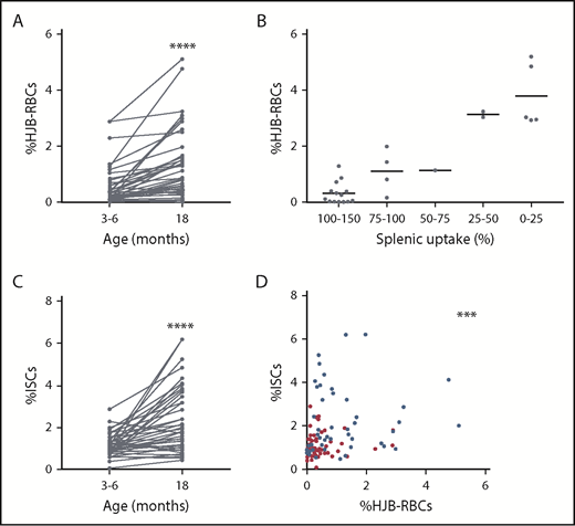
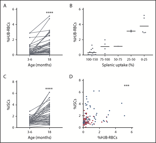
The median %HJB-RBCs in 45 patients at enrollment was low (0.3%; range, 0.01%-2.9%) and comparable to that in an adult healthy control group, albeit with a different distribution range (0.3%; range, 0.01%-0.6%; n = 7; P = .93; Figure 1A; supplemental Table 1).
HJB-RBCs and ISCs in children with SCA. (A) %HJB-RBCs determined by IFC in 45 children at 3 to 6 and 18 months. ****P < .0001, Wilcoxon paired test. (B) Distribution of %HJB-RBCs with respect to splenic uptake as measured by 99mTc heated RBCs spleen scintigraphy. Splenic uptake of 100% to 150% (n = 15), 75% to 100% (n = 4), 50% to 75% (n = 1), 25% to 50% (n = 2), and 0% to 25% (n = 5). (C) %ISCs determined by IFC in 45 children at 3 to 6 and 18 months. ****P < .0001, Wilcoxon paired test. (D) Spearman correlation between %ISCs and %HJB-RBCs at 3 to 6 (red dots) and 18 months (blue dots). n = 99; R2 = 0.69; ***P < .001.
HJB-RBCs and ISCs in children with SCA. (A) %HJB-RBCs determined by IFC in 45 children at 3 to 6 and 18 months. ****P < .0001, Wilcoxon paired test. (B) Distribution of %HJB-RBCs with respect to splenic uptake as measured by 99mTc heated RBCs spleen scintigraphy. Splenic uptake of 100% to 150% (n = 15), 75% to 100% (n = 4), 50% to 75% (n = 1), 25% to 50% (n = 2), and 0% to 25% (n = 5). (C) %ISCs determined by IFC in 45 children at 3 to 6 and 18 months. ****P < .0001, Wilcoxon paired test. (D) Spearman correlation between %ISCs and %HJB-RBCs at 3 to 6 (red dots) and 18 months (blue dots). n = 99; R2 = 0.69; ***P < .001.
HJB-RBCs counts by classic May-Grünwald Giemsa blood smears were only slightly elevated in a very small proportion of patients and did not allow further interpretation by lack of sensitivity.
Spleen scintigraphy with 99mTc heated RBCs was performed in a subgroup of 25 patients (with characteristics comparable to the rest of the cohort; data not shown) at a median age of 6.2 months (range, 4.9-8.0). Splenic uptake was normal in 17 cases (68%) and decreased in 8 (32%; Table 1). All scans were qualified as homogenous. Median splenic volume was 45 mL (range, 0-100), an increase compared with that of healthy age-matched controls (median, 21 mL; range, 14-42).27 Only 28% of the patients had a splenic volume within the 5th to 95th percentiles of age-matched healthy controls. There was no predictive value between routine laboratory parameters, including RBC parameters (Hb, mean corpuscular volume, and mean corpuscular hemoglobulin concentration), hemolysis markers, %HbF measured at enrollment, and the result of spleen scans at this age (supplemental Table 2).
Between enrollment and 18 months of age, the median %HJB-RBCs increased significantly (from 0.3% to 0.74%; range, 0.01-5.11) illustrating the expected decline in splenic function with time. Of note, excluding the patients who were transfused during follow-up (n = 7) did not yield any significant changes in the results. Spleen scintigraphy performed sequentially in 17 patients at a median age of 18.3 months (range, 16.6-19.5) showed a decreased splenic uptake in 7 patients (41.7%) and a stable scan in 9 (53%), further illustrating the decline of function with age, except for 1 patient who had an unexpected increase in splenic uptake (Table 1). Altogether, there was no evidence of a significant positive effect of transfusion on spleen scintigraphy results (data not shown).
At 18 months of age, the median splenic volume was 70 mL (range, 0-115), an increase compared with the 31 mL (range, 10.59-65.4) in healthy controls. At this time point, only 17% of the patients had a splenic volume within the 5th to 95th percentiles of age-matched healthy controls.27 Whereas 56% had an increased volume, 28% had a decreased volume. There was no correlation between splenic uptake and volume, altogether or independently, at each time point. Similarly, there was no correlation between %HJB-RBCs and splenic volume, demonstrating that volume is not predictive of splenic function. Importantly, at all time points, a significant correlation was found between %HJB-RBCs and splenic uptake (Figure 1B; R2 = 0.69; P < .0001).
Impaired deformability of RBCs increases with time and impacts splenic function
At enrollment, ISCs were found in the circulation (median, 0.96%; range, 0.08% to 2.9%; n = 45) and increased significantly at 18 months of age (1.71%; range, 0.47-6.21; Figure 1C; supplemental Table 1), similar to %HJB-RBCs (Figure 1A). Pooling of all time point measurements showed a positive correlation between %ISCs and %HJB-RBCs (Figure 1D), suggesting a relationship between altered RBC deformability and splenic loss of function. As expected, there was a positive correlation between %ISCs and the splenic uptake measured by scintigraphy (R2 = 0.3; ***P = .0005; supplemental Figure 2A). Because HbF modulates the polymerization of HbS and hence RBC deformability,28 we looked at the relationship between ISCs and HbF and found a significant correlation (R2 = 0.16; ***P < .0001; supplemental Figure 2B).
RBC adhesion increases with time in young infants and plays a role in loss of splenic function
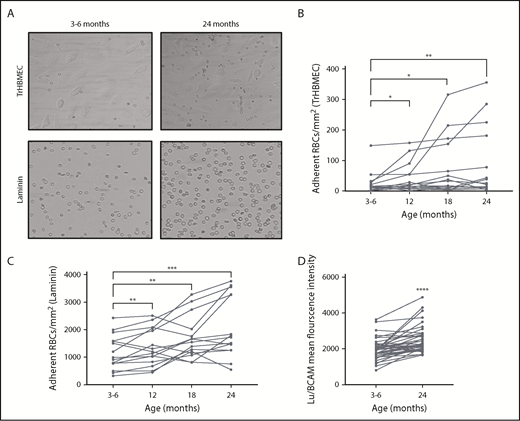
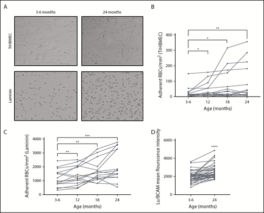
RBC adhesive properties were investigated in a subgroup of 15 infants by first performing adhesion assays on tumor necrosis factor-α–activated endothelial cells. RBCs were adherent at 3 to 6 months of age, with a significant progressive increase of the adhesion level at 12, 18, and 24 months (Figure 2A-B), indicating an early triggering of the RBC adhesive phenotype and subsequent increase during the first 2 years of life.
Lu/BCAM expression and mediated RBC adhesion. (A) Microscopic images of RBCs adhering to TrHBMEC monolayers and laminin 521-coated microchannels at 3 to 6 and 24 months. Original magnification ×20. (B-C) The amount of adherent RBCs/mm2 on TrHBMEC-coated channels (n = 15) (B) and laminin-coated channels (n = 15) (C) at 3 to 6, 12, 18, and 24 months. *P < .05, **P < .005, ***P < .001, Wilcoxon test. (D) Mean fluorescence intensity of Lu/BCAM on mature RBCs. ****P < .0001, Wilcoxon test.
Lu/BCAM expression and mediated RBC adhesion. (A) Microscopic images of RBCs adhering to TrHBMEC monolayers and laminin 521-coated microchannels at 3 to 6 and 24 months. Original magnification ×20. (B-C) The amount of adherent RBCs/mm2 on TrHBMEC-coated channels (n = 15) (B) and laminin-coated channels (n = 15) (C) at 3 to 6, 12, 18, and 24 months. *P < .05, **P < .005, ***P < .001, Wilcoxon test. (D) Mean fluorescence intensity of Lu/BCAM on mature RBCs. ****P < .0001, Wilcoxon test.
We then focused on the erythroid adhesion protein CD239/Lu/BCAM for its known role in abnormal RBC adhesion in SCA,14,29,30 in particular to laminin,31 an important structural component of the splenic extracellular matrix. In addition, Lu/BCAM has an early increased expression on RBCs of infants with SCA, compared with age-matched controls.13 RBC adhesion to immobilized laminin was higher than control RBCs (data not shown) with a significant increase at 12, 18, and 24 months (n = 15; Figure 2A,C). This increase was associated with increased Lu/BCAM expression per cell with age (Figure 2D). To test whether the increase in RBC adhesion was secondary to the activation of Lu/BCAM, we determined the phosphorylation level of Lu/BCAM in the same blood samples at the 4 different time points. Lu phosphorylation began as early as 3 to 6 months of age, in accordance with the high adhesion levels observed at this stage. Lu phosphorylation ratio did not differ significantly between 3 to 6 and 24 months of age, indicating that both phospho-Lu and total Lu/BCAM were increasing at the same rate within this time frame (supplemental Figure 3).
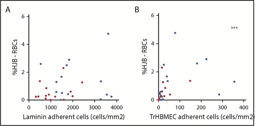
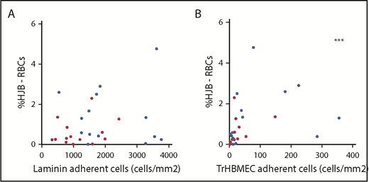
There was no correlation between the number of adherent RBCs on laminin and splenic function measured by %HJB-RBCs (P = .39; Figure 3A). Conversely, a positive correlation was found between the number of adherent cells on TrHBMECs and the %HJB-RBCs (Spearman correlation R2 = 0.42; P < .0001; Figure 3B) suggesting a role of adhesion, beyond Lu-BCAM, in the occurrence of decline of splenic function.
RBC adhesion and HJB-RBCs in children with SCA. Spearman correlation between the amount of adherent RBCs/mm2 on laminin (A) and TrHBMEC and %HJB-RBCs (B). At 3 to 6 (red dots) and 18 months (blue dots). n = 28; R2 = 0.43; ***P < .001.
RBC adhesion and HJB-RBCs in children with SCA. Spearman correlation between the amount of adherent RBCs/mm2 on laminin (A) and TrHBMEC and %HJB-RBCs (B). At 3 to 6 (red dots) and 18 months (blue dots). n = 28; R2 = 0.43; ***P < .001.
Incidence, determinants, and consequence of ASS
As previously reported,16 during the study period, 8 (17%) infants experienced at least 1 episode of ASS, at a median age of 13.4 months (8.0-15.9), an incidence rate within the expected range.32,33 Thirty-five infants remained asymptomatic during the follow-up period, and 14 experienced other SCD-related complications.
Analysis of routine laboratory parameters and more specific SCA-related biomarkers measured at enrollment showed that the level of HbF was the only factor that was prognostic of ASS.16 When the analysis was extended to %ISCs, the %HJB-RBCs and Lu/BCAM expression levels (in 7 and 22 patients of the ASS and asymptomatic groups, respectively, for whom samples were available) did not reveal an additional significant prognostic value of these markers.
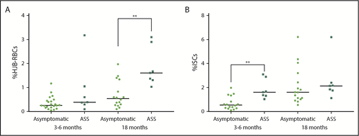
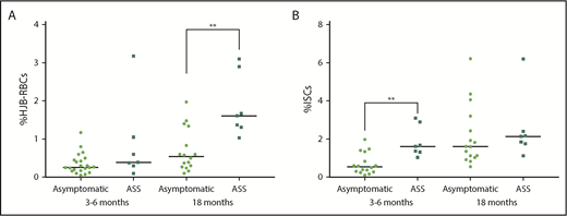
Moving on to comparing %HJB-RBCs at enrollment in infants who later experienced ASS with those who remained asymptomatic showed no significant difference, demonstrating similar splenic function in both groups at 3 to 6 months of age and excluding intrinsic abnormalities of the spleen filtration function in the ASS group at this stage (Figure 4A). Likewise, no significant difference was noted between Lu/BCAM expression and Lu/BCAM-mediated adhesion levels in the 2 groups. Conversely, patients from the ASS group had a significantly higher %ISCs at enrollment than did those from the asymptomatic group (median: 1.61% vs 0.54%; P = .0025; Figure 4B), suggesting that high levels of ISCs in the circulation may be a contributing factor to ASS.
HJB-RBCs and ISCs in asymptomatic and ASS-affected infants. Comparison of %HJB-RBCs (A) and %ISCs (B) at 3 to 6 and 18 months in asymptomatic (n = 22) and ASS infants (n = 7, for whom samples were available for analysis). **P < .005, Wilcoxon test.
HJB-RBCs and ISCs in asymptomatic and ASS-affected infants. Comparison of %HJB-RBCs (A) and %ISCs (B) at 3 to 6 and 18 months in asymptomatic (n = 22) and ASS infants (n = 7, for whom samples were available for analysis). **P < .005, Wilcoxon test.
At 18 months, there was no difference in ISC level, Lu/BCAM expression, and Lu/BCAM-mediated adhesion levels, in both groups (data not shown). Importantly, %HJB-RBCs was significantly higher in the ASS group at 18 months, as compared with the asymptomatic group (Figure 4A), indicating altered splenic function after the occurrence of ASS.
Discussion
SCA is known to result in splenic dysfunction, with the spleen being the first organ to be severely injured, causing substantial morbidity and mortality. However, in clinical practice, exploration of splenic function is limited by the lack of easy, high-throughput, noninvasive tools, so that little is known about the timing and extent of splenic injury in SCA. In addition, direct access to spleen histology is rare because autosplenectomy is the natural outcome in SCA, and surgical splenectomy is therefore infrequently necessary.
A previous study, relying on pitted cell counts, an indirect peripheral measure of vesicle-containing RBCs, showed that splenic function was lost within the first 5 years of life in 90% of children with SCA.34 More recently, the BABYHUG study using liver/spleen 99mTc colloid scans showed that splenic function was impaired in 75% of the cases,35 specifically in infants <12 months of age with SCA (n = 12, with the youngest aged 8 months). The present study went further by exploring longitudinally and specifically the filtration function of the spleen in a younger cohort. In fact, this splenic exploration could hardly be undertaken earlier (ie, before 6 months of age), given the time lapse between neonatal diagnosis and enrollment in the study, in addition to the difficulty in obtaining consent for an invasive spleen exploration in otherwise asymptomatic very young infants. Importantly, the longitudinal follow-up allowed insight into the dynamic dimension of splenic decline of function. Furthermore, splenic 99mTc heated RBC scintigraphy specifically explored the filtration function of the spleen (as opposed to 99mTc colloid spleen scanning that measures the phagocytic uptake by splenic macrophages), a relevant exploration given the major RBC deformability impairment occurring in SCA.
Our results show that at enrollment, 32% of patients had decreased filtration function evidenced by decreased splenic uptake. A previous study by Adekile et al,36 based on a sequential use of colloid and heated RBC scintigraphy in a cohort of older patients (n = 17; mean age, 7 ± 3.5 years), suggested a temporal loss of splenic function, with an initial loss of the phagocytic function followed by the loss of the filtration function. If this temporal sequence is true, our results suggest that the onset of splenic phagocytic dysfunction may start earlier than 6 months and agree with the results from the baseline splenic exploration of the BABYHUG trial mentioned above. In addition to the known immaturity of splenic function in all infants below 2 years of age, which increases the risk of pneumococcal infection in this age group, our data support the early onset of an additional increased susceptibility to pneumococcal infections in infants with SCA and argues for very early penicillin prophylaxis therapy in infants with SCA.
The longitudinal follow-up further allowed insights into the natural history of splenic decline of function and its relationship with splenic volume. Almost half of patients (42%) showed a decrease in splenic function at 18 months—that is, within a year (increase in %HJB and decrease in splenic uptake). Furthermore, splenic volume measured by scintigraphy showed an increased volume in the majority of the children and, importantly, did not correlate with function, a finding not only illustrating functional asplenia,37 but also demonstrating that splenic volume is not predictive of splenic function. Importantly, the very significant correlation between %HJB-RBCs and results of spleen scintigraphy demonstrates that the measurement of %HJB-RBCs by flow cytometry may allow accurate evaluation of the spleen’s filtration function in future studies. In clinical practice, this measurement could help monitor spleen function and guide the appropriate timing of splenectomy in very young children with recurrent ASS, for instance, by demonstrating the absence of function and hence no additional infectious risk related to the surgical removal of the spleen. Beyond SCA, easily available measurement of HJB is a significant improvement, given the growing suspected role of the spleen in the occurrence of severe complications, such as autoimmune diseases, neoplasia, thromboembolic events, and pulmonary hypertension in other patients with hemolytic anemia and in healthy subjects.
We chose to focus on sickle RBC adhesion and deformability properties, as potential contributors to splenic injury, because determinants of splenic injury in SCD are not known and because the splenic microcirculation specifically challenges these properties. Increased adhesion may play a role in splenic injury because the increased adhesive properties of sickle RBCs prolong transit time in the red pulp, favor HbS polymerization, and hence promote both sickling (and subsequent congestion) and increased cell–cell interaction (and subsequent phagocytosis) within the filtering beds. In this study, we confirmed the increase with time of the expression of Lu/BCAM on RBCs. We also showed that Lu/BCAM is activated. We further showed that this expression functionally translates into a significant increase in adhesion of RBCs on laminin-coated capillaries. Yet, no correlation (positive or negative) was observed between the percentage of adherent RBCs on laminin and the spleen function measured by %HJB-RBCs. This finding may pertain to the restricted interaction of Lu/BCAM with laminin 521, which may not be the major type of laminin present in the extracellular matrix of the spleen. Conversely, when adhesion assays were performed on TrHBMECs, we not only observed a significant increase in the number of adherent RBCs with age, we also observed a significant correlation with the %HJB-RBCs, implying a relation between splenic function and RBC adhesion. The activation of TrHBMECs leads to the expression of ICAM-1 and VCAM-1, which mediates cell adhesion,23 and previous studies have indeed shown the presence of VCAM-1 in the red pulp of the spleen.38,39 Further immunohistological studies are needed to explore VCAM-1–mediated endothelial–sickle RBC adhesion within the human spleen.
In SCA, repeated HbS polymerization results in circulating ISCs. They have a short life span, correlate with hemolysis in vivo, and contribute to the pathophysiology of vaso-occlusion.18 We recently showed that ISCs are poorly deformable and prone to blocking capillaries in vitro.20 We therefore chose to explore the role of ISCs in splenic function because we hypothesized that ISCs get trapped in the filtering beds of the red pulp, notably at the interendothelial slit barrier, because of their lack of deformability. In the present study, ISCs were indeed present in the circulation at a very young age and increased significantly with time, similar to %HJB-RBCs. Furthermore, the %ISCs correlated with splenic uptake, indicating that impaired deformability negatively affects splenic filtration.
Because ASS is the most life-threatening splenic complication in infants with SCA and is probably the extreme acute expression of spleen dysfunction, we specifically analyzed infants who experienced ASS and compared them to a selected subgroup of the cohort that remained asymptomatic throughout the follow-up. ASS caused further splenic dysfunction, as illustrated by the increase of %HJB-RBCs in those who experienced ASS, an unsurprising finding, yet one that is so far undemonstrated. Regarding determinants of ASS, while keeping in mind the small number of events, we found no correlation between increased RBC adhesion properties and the occurrence of this event. Conversely, the %ISCs was significantly elevated at 3 to 6 months of age in infants who later experienced ASS, a finding suggesting that ISCs, and hence decreased deformability, may be an important contributor to ASS. Trapping of ISCs may cause a decreased outflow, favoring further sickling and ultimately, ASS. Further studies on larger samples are necessary to confirm these findings.
In conclusion, our study demonstrated that flow cytometry analysis of %HJB-RBCs alone may accurately reflect spleen filtration function in very young children with SCA. Using the analysis, together with spleen scintigraphy, we showed that splenic loss of function is present very early in life (at 3 to 6 months of age) in SCA-affected infants, declines further, and is unrelated to splenic volume. Hyposplenism results from both increased RBC adhesive properties and, critically, loss of deformability. ISCs are additionally a potential contributor to ASS, which in turn results in further loss of spleen function.
The full-text version of this article contains a data supplement.
Acknowledgments
The authors thank Geneviève Milon, Peter David, Pierre Buffet, Gil Tchernia, Jacques Elion, and Narla Mohandas for intellectual contribution to the study; Thierry Peyrard, Dominique Gien, Eliane Vera, and Sirandou Tounkara at the Centre National de Référence pour les Groupes Sanguins for the management of blood samples; Frédérique Archambault for input into the design of the study and the acquisition and analysis of the spleen scintigraphy; Julien Picot and Catia Pereira for flow cytometry analyses; and Jean-Philippe Semblat for assistance.
This work was supported by the Projet Hospitalier de Recherche Clinique (Ministry of Health, Projet Hospitalier de Recherche Clinique 2008 AOM08171), INSERM, the Institut National de la Transfusion Sanguine, and the Laboratory of Excellence GR-Ex (grant ANR-11-LABX-0051). GR-Ex is funded by the program “Investissements d’avenir” of the French National Research Agency (grant ANR-11-IDEX-0005-02). S.E.H. was funded by the Ministère de l’Enseignement Supérieur et de la Recherche (Ecole Doctorale BioSPC) and received financial support from Club du Globule Rouge et du Fer and Société Française d’Hématologie.
Authorship
Contribution: S.E.H. conducted the experiments, acquired and analyzed the data, and wrote and edited the manuscript; S.C., M.M., C.L., and M.D. conducted the experiments and acquired the data; N.B. and C.E. performed the statistical analysis on the spleen scintigraphic data; M.d.M., C.A., C.G., B.P., M.H.O., and V.B. enrolled and were in charge of the patients; F.M. performed the spleen scintigraphy acquisition and analysis; C.L.V.K. and Y.C.A. edited the manuscript; W.E.N. contributed significantly to the design of the study, analyzed the data, and wrote and edited the manuscript; and V.B. designed and was responsible for the study, conducted the experiments, acquired and analyzed the data, and wrote and edited the manuscript.
Conflict-of-interest disclosure: The authors declare no competing financial interests.
Correspondence: Valentine Brousse, Service de Pédiatrie et Maladies Infectieuses, Hôpital Universitaire Necker-Enfants Malades, 149 rue de Sèvres, 75015 Paris, France; e-mail: valentine.brousse@gmail.com.
References
Author notes
V.B. and W.E.N. contributed equally to this work.

