Abstract
Pyrimidine 5′ nucleotidase-I (P5N-I) deficiency is a rare autosomal recessive disorder associated with hemolytic anemia, marked basophilic stippling, and accumulation of high concentrations of pyrimidine nucleotides within the erythrocyte. Recently, the structure and location of the P5N-I gene have been published. This paper presents the results of a study characterizing the molecular pathologies of P5N-I deficiency in a total of 6 Turkish patients from 4 unrelated families of consanguineous marriages. Mutation analysis in the P5N-I gene led to the identification of 3 novel mutations in these patients. In 4 patients from 2 families, a homozygous insertion of double G at position 743 was detected in exon 9 (743-744insGG), leading to premature termination of translation 23 bp downstream. In one family, a homozygous T to G transition at position 543 (543T>G) in exon 8 resulted in the replacement of tyrosine (Tyr) with a stop codon (Tyr181Stop). In another family, a homozygous insertion of a single A in exon 7 (384-385insA) created a stop signal at the codon nearby. In all families, the parents were heterozygous for the relevant mutations. None of these changes was detected in 200 chromosomes from a healthy Turkish population. These mutations were not correlated with any particular phenotype.
Introduction
Erythrocyte maturation is accompanied by RNA degradation and release of mononucleotides. Pyrimidine 5′ nucleotidases (P5N), often called uridine 5′ monophosphate hydrolase, are enzymes involved in the dephosphorylation of pyrimidine nucleotides. Isolated from the human erythrocytes were 2 cytoplasmic forms of the enzyme. These enzymes, namely P5N-I and P5N-II, have different molecular properties, substrate specifities, and genetic basis for disease associations.1,2 The deficiency of P5N-I is an autosomal recessive disorder characterized by hemolytic anemia, marked basophilic stippling, and accumulation of high concentrations of pyrimidine nucleotides within the erythrocyte.3,4 The disease has a wide geographic distribution and has been described in many parts of the world. To date, 40 patients with P5N-I deficiency have been reported, while presumably large numbers go undetected.5 It has been suggested that after glucose-6-phosphate dehydrogenase and pyruvate kinase deficiencies, it is the third most common red blood cell (RBC) enzymopathy causing hemolysis.6
The P5N-I cDNA was cloned and expressed. P5N-I peptide sequences revealed substantial homology with p36—an alpha interferon–induced protein in cells forming lupus inclusion formation.7 Recently, identification of the genomic DNA sequence corresponding to the cDNA revealed that the P5N-I gene is located on chromosome 7p15-p14, consists of 10 exons, and codes for 286 and 297 amino acid (aa)–long proteins.5 P5N-I deficiency is associated with the shorter (286 aa) protein, which is produced by alternative splicing of exon 2. Reported were 3 different homozygous mutations in the 4 affected patients from 1 Norwegian and 2
The purpose of this study was to elucidate the molecular defects responsible for the pathologies observed in Turkish patients with P5N-I deficiency. To this end, 6 patients from 4 unrelated families were characterized, and 3 novel mutations in the gene were described.
Patients and methods
Subjects
The subjects of this study were 6 Turkish patients from 4 unrelated families (Families I-IV) who were admitted to Ihsan Dogramaci Children's Hospital at Hacettepe University (Ankara, Turkey) for evaluation of chronic congenital hemolytic anemia. All patients were the product of first-degree cousin marriages. The ages of patients at diagnosis ranged from 6 months to 14 years (mean, 6.5 years). Originally, there were a total of 8 patients in 4 families. The patients in Families IV were previously reported.8 All patients underwent splenectomy at the ages of 10 to 15 years. However, long before this study, one patient in each of Family III and IV died of an acute illness associated with high fever without having any medical attention. The clinical diagnosis was based on typical findings, including hemolytic anemia, marked basophilic stippling, increased reticulocyte count, decreased purine-pyrimidine ratio, and low enzyme activity (Table 1). The control group consisted of 100 unrelated, apparently healthy blood bank donors without any evidence of hemolytic anemia. This study was approved by the Hacettepe University Review Board and informed consent was obtained from all individuals participating in the study.
Hematologic analysis
Mutation analysis
Genomic DNA was isolated from peripheral blood using a standard method. Primers were generated from intron sequences flanking the individual exons of the P5N-I gene using ensemble human genome database and published genomic DNA sequence (Table 2). The P5N-I gene (exons 3 to 10) was screened for mutations by polymerase chain reaction (PCR) amplification and combined single-strand conformation polymorphism (SSCP)/heteroduplex (HD) analysis using GeneGel Excel 12.5/24 gels (Amersham Biosciences, Uppsala, Sweden) and the Genephor DNA Electrophoresis System (Pharmacia Biotech, Uppsala, Sweden). Upon detection of an aberrant SSCP and/or HD pattern, underlying mutations were established by sequencing (ABI Prism 310 Genetic Analyser; Perkin Elmer Applied Biosciences, Foster City, CA) the gel-purified DNA fragments (Wizard PCR Preps DNA Purification System; Promega, Madison, WI) as recommended by the suppliers. The identified mutations were further confirmed by developing alternative methods according to the type and sequence environment of the mutations. The methods were as follows: denaturing polyacrylamide gel electrophoresis (PAGE) (6%) for the 2-bp insertion, PCR–restriction fragment length polymorphism (RFLP) analysis using SfaNI enzyme digestion for the nonsense mutation, and HD assay on GeneGel HyRes Native gels (Amersham Biosciences) for the 1-bp insertion (see “Results”). The family members and 200 control chromosomes were also screened for each variation by means of these methods.
Haplotype analysis
There were 3 microsatellite markers, namely D7S817, D7S683, and D7S2846 (Research Genetics, Huntsville, AL), used for the haplotype analysis of the region 7p14.3-p14.1. All members of the families were genotyped using site-specific PCR and denaturing PAGE (6%).
TATA-box genotype (Gilbert Syndrome) analysis
The A[TA]6/7TAA repeat motif in the promoter region of the UDP–glucuronosyltransferase (UGT-1A) gene was determined as described before.11
Results
Some of the clinical features and hematologic data of the patients are given in Table 1. Peripheral blood smears revealed moderate anisocytosis, mild polychromasia, a few acanthocytic spherocytes, and heavy basophilic stippling in most RBCs in all patients. Red cell purine-pyrimidine ratios and P5N-I enzyme activities were compatible with the P5N-I deficiency (Table 1). In the patients with thalassemic face and bony changes, β-thalassemia was excluded by normal HbF (< 1%) and HbA2 (< 3.5%) values, and the absence of hypochromia or microcytosis in the patients and parents. These patients received blood transfusions twice prior to splenectomy.
In the mutation analysis, the complete coding sequence of the P5N-I gene (exon 3-10) in the affected patients was amplified from genomic DNA and screened for mutations by SSCP/HD analysis. Detected in exons 7, 8, and 9 of the P5N-I gene were 3 different types of aberrant bands (data not shown). Characterization of abnormal bands by sequencing led to the identification of 3 novel mutations responsible for the pathologies observed in 6 patients from 4 unrelated families.
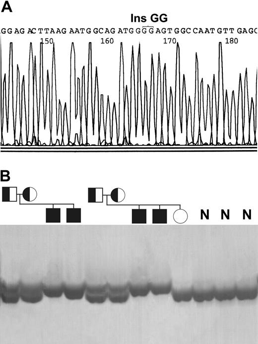
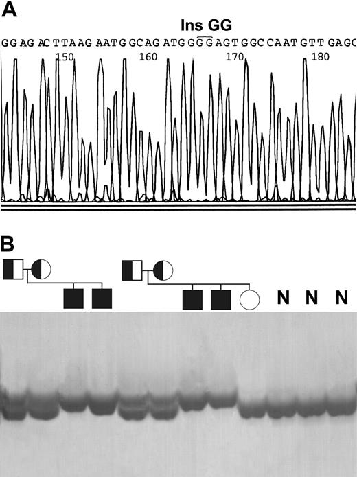
A homozygous insertion of double G at position 743 in exon 9 (743-744insGG) was observed in 4 patients from Families I and II (Figure 1A). This mutation led to a shift in the open reading frame and to premature termination of translation 23 bp downstream in the same exon. Haplotype analysis with 3 microsatellite markers showed that this mutation was associated with the same haplotype in both families (data not shown). Analysis of the local DNA sequence environment surrounding the mutation indicated that the mutation site is flanked by many overlapping short direct and inverted repeats (2-4 bp). Studying the mutation in the family members by denaturing PAGE indicated that parents in both families were heterozygous for the mutation, while the only healthy sibling in Family II was normal (Figure 1B). This mutation was not detected in 200 chromosomes studied from the healthy Turkish population.
Identification of the homozygous 743-744insGG mutation in exon 9. (A) DNA sequence analysis indicating the site of the insertion. (B) Denaturing polyacrylamide gel electrophoresis showing the mutation in the family members and in the healthy individuals (N).
Identification of the homozygous 743-744insGG mutation in exon 9. (A) DNA sequence analysis indicating the site of the insertion. (B) Denaturing polyacrylamide gel electrophoresis showing the mutation in the family members and in the healthy individuals (N).
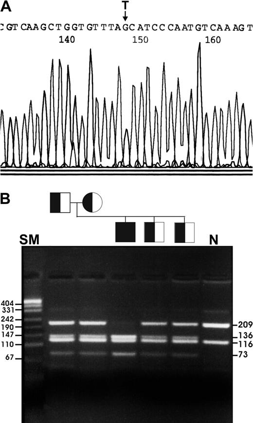
A homozygous T to G transition at position 543 (543T>G) in exon 8 led to the substitution of tyrosine (Tyr) (TAT) with a stop codon (TAG) and, consequently, to premature truncation of the protein at codon 181 (Tyr181Stop) in the affected patient of Family III (Figure 2A). Many overlapping short direct and inverted repeats (2-4 bp) were present at and around the mutation. The transition of the sequence TCATC to GCATC created a recognition site for SfaNI restriction enzyme, which generated digestion products of 209- and 116-bp DNA fragments in normal and 136, 116, and 73 bp in homozygous mutant subjects. Analysis of the family members using this enzyme showed that both parents and 2 healthy siblings carried the substitution (Figure 2B). This mutation was not observed in the control group of this study.
Identification of the homozygous 543T>G mutation in exon 8. (A) DNA sequence analysis showing the site of the substitution. (B) SfaNI digestion showing the mutation in the family members. N indicates normal control; SM, pUC19 DNA/MspI size marker. Numbers indicate bp.
Identification of the homozygous 543T>G mutation in exon 8. (A) DNA sequence analysis showing the site of the substitution. (B) SfaNI digestion showing the mutation in the family members. N indicates normal control; SM, pUC19 DNA/MspI size marker. Numbers indicate bp.
A homozygous insertion of single A to the 4 A tract in exon 7 (384-385insA) created a stop signal at the codon nearby in the patient of Family IV (data not shown). The mutation site is surrounded by many overlapping inverted and direct repeats 2 to 5 bp in size. Mutation-specific HD analysis of the family members showed that both parents and 3 of the 4 siblings were heterozygous for the mutation (data not shown). The mutation was not detected in the 200 control chromosomes.
In an attempt to provide a possible explanation for the heterogeneity in the bilirubin levels, the patients and the family members were screened for the common TATA-box repeat variation in the UGT-1A gene promoter (Gilbert Syndrome). This analysis revealed that the patients of the first 3 families were all heterozygous (6/7) and the patient of the Family IV was homozygous for the rarer 7-repeat allele (7/7).
Discussion
Recently, the discovery of the chromosomal localization and detailed sequence information for the P5N-I gene has permitted exon-by-exon screening of genomic DNA as a means of determining the molecular pathologies responsible for the phenotype in patients with P5N-I deficiency.5 This paper presents the results of the study characterizing the molecular pathologies of 6 Turkish patients coming from 4 unrelated families. Mutation analysis of the P5N-I gene led to the identification of 3 novel mutations. As expected from parental consanguinity, all of the patients proved to have homozygous mutations. The types of the detected mutations were microinsertion and nonsense mutations, which would be predicted to generate null alleles, although residual function for part of the protein cannot be excluded.
Mutation screening by combined SSCP/HD analysis of amplified exons in this study has proved to be highly efficient for detection of mutations in the P5N-I gene. The exon sequences of the gene do not appear to be very polymorphic and hypermutable since all of the exons were screened in all patients and no other variant bands were detected. Similar to the previous report, no deletions and common polymorphisms were observed in the exons of the gene.5 The identification of 3 different mutations in 4 families suggested that the mutations of the P5N-I gene are quite heterogeneous in Turkey. A preliminary screening of 200 control chromosomes using the mutation-specific assays did not detect any mutant alleles, suggesting that as expected these alleles are not likely to be common in the Turkish population. The 2 families (I and II) with the mutation 743-744insGG originated from the same town in the eastern part of the Black Sea region. Although there is no apparent relation between these families, the results of the haplotype analysis indicated that they most probably carry a common mutant chromosome, and therefore, are likely to be descendants of the same ancestor.
Recent studies on the mechanism of short gene deletion/insertion mutations causing human disease indicated that homonucleotide and short direct/inverted repeats (2-8 bp) around the site of mutations are associated with a high mutation frequency in human genes.12,13 The regions flanking the mutations described here essentially contain a high number of short direct and inverted repeats (also homonucleotide in one), suggesting that these repeat elements may also play a role in the generation of base substitution as well as microinsertion mutations in the P5N-I gene. Even though no obvious mutational hot spot consensus sequence was found at the site of the mutations identified, given that the nonsense mutation identified in exon 8 (Tyr181Stop) is located only 13 bp downstream from the one reported (Gln177Stop) for the South African family, there is reason to believe that there may be additional mutational hot spots in the local DNA surrounding these 2 mutations.5
The mutations described here are not related to a particular clinical phenotype. However, all of the patients had moderate to severe clinical outcome, which may be attributed to their null mutations. The patients in Family I showed relatively low serum bilirubin levels, while the patients in Family II with the same mutation (743-744insGG) had higher levels (Table 1). The possible association of the heterogeneity in bilirubin levels with the common TATA-box repeat variation in the UGT-1A gene promoter (Gilbert Syndrome) was excluded by the observation that all 4 patients in these 2 families were heterozygous (6/7) for this variant.11 One patient in each of Families III and IV probably died of acute infections due to complications of splenectomy. Therefore, the decision for splenectomy might be restricted only to patients who have signs of hypersplenism.
The patients in Family III with the mutation 543T>G (Tyr181Stop) had mild mental retardation. This is the first report indicating the presence of mild mental retardation in patients with P5N-I deficiency. Therefore, it is difficult to make a direct correlation with this clinical observation and the specific mutation. In addition, the history of neonatal hyperbilirubinemia reported by the parents might be implicated as a cause of mental retardation in this family. The patients in Family IV with 383-384insA mutation had thalassemic face and bony changes. β-Thalassemia in this family was excluded by the absence of any parameters compatible with this diagnosis. The variations observed in the phenotypic expression of the disease might have resulted from other undefined genetic and/or environmental factors.
In conclusion, this study represents the start of the efforts to describe pathologic sequence variations of the P5N-I gene in different populations. Further studies are needed to define the mutational spectrum of the P5N-I gene. Attention can then be given to the clinical phenotypes associated with specific mutations, since this may have prognostic significance, and thus affect patient management in the future. Mutation detection in the P5N-I gene will also be of considerable diagnostic importance since knowledge of the causative mutations will permit rapid prenatal diagnosis on the basis of a molecular test. It will also help in diagnosing patients who have P5N-I deficiency in cases where a diagnostic test is not available or atypical clinical phenotype leads to misdiagnosis.
Prepublished online as Blood First Edition Paper, April 24, 2003; DOI 10.1182/blood-2003-02-0628.
Supported by grants from Hacettepe University Research Foundation (02G116) and the Turkish Academy of Science.
Presented in abstract form at the 44th annual meeting of the American Society of Hematology, Philadelphia, PA, December 9, 2002.
The publication costs of this article were defrayed in part by page charge payment. Therefore, and solely to indicate this fact, this article is hereby marked “advertisement” in accordance with 18 U.S.C. section 1734.
We would like to express our gratitude to Prof Dr Omer Kalayci for his critical review of the manuscript and to Oya Ekici for her technical support.


