Abstract
Immunodeficiency, centromeric region instability, and facial anomalies (ICF) syndrome is a rare autosomal recessive disease. Mutations in the DNA methyltransferase 3B (DNMT3B) gene are responsible for most ICF cases reported. We investigated the B-cell defects associated with agammaglobulinemia in this syndrome by analyzing primary B cells from 4 ICF patients. ICF peripheral blood (PB) contains only naive B cells; memory and gut plasma cells are absent. Naive ICF B cells bear potentially autoreactive long heavy chain variable regions complementarity determining region 3's (VHCDR3's) enriched with positively charged residues, in contrast to normal PB transitional and mature B cells, indicating that negative selection is impaired in patients. Like anergic B cells in transgenic models, newly generated and immature B cells accumulate in PB. Moreover, these cells secrete immunoglobulins and exhibit increased apoptosis following in vitro activation. However, they are able to up-regulate CD86, indicating that mechanisms other than anergy participate in silencing of ICF B cells. One patient without DNMT3B mutations shows differences in immunoglobulin E (IgE) switch induction, suggesting that immunodeficiency could vary with the genetic origin of the syndrome. In this study, we determined that negative selection breakdown and peripheral B-cell maturation blockage contribute to agammaglobulinemia in the ICF syndrome. (Blood. 2004;103:2683-2690)
Introduction
In the bone marrow (BM), B-cell differentiation leads to generation of a broad repertoire of antibody-bearing cells in an antigen-independent manner. After ordered rearrangement of the heavy and light chain loci, immature B cells are the first B-cell subset to express a B-cell receptor (BCR), composed of a surface immunoglobulin M (IgM) and the Igα/Igβ signaling complex (reviewed in Meffre et al1 ). This process yields a number of potentially self-reactive clones, which can be eliminated by apoptosis (clonal deletion), modified by secondary rearrangements (receptor editing), or rendered hyporesponsive (anergy).2-5 In the mouse, 2 × 10⁷ immature B cells are generated every day, of which around 90% undergo negative selection.6 Before migrating to the periphery, immature B cells develop into IgMhiIgDloCD21-CD23- type 1 (T1) transitional B cells.7 They differentiate in the spleen into IgMhiIgD+CD21+CD23+ type 2 (T2) transitional B cells and then into either IgMhiIgD-CD21+CD23- marginal zone or IgMloIgDhiCD21+CD23+ follicular mature B cells.8 Both transitional B-cell subsets appear to be short lived (3 to 4 days)6 and nondividing in vivo,9 although recent studies show that T2's proliferate and up-regulate survival signals, whereas T1's die after in vitro BCR engagement.10 Moreover, T1's express CD95 but not the antiapoptotic molecule bcl-2,7 further suggesting that T1 might be the target of the B-cell-negative selection occurring in the periphery.11 Such transitional B-cell subsets have not yet been defined in humans. Current characterization of human peripheral blood (PB) B cells is based on the expression of the tumor necrosis factor family member CD27, which distinguishes unmutated IgM+IgD+CD27- naive cells (approximately 60% in adults) from somatically hypermutated CD27+ memory cells (40%, of which approximately 40% are IgM+IgD+).12 Naive B cells may be the target of negative selection, since heavy chain variable region (VH) usage as well as the VH complementarity determining region 3 (CDR3) length changes in peripheral mature B cells.13-15
The analysis of primary immune deficiencies is a powerful tool for the study of normal human B-cell differentiation. Immunodeficiency, centromeric region instability, and facial anomalies (ICF) disease is a rare autosomal recessive syndrome, characterized by chromosomal instability and humoral immune deficiency. Patients present recurrent respiratory infections and diarrhea as consequence of hypogammaglobulinemia or agammaglobulinemia, sometimes associated with defective cell-mediated immunity.16 Developmental defects such as delayed developmental milestones, facial dismorphy (eg, roundness, hypertelorism, macroglossia), and mental retardation have also been observed. Cytogenetic abnormalities include elongation of centromeric or juxtacentromeric heterochromatin of chromosomes 1, 9, and 16, leading to formation of multiradiate figures involving mainly chromosomes 1 and 16.17 ICF patients show marked hypomethylation of classical satellites II and III, leading to centromeric instability.18,19 Most ICF patients (around 65%)20-22 carry mutations in the catalytic domain of the DNA methyltransferase 3B (DNMT3B) gene. This enzyme is a de novo methyltransferase required for genomic methylation following embryo implantation and for transposon and endogenous retrovirus methylation (reviewed in Bestor23 ). Dnmt3B-null mice are not viable, and mutant embryos present developmental defects and growth retardation.24
In this paper, we have studied B-cell defects associated with agammaglobulinemia in ICF patients. Detailed phenotypic and functional analysis of primary ICF B cells reveals impaired B-cell-negative selection and defective peripheral terminal B-cell differentiation, contributing to the agammaglobulinemia associated with the ICF syndrome.
Patients, materials, and methods
Patients
Four patients with clinical and cytogenetic diagnosis of ICF syndrome were studied. Patients P1 and P4 are 3- and 4-year-old girls (I.T. et al, unpublished data, 2003); patients P2 and P3 are a 16-year-old boy25 and a 9-year-old girl.26 At the time of diagnosis, P1 presented bacterial infections of the appendix and the vulva, and P4 had viral myocarditis and acute otitis, which will be described in detail (I.T. et al, unpublished data, 2003). Mutations in the coding region of the DNMT3B were not found in P1 and P4 (Y.L.J. et al, unpublished data, 2003). P2 had digestive tract candida and recurrent broncho-pulmonary bacterial infections. He died during this study. P3 had bacterial colitis and respiratory tract infections. P2 and P3, previously referred as G and R, respectively, present mutations on the DNMT3B gene.20,26 Parents gave informed consent for this work.
Antibodies and flow cytometry
Flow cytometry analyses on FACSCalibur (Becton Dickinson, San Jose, CA) were processed by means of CellQuest software (Becton Dickinson). The peripheral blood lymphocyte (PBL) population was defined as greater than 95% CD45+ and less than 5% CD14+. Cells (2 to 5 × 10⁵) were stained as described27 with the following antibodies: fluorescein isothiocyanates (FITC)-anti-CD19, anti-CD21, anti-CD23, anti-CD38, anti-CD69 (Immunotech, Marseilles, France), anti-CD86 (Pharmingen, San Diego, CA), anti-CD27 (Becton Dickinson), biotinylated anti-CD10 (Dako, Glostrup, Denmark), phycoerythrin (PE)-anti-δ (Pharmingen) anti-CD19 and anti-CD27 (Immunotech), allophycocyanin (APC)-anti-CD19, and biotinylated anti-μ (Pharmingen) revealed by peridinin chlorophyll-alpha protein (PerCP)-streptavidin (Becton Dickinson).
B-lymphocyte purification
Peripheral blood mononuclear cells (PBMCs) from patients and healthy donors were separated by means of Ficoll-Paque (Pharmacia Biotech, Uppsala, Sweden). B cells were enriched to greater than 90% by means of the StemSep B-cell enrichment kit or RosetteSep (StemCell Technologies, Vancouver, BC, Canada), allowing negative selection by depletion of CD2+, CD3+, CD16+, CD36+, and CD56+ cells. For activation assays, naive B cells from controls were enriched by CD27+ cell depletion, using CD27 microbeads and MiniMACS magnetic sorter (Miltenyi Biotech, Auburn, CA). For amplification of VH-heavy chain constant region 1μ (CH1μ) sequences, control cells were sorted on a FACSVantage SE (Becton Dickinson), leading to 99% sample purity: naive B cells were sorted with the use of APC-anti-CD19, PE-anti-δ, and FITC-anti-CD27 monoclonal antibodies (mAbs), whereas transitional and mature B cells were separated with APC-anti-CD19, PE-anti-CD27, and FITC-anti-CD21 mAbs.
In vitro B-cell activation and apoptosis/cell-cycle analysis
PBMCs (106) were cocultured with 105 irradiated (7000 rad) human CD40 ligand (hCD40L)-transfected Ltk-fibroblasts at 37°C on 24-well plates in RPMI 1640 supplemented with 10% fetal calf serum (FCS), 10 ng/mL recombinant human interleukin 10 (rhIL-10) (PeproTech, Rocky Hill, NJ), 20 U/mL rhIL-2 (a kind gift from E. Vivier, Marseilles, France), and 12.5 μg/mL F(ab′)2 antihuman IgM (ICN Biomedicals, Eschwege, Germany). After 20 hours, cells were harvested for monitoring of CD27, CD69, and CD86 expression. For cell-cycle analysis, enriched CD19+CD27- naive B cells were activated with the use of 1 μg/mL recombinant soluble human CD40L (rshCD40L), 1 μg/mL ligand enhancer (Alexis Biochemicals, Illkirch, France), and 100 U/mL rhIL-4 (PeproTech). Cells were harvested at indicated times and fixed overnight in 70% ethanol, washed twice in citrate-phosphate buffer (0.2 M Na2PO4, 0.1 M citric acid), and resuspended in phosphate-buffered saline (PBS). To ensure that only DNA was stained, cells were treated with RNAse A (100 μg/mL) before staining with 50 μg/mL propidium iodide (Molecular Probes, Leiden, the Netherlands). Percentages of apoptotic (sub-G0) and cycling (S and G2/M) cells were analyzed in a FACScan flow cytometer on FL2 wavelength after cell aggregates were gated out.
In vitro immunoglobulin-switch induction
Enriched CD19+CD27- naive B cells (5 × 10⁴) were cocultured with irradiated hCD40L fibroblasts (5 × 10³) and 100 U/mL rIL-4 on 96-well flat-bottom plates in RPMI 1640 with 10% FCS, in 200 μL total volume per well. Cells and supernatants were harvested after 5 or 10 days' culture and processed for isotype switch and immunoglobulin secretion, respectively. For all studies, patients' blood was collected before immunoglobulin replacement.
RNA and RT-PCR
Total RNA was isolated from 2.5 × 10⁵ cells by means of TRIzol reagent (Gibco BRL, Cergy Pontoise, France) and was reverse transcribed by means of Superscript II (Invitrogen, Cergy Pontoise, France) in 20 μL total volume, according to manufacturer's instructions. Reverse transcription-polymerase chain reaction (RT-PCR) amplification cycles consisted of 30-second denaturation at 94°C, 30-second annealing at the indicated temperature, and 1-minute elongation at 72°C; followed by a final 7-minute extension cycle at 72°C, with the use of Taq polymerase (Life Technologies) in a Peltier Thermal cycler 200 (MJ Research, Boston, MA).
For isotype switch analysis, 3, 2, and 1 μL cDNA were amplified for 28 cycles of annealing at 70°C, with 5′-Iϵ 5′-AGGCTCCACTGCCCGGCACAGAAAT-3′ (sense) and 3′-CH1ϵ 5′-ACGGAGGTGGCATTGGAGGGAATGT-3′ (antisense) primers; and 28 cycles of annealing at 62°C with hypoxanthine phosphoribosyltransferase (HPRT) 5′-CCTTGGTCAGGCAGTATAATCC-3′ (sense) and HPRT 5′-TTGTATTTTGCTTTTCCAGTTTCAC-3′ (antisense) primers. Postswitch VH-CH1ϵ and VH-CH1γ transcripts were amplified for 32 cycles of annealing at 58°C with 3 μL cDNA and a VH consensus 5′-GACACGGCCGTGTATTACTG-3′ (sense) and the 3′-CH1ϵ or 3′-CH1γ 5′-CGGGACCCGACGGACCAGTTCCTGATGAAG-3′ (antisense) primers. Activation-induced cytidine deaminase (AID) and Igβ amplification were previously described.28,29
For sequence analysis, VH1-CH1μ and VH5-CH1μ transcripts were amplified for 38 cycles from cDNA of 5 × 10⁵ to 106 sorted control CD19+IgD+CD27-, CD19+CD27-CD21-, and CD19+CD27-CD21+ cells or enriched ICF CD19+ cells with the use of VH5-leader 5′-AAAGCGGCCGCATGGGGTCAACCGCCATCCTCG-3′ or VH1-leader 5′-AAAGCGGCCGCATGGACTGGACCTGGAGGGTC-3′ (sense) and 3′-CH1μ 5′-GTCAGAGTTGTTCTTGTATTTCCAG-3′ primers (antisense), with annealing at 60°C. VH(1/5)-CH products were cloned into pGEM-T vector by means of TA cloning (Promega, Charbonnières, France) and transformed into competent bacteria. Individual clones were sequenced with Sp6 and T7 primers on an ABI Prism 310 sequencer (Applied Biosystems, Streetsville, ON, Canada) with the use of the Big Dye Terminators protocol.
Statistical analysis
Analysis of the positive-charge content of CDR3 sequences was performed by Student t test with GraphPad Prism 3.0 software (GraphPad Software, San Diego, CA), with the mean number of positive charges per sequence considered the relevant parameter. The same confidence coefficient was obtained under the assumption of either equal or different variances.
Dosage of immunoglobulin secretion
After in vitro switch induction, culture supernatants were kept at -20°C until they were assayed for IgG and IgM secretion by enzyme-linked immunosorbent assay (ELISA). Plates (Nunc, Roskilde, Denmark) were coated with goat antihuman IgA plus IgG plus IgM (heavy [H] plus light [L]) (Jackson ImmunoResearch Laboratories, West Grove, PA). Diluted supernatants and immunoglobulin standard (N/T Protein Control SL/M; Dade Behring, Marburg, Germany) were added to plates and incubated overnight at 4°C. Alkaline phosphatase-conjugated goat antihuman IgG and IgM (Jackson ImmunoResearch Laboratories) were used for detection of IgG and IgM, respectively, and were revealed by means of phosphatase substrate tablets (Sigma, Lyons, France).
Immunohistochemistry
Paraffin-embedded formalin-fixed jejunal biopsies were cut at 5-μm-thick intervals for immunohistochemistry. Immunodetections were performed by means of a Ventana automat (Ventana Medical Systems, Illkirch, France). For antigen unmasking, Ventana protease containing approximately 0.5 U alkaline protease was used. Staining was performed with a 1:2000 dilution of peroxidase-conjugated anti-IgA monoclonal antibody (Dako) and aminoethylcarbazol as chromogen for revelation. Hematoxylin counterstaining was used for plasma cell identification.
Results
ICF syndrome was diagnosed for 4 patients by karyotype analysis of phytohemagglutinin (PHA)-stimulated lymphocytes. This analysis revealed the hallmarks of the syndrome: heterochromatin decondensation and multibranched configuration of chromosomes 1, 9, and 16 (I.T. et al, unpublished data, 2003; Turleau et al25 ; Miniou et al26 ). For these patients, immunodeficiency was diagnosed in early childhood following recurrent bacterial infections. Serum immunoglobulin levels of all immunoglobulin isotypes, including IgM, were below the detection threshold in all patients except P2, in whom IgG levels reached 4.4 g/L (Table 1). Phenotypic analysis revealed that absolute and relative numbers of T (CD3+), B (CD19+), and natural killer (NK) (CD56+CD3-) lymphocytes in patients' PB were within the normal range for their ages (Table 1). An inversion of the CD4-CD8 ratio was observed for P2, as it has already been reported in the ICF syndrome.32 In general, T cells proliferated normally to PHA or PWM (Table 1) and up-regulated CD40L following phorbol 12-myristate 13-acetate (PMA) and ionomycin in vitro stimulation (data not shown). Only P3 T cells responded significantly to tetanus toxoid and candidin in vitro. For these 4 ICF patients, we investigated the contribution of B cells to the pathology by a detailed phenotypic and functional analysis of their primary B cells.
Bone marrow emigrants accumulate, but memory B cells and plasma cells are absent in ICF patients
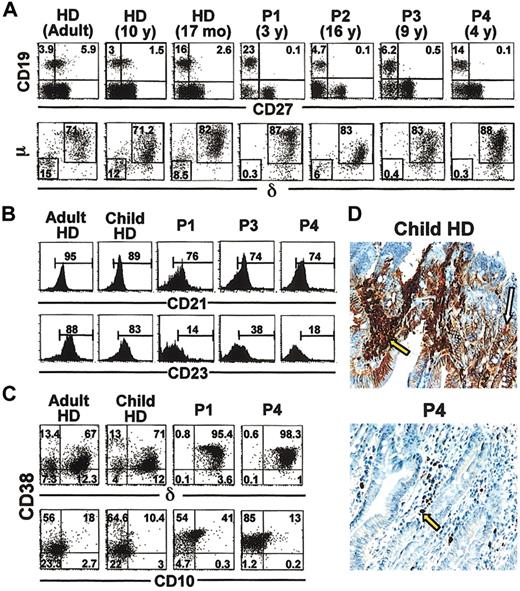
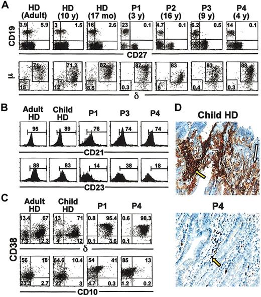
Flow cytometry analysis of CD19+ cells showed complete lack of the CD19+CD27+ memory B-cell compartment in ICF patients in contrast to controls, in which this population was present and increased with age, ranging from 14% of B cells in children to 60% in adults (Figure 1A, top). Similar patterns of IgM and IgD expression on B cells and absence of the class-switched B-cell population were observed in 3 of 4 patients, compared with controls (Figure 1A, bottom). Only P2 lacked IgMhiIgD+ cells and presented a small fraction of class-switched B cells, in accordance with the presence of serum IgG in this patient.
Absence of circulating memory B cells and gut plasma cells and accumulation of newly generated B cells in PB of ICF patients. PBMCs from 4 ICF patients (P1 to P4) and HDs were analyzed by flow cytometry (panels A-C). In panels A to C, representative dot plots and histograms are shown for 1 adult and 1 child HD of 5 adults and 5 children. (A) Four-color flow cytometry analysis of anti-CD19, anti-CD27, anti-δ, and anti-μ staining. Expression of CD19 and CD27 on PBLs and of surface μ and δ within the CD19+ gate are represented in the top and bottom subpanels, respectively. (B) PBMCs from P1, P3, P4, and HDs were stained with anti-CD19, anti-CD27, and anti-CD21 or anti-CD23 and analyzed within the CD19+CD27- gate. (C) PBMCs from P1, P4, and HDs were stained with anti-CD19, anti-CD38, anti-δ, and anti-CD10 and were then analyzed within the CD19+ gate. Numbers in quadrants (A,C) and brackets (B) indicate percentage of cells within the corresponding gate. (D) Jejunal sections from a healthy child and P4 were paraffin embedded and formalin fixed. IgA staining is shown in red, hematoxylin counterstaining in blue; magnification, × 250. IgA+ plasma cells (PCs) are indicated by yellow arrows; secretory IgA is indicated by a white arrow. Results are representative of P2, P3, P4, and 10 age-matched controls.
Absence of circulating memory B cells and gut plasma cells and accumulation of newly generated B cells in PB of ICF patients. PBMCs from 4 ICF patients (P1 to P4) and HDs were analyzed by flow cytometry (panels A-C). In panels A to C, representative dot plots and histograms are shown for 1 adult and 1 child HD of 5 adults and 5 children. (A) Four-color flow cytometry analysis of anti-CD19, anti-CD27, anti-δ, and anti-μ staining. Expression of CD19 and CD27 on PBLs and of surface μ and δ within the CD19+ gate are represented in the top and bottom subpanels, respectively. (B) PBMCs from P1, P3, P4, and HDs were stained with anti-CD19, anti-CD27, and anti-CD21 or anti-CD23 and analyzed within the CD19+CD27- gate. (C) PBMCs from P1, P4, and HDs were stained with anti-CD19, anti-CD38, anti-δ, and anti-CD10 and were then analyzed within the CD19+ gate. Numbers in quadrants (A,C) and brackets (B) indicate percentage of cells within the corresponding gate. (D) Jejunal sections from a healthy child and P4 were paraffin embedded and formalin fixed. IgA staining is shown in red, hematoxylin counterstaining in blue; magnification, × 250. IgA+ plasma cells (PCs) are indicated by yellow arrows; secretory IgA is indicated by a white arrow. Results are representative of P2, P3, P4, and 10 age-matched controls.
A detailed analysis of naive CD19+CD27- B-cell subsets was undertaken in terms of their CD21 and CD23 levels. As shown in Figure 1B, most naive B cells from healthy donors present a mature CD21+CD23+ phenotype, whereas B cells from 3 ICF patients tested (P1, P3, and P4) show decreased CD21 expression and a greatly reduced proportion of CD23+ cells. This phenotype suggests that ICF B cells are immature. Analysis of CD38 and IgD expression in B cells from 2 patients tested (P1 and P4) confirmed the absence of switched IgD-CD38+ memory B cells and revealed high levels of CD38 expression in the IgD+ cells of patients compared with controls (Figure 1C). Moreover, an increased number of CD38+ cells expressing CD10 was observed in patients. We confirmed the stability of the CD38hiCD10+ population for P4 after 6 months. CD10 expression on ICF naive B cells is reminiscent of the phenotype of the bone marrow CD10+ B cells that emerge in the periphery 7 weeks after bone marrow transplantation.33 Furthermore, in addition to the IgD+CD10- population of recirculating mature B cells,34 newly generated IgD+CD10+ cells are also observed in normal adult bone marrow (our observations).
To determine whether ICF B cells were capable of generating plasma cells (PCs) in vivo, we looked for IgA-secreting PCs in gut sections from patients and healthy children. In P2, P3, and P4, very few or no IgA+ PCs (yellow arrows) were detected in the lamina propria, as shown in Figure 1D for P4 and a healthy donor. Further, in contrast to healthy donors, no diffuse staining of secretory IgA (white arrow) was observed in patients. In all cases, IgG+ and IgM+ PCs were only weakly detectable, whatever the gut section tested (data not shown).
Overall, ICF patients' PB contains only naive B cells presenting an immature phenotype, with an accumulation of bone marrow B-cell emigrants and a lack of memory B and plasma cells. These data indicate a terminal B-cell differentiation blockage in ICF patients.
ICF B cells respond to BCR stimulation
Since arrest in terminal differentiation of ICF B cells may be due to defective BCR signaling, we investigated the ability of these cells to up-regulate activation markers in response to BCR stimulation in vitro. Induction of the early activation marker CD69 and the costimulatory molecule CD86 was assessed on CD19+ cells from P1, P3, P4, and healthy donors following in vitro stimulation. At time zero, a small proportion of B cells from patients were CD86+ (11.4% ± 3.1%), and a variable proportion (reaching up to 70% in P3) were CD69+ (Figure 2 and data not shown). These markers were strongly up-regulated in roughly all ICF B cells after 20 hours' stimulation with anti-μ, cytokines (IL2 plus IL10), and irradiated CD40L-transfected fibroblasts (Figure 2). Activation induced lymphoblast differentiation as shown by forward scatter shift (data not shown). ICF B cells seem to respond more robustly than controls, especially in terms of CD86 up-regulation, since after 20 hours' stimulation, the mean fluorescence was 10-fold higher in patient cells than in control cells. Since CD27 and its ligand CD70 have been proposed to be key elements of terminal B-cell differentiation into antibody-secreting cells,35 we also examined CD27 expression in ICF B cells upon activation. Indeed, up to 65% of ICF B cells express CD27 at the cell surface after 20 hours (Figure 2). These results were also observed with the use of cytokines and either anti-μ or CD40L stimulation exclusively, although combination of both stimuli leads to enhancement of the response (data not shown).
Response of ICF B cells to BCR stimulation and CD40 cross-linking. Flow cytometry analysis of PBMCs from a healthy child (HD) and patient P1 stained with anti-CD19, anti-CD69, anti-CD86, and anti-CD27, gated on CD19+ cells at time zero or after 20 hours' stimulation. Numbers in brackets indicate the percentage of strained cells within the CD19+ cells. Cells were stimulated by coculture with irradiated hCD40L-transfected fibroblasts in the presence of F(ab) antihuman μ, rIL-2, and rIL-10. P1 data are representative of P1, P3, and P4; HD data are representative of 3 healthy children.
Response of ICF B cells to BCR stimulation and CD40 cross-linking. Flow cytometry analysis of PBMCs from a healthy child (HD) and patient P1 stained with anti-CD19, anti-CD69, anti-CD86, and anti-CD27, gated on CD19+ cells at time zero or after 20 hours' stimulation. Numbers in brackets indicate the percentage of strained cells within the CD19+ cells. Cells were stimulated by coculture with irradiated hCD40L-transfected fibroblasts in the presence of F(ab) antihuman μ, rIL-2, and rIL-10. P1 data are representative of P1, P3, and P4; HD data are representative of 3 healthy children.
These data demonstrate that ICF B cells are able to integrate maturation signals through the BCR and CD40 in terms of up-regulation of activation markers and differentiation into lymphoblasts.
ICF B cells are able to switch and secrete immunoglobulins in vitro
We further tested the ICF B cells' ability to undergo class-switch recombination and to secrete immunoglobulins in vitro. Enriched naive B cells from ICF patients and from healthy donors (Figure 3A) were cocultured with irradiated CD40L-transfected fibroblasts in the presence of IL-4. Comparable levels of total immunoglobulin were secreted in culture supernatants of ICF and control cells after 10 days' stimulation, although a variation among patients in IgM and IgG secretion was found (Figure 3B, bottom). Consistent with serum IgG titer, VH-CHγ transcripts were detected in P2 B cells before stimulation. After stimulation, VH-CHγ transcripts were induced in all patients, confirming that IgGs in culture supernatants result from in vitro-induced class switch (Figure 3B, top).
ICF B-cells switch and secrete immunoglobulins after in vitro CD40L and IL-4 stimulation. (A) Enrichment of naive B cells from P1 and an HD. Naive CD19+CD27- B cells were enriched up to 95% by means of the StemSep B-cell enrichment system, and controls were depleted of memory B cells with the use of CD27 microbeads on a MiniMACS magnetic sorter. Results are representative of all patients and HDs. (B) (C) (D) Enriched naive B cells of patients and 4 HDs were stimulated in vitro with irradiated CD40L-transfected fibroblasts and rIL-4. Panel B (top) shows RT-PCR on VH-CH1γ and Igβ transcripts at time zero or after 10 days' stimulation. Bottom histogram in panel B shows total immunoglobulin production after 10 days' stimulation expressed in nanograms per milliliter. ▦ represents IgM fraction; ▪, IgG fraction. Panel C shows Iϵ-CH1ϵ and HPRT transcripts amplified by PCR from decreasing cDNA amounts (3, 2 and 1 μL) from patients and HDs. Panel D shows PCR amplification of AID, VH-CH1ϵ, and HPRT transcripts with the use of 3 μL cDNA. Histograms in panels C and D show transcription rate values normalized to HPRT values after quantification by means of MacBAS software (Fuji Film, Tokyo, Japan). Ratios are expressed on an arbitrary unit. Bars indicate SD. NS indicates unstimulated.
ICF B-cells switch and secrete immunoglobulins after in vitro CD40L and IL-4 stimulation. (A) Enrichment of naive B cells from P1 and an HD. Naive CD19+CD27- B cells were enriched up to 95% by means of the StemSep B-cell enrichment system, and controls were depleted of memory B cells with the use of CD27 microbeads on a MiniMACS magnetic sorter. Results are representative of all patients and HDs. (B) (C) (D) Enriched naive B cells of patients and 4 HDs were stimulated in vitro with irradiated CD40L-transfected fibroblasts and rIL-4. Panel B (top) shows RT-PCR on VH-CH1γ and Igβ transcripts at time zero or after 10 days' stimulation. Bottom histogram in panel B shows total immunoglobulin production after 10 days' stimulation expressed in nanograms per milliliter. ▦ represents IgM fraction; ▪, IgG fraction. Panel C shows Iϵ-CH1ϵ and HPRT transcripts amplified by PCR from decreasing cDNA amounts (3, 2 and 1 μL) from patients and HDs. Panel D shows PCR amplification of AID, VH-CH1ϵ, and HPRT transcripts with the use of 3 μL cDNA. Histograms in panels C and D show transcription rate values normalized to HPRT values after quantification by means of MacBAS software (Fuji Film, Tokyo, Japan). Ratios are expressed on an arbitrary unit. Bars indicate SD. NS indicates unstimulated.
The IgE switch in ICF B cells was monitored by induction of germ line Iϵ-Cϵ and functional VH-Cϵ transcripts after 5 days' stimulation. Semiquantitative RT-PCR performed on dilutions of cDNA revealed that cells from all patients analyzed expressed levels of germ line Iϵ-Cϵ transcripts that were comparable to those of control cells (Figure 3C). Nonetheless, normal levels of VH-Cϵ transcripts were observed only for P4 compared with controls, while P1 and P3 showed little or no transcript induction, respectively (Figure 3D). The deficient IgE switch in P1 and P3 is not due to differences in AID induction, since AID transcripts were induced equally in all patients (Figure 3D).
These data indicate that ICF B cells are competent for class-switch recombination and immunoglobulin secretion in vitro. Nevertheless, intrinsic B-cell defects concerning IgE switch induction may account for the lack of IgE production in some patients.
ICF B cells present increased apoptosis following in vitro activation
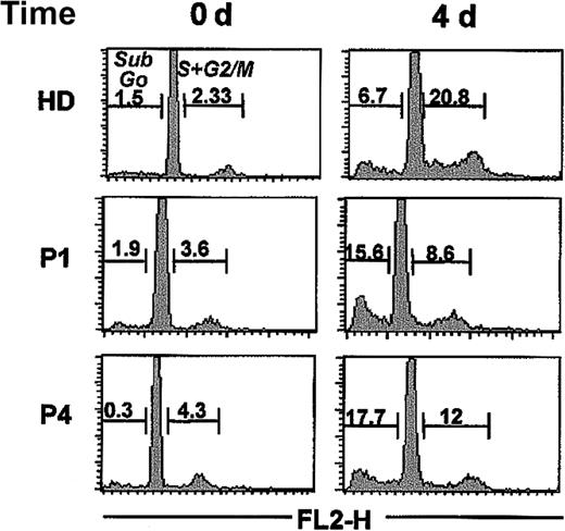
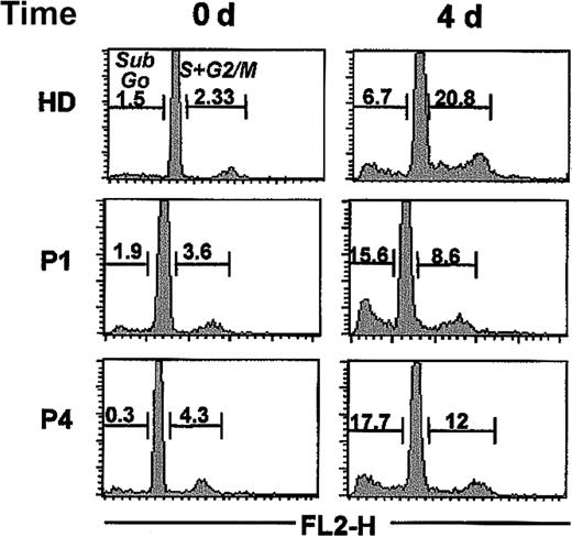
We tested the ability of ICF B cells to proliferate and their susceptibility to apoptosis, following in vitro stimulation. Cell-cycle analysis by propidium iodide staining on freshly isolated ICF B cells and enriched naive B cells from healthy children showed a predominant resting state (G0/G1 phase) of these cells before stimulation (Figure 4). After 2 days' stimulation in the presence of rshCD40L and IL4, cells entered the cell cycle (S and G2/M phases) at similar rates in ICF and control cells (data not shown). Nevertheless, after 4 days' stimulation (Figure 4), there were 2 to 3 times more apoptotic cells (sub-G0/G1 phase) in B cells from P1 and P4 (15.6% and 17.7%, respectively) than in naive B cells from controls (7.5% ± 1.98%). Moreover, after 4 days' stimulation, ICF B cells proliferated half as much (8.6% and 12% for P1 and P4, respectively) as normal naive B cells (20.06% ± 4.49%). When this experiment was repeated 6 months later for P4, similar proportions of mitotic (11%) and apoptotic (23.5%) cells were observed.
Increased ICF B-cell mortality after in vitro activation. Analysis of apoptotic and cycling cells from a healthy child (HD), P1, and P4 was done by propidium iodide staining on freshly isolated CD19+CD27- naive B cells (time 0) or after 4 days' stimulation with rshCD40L and IL-4. Numbers in histograms indicate the percentage of apoptotic (sub-G0) and cycling (S and G2/M) cells analyzed on the FL2 wavelength on a FACScan flow cytometer after cell aggregates were gated out. Data shown for HD are representative of 5 healthy children.
Increased ICF B-cell mortality after in vitro activation. Analysis of apoptotic and cycling cells from a healthy child (HD), P1, and P4 was done by propidium iodide staining on freshly isolated CD19+CD27- naive B cells (time 0) or after 4 days' stimulation with rshCD40L and IL-4. Numbers in histograms indicate the percentage of apoptotic (sub-G0) and cycling (S and G2/M) cells analyzed on the FL2 wavelength on a FACScan flow cytometer after cell aggregates were gated out. Data shown for HD are representative of 5 healthy children.
The high sensitivity of ICF B cells to apoptosis after in vitro stimulation might reflect an increased propensity of these cells to undergo apoptosis in vivo. Furthermore, increased mortality correlates with the immature phenotype of ICF B cells.
ICF B cells display unselected VH sequences
As normal immature B cells display a diverse VH repertoire and a broad range of VH CDR3 lengths,15,36 we analyzed the B-cell repertoire of ICF patients. VH1 and VH5 gene families were amplified, and 75 sequences from 3 ICF patients (P1, P2, and P4) were compared with 108 control sequences obtained from sorted CD19+IgD+CD27- naive B cells from 2 healthy children.
Although circulating ICF B cells do not express CD27, which is a marker of somatically mutated B cells,12 we screened ICF VH5-51-(D)-JH sequences for somatic hypermutations. We observed 0 to 2 nucleotide changes per sequence, outside CDRs, with a mutation frequency of 0.09% ± 0.02% (data not shown), which is comparable to the error rate of the Taq polymerase used for PCR amplification, confirming the naive status of ICF B cells.
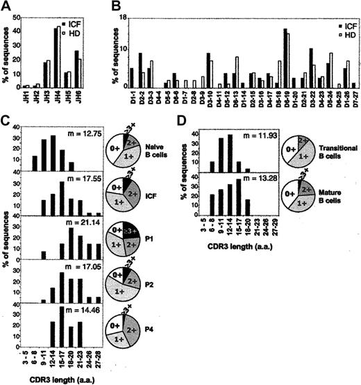
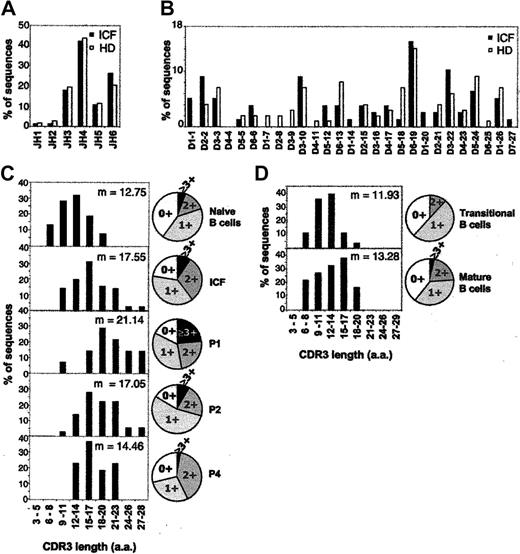
B cells from all patients displayed a normal JH and DH gene usage: preferential JH4, JH3, and JH6 gene usage and representation of DH segments all through the IgH locus (Figure 5A and 5B, respectively). However, the lengths of VH CDR3 sequences in ICF B cells (Figure 5C) show an overall pattern skewed to longer CDR3's (9 to 28 amino acids [aa]) than control sequences (6 to 20 amino acids). Long CDR3's do not result from extended N regions or from D-D fusions but rather from use of full-length DH gene segments (data not shown). The CDR3 length pattern of control naive B cells was confirmed when VH sequences were amplified with different VH1/5 primers33 and shown to be independent of the age (3, 11, or 33 years old) of the donor (E. Meffre, personal oral communication, 2003). Moreover, we observed an enrichment of positively charged residues in ICF CDR3's. Indeed, 76.6% of ICF CDR3's bear positive charges, of which 40% contain at least 2 positive charges, in contrast to CDR3 sequences from controls of which 60% bear positive charges and only 20% present at least 2 positive charges (Figure 5C, pie charts). The positive-charge content of CDR3 sequences is significantly different in patients and healthy donors, as shown by Student t test (P < .05).
Defective selection of VHDHJH CDR3's in ICF patients. VHDHJH of the VH1 and VH5 families from P1, P2, and P4 (17, 23, and 35 sequences, respectively) were compared with 108 sequences from sorted naive CD19+IgD+CD27- B cells from 2 healthy children. (A-B) Histograms show JH gene usage (panel A) and DH gene usage (panel B) for total ICF and control sequences. (C) Histograms show CDR3 length distribution for sequences from sorted naive CD19+IgD+CD27- B cells, total ICF cells, and P1, P2, and P4 B cells. (D) VH5-51-DHJH sequences from control CD19+CD27-CD21- transitional (26 sequences) and mature CD19+CD27-CD21+ (25 sequences) B cells were analyzed. CDR3 length was determined by counting amino acid residues between codon 94 and the conserved Trp in JH segments at position 102. Pie charts represent the corresponding distribution of positively charged residueswithin CDR3's for each sample; m indicates mean.
Defective selection of VHDHJH CDR3's in ICF patients. VHDHJH of the VH1 and VH5 families from P1, P2, and P4 (17, 23, and 35 sequences, respectively) were compared with 108 sequences from sorted naive CD19+IgD+CD27- B cells from 2 healthy children. (A-B) Histograms show JH gene usage (panel A) and DH gene usage (panel B) for total ICF and control sequences. (C) Histograms show CDR3 length distribution for sequences from sorted naive CD19+IgD+CD27- B cells, total ICF cells, and P1, P2, and P4 B cells. (D) VH5-51-DHJH sequences from control CD19+CD27-CD21- transitional (26 sequences) and mature CD19+CD27-CD21+ (25 sequences) B cells were analyzed. CDR3 length was determined by counting amino acid residues between codon 94 and the conserved Trp in JH segments at position 102. Pie charts represent the corresponding distribution of positively charged residueswithin CDR3's for each sample; m indicates mean.
All together, the diverse JH and DH gene usage indicates that V-D-J rearrangements proceed normally in ICF patients, while the presence of long and positively charged CDR3's suggests a defect of B-cell selection.
To determine if this defect takes place during the early steps of peripheral B-cell differentiation, we sorted normal transitional CD19+CD27-CD21- and mature CD19+CD27-CD21+ B cells according to the current murine classification8 and analyzed their VH5-51-(D)-JH sequences. As reported in Figure 5D, no significant differences (P > .05) were observed between these 2 subpopulations or with total naive B cells in terms of CDR3 length and number of sequences bearing positive charges: that is, CDR3 sequences are short and a reduced proportion of them bear at least 2 positive charges. These results indicate that CDR3 selection in healthy individuals occurs before the transitional B-cell stage and that the B-cell selection defect in ICF patients may occur before emigration of B cells from the bone marrow.
Discussion
Patients with ICF syndrome are very rare (around 35 cases reported worldwide). Strikingly, although one of the main clinical features of the disease is hypogammaglobulinemia or agammaglobulinemia, associated in some cases with defective cell-mediated immunity, most studies performed to date concern genetic defects related to hypomethylation. To investigate the B-cell defects associated with this syndrome, we performed a complete phenotypic and functional analysis of primary B cells from 4 ICF patients with severe agammaglobulinemia.
All patients presented normal numbers of B lymphocytes in PB, containing only naive CD19+CD27- but no memory B cells or plasma cells in the gut (Figure 1). In mice, the naive B-cell population is composed of T1, T2, and mature B cells. T1 cells emigrate from the bone marrow to the spleen, where they develop into T2 and mature B cells as they acquire CD21 and CD23.7-10 In humans, we have analyzed the expression of CD21 and CD23 within the naive B-cell population of healthy donors, providing the first analysis of human transitional B cells. Healthy adult and child PB CD19+CD27- cells contain a majority of CD21+CD23+ mature B cells (more than 80%), whereas low numbers of CD21-CD23- T1 cells are detectable (fewer than 20%). By contrast, in ICF patients, although a majority of B cells express low to normal levels of CD21, CD23 expression is severely decreased (Figure 1B), indicating that the generation of mature B cells in the periphery is impaired. In addition, CD38hiIgD+CD10+ B cells accumulate in 2 patients tested (Figure 1C). On the basis of phenotypic similarities with a tonsillar B-cell subpopulation, these cells were proposed to be recirculating germinal center (GC) founders.37 Tonsillar GC founders express CD27 and present somatic hypermutations in their CDRs, although to a lesser extent than class-switched GC B cells.37,38 As ICF B cells and the so-called recirculating GC founders are CD27- and their VH sequences are devoid of somatic hypermutations, we consider that CD38hiIgD+CD10+ B cells do not correspond to GC founder cells but to newly generated bone marrow emigrants. Supporting this statement, CD19+IgD+CD38hiCD10+ cells are present in normal human bone marrow (our observations), and CD10+ B cells have been observed to emerge in PB following bone marrow transplantation.33
When VH-DH-JH rearrangements of ICF B cells were compared with those of normal naive B cells, we did not observe any bias in the JH and DH gene segment usage (Figure 5). However, VH CDR3 sequences from ICF patients are long (mean, 17.6 aa), and although they come from PB, their length pattern is similar to those of normal bone marrow pro-B, pre-B, and early immature B cells (mean, 16.5, 15.6, and 16.1 aa, respectively) and not to that of peripheral naive B cells (mean, 13.5 aa).14,15 Moreover, ICF CDR3's contain a higher proportion of positively charged amino acids than control naive B cells (Figure 5C). Both long length and positive charges in VH CDR3's have been associated with poly- and self-reactivity in normal and pathological circumstances,15,39-43 and in consequence, such CDR3's are counterselected in the normal mature B-cell pool.14,15,36,44 Therefore, it appears that deletion of potentially autoreactive CDR3's preceding the mature B-cell stage has not occurred in ICF patients. Clonal deletion happens at the immature B-cell stage in normal bone marrow, although peripheral T1 cells have also been proposed as a target for negative selection in mouse spleen.10,45 As VH CDR3 sequences of sorted PB transitional and mature B cells from healthy children display short CDR3's (mean, 11.9 and 13.3 aa, respectively) with a restricted content of positive charges (Figure 5D), the failure of clonal deletion in ICF B cells is likely to occur in the bone marrow. This could be related to a defect affecting either ICF B cells themselves or other components of the bone marrow microenvironment.46
Self-reactive clones generated in the bone marrow are dealt with by deletion, anergy, or receptor editing.2-5 However, a fraction of autoreactive clones may emigrate to the periphery, in which case they are anergized, blocked in their maturation, and excluded from splenic follicles, preventing autoantibody production.47 In the case of ICF patients, PB B cells are enriched with potentially self-reactive cells owing to the absence of negative selection. Nevertheless, they do not develop autoimmunity, suggesting that autoreactive ICF B cells are silenced in vivo. In fact, ICF B cells present characteristics of anergic B cells. First of all, peripheral differentiation of immature B cells is blocked in patients as is the case in murine models of anergy in which B cells express antinuclear and anti-dsDNA BCRs.47,48 Second, a fraction of ICF B cells exhibit activation markers such as CD69 and CD86, suggesting that these cells have encountered antigen in vivo (Figure 2). Third, ICF B cells show reduced proliferation and increased apoptosis following activation in vitro (Figure 4), as reported for murine anergic B cells.5 Nevertheless, in contrast to anergic B cells,49 ICF B cells can undergo class-switch recombination and secrete immunoglobulin after in vitro stimulation by the BCR and by CD40 in the presence of cytokines. Furthermore, ICF B cells strongly up-regulate CD86 costimulatory molecule (Figure 2), unlike murine anergic B cells.50 Finally, since in transgenic models of anergy the pool of B cells bearing endogenous nonautoimmune receptors is able to re-establish serum antibody titers, silencing of self-reactive clones seems not sufficient to explain the complete agammaglobulinemia in ICF patients.
Cytogenetics studies on ICF syndrome have revealed marked DNA hypomethylation affecting classical satellites and silencing of genes on the inactive X and Y chromosomes.18,19,51 These anomalies are related in 65% of ICF cases reported to mutations in the DNMT3B gene encoding the de novo Dnmt3b methyltransferase.20-22,52 Although the link between Dnmt3b and immune deficiency remains obscure, methylation has been shown to play an essential role in key processes of B-cell differentiation such as V(D)J, allelic exclusion and class-switch recombination.53-55 Moreover, DNMT3B transcripts are expressed at low levels in all hematopoietic cells and up-regulated 2-fold in activated T cells,56 suggesting that ICF hypomethylation may concern other cell lineages than B lymphocytes, affecting them indirectly. In ICF patients from our cohort, T cells are present at normal levels, up-regulate CD40L and CD69 upon PMA and ionomycin (data not shown), and proliferate normally in response to mitogens but poorly to antigens (Table 1). This deficient T-cell proliferation to antigen opens the possibility that hypomethylation also affects T-lymphocyte function, compromising T-cell help. Therefore, combined immune deficiency could be responsible for agammaglobulinemia in ICF patients.
The fact that mutations in the DNMT3B gene account for only a fraction of ICF cases20-22 (2 of 4 in our cohort) shows that the genetic origin of the disease is heterogeneous. In our study, although ICF B cells from all patients have a homogeneous naive phenotype and a similar response to in vitro stimulation, only B cells from P4, who does not present mutations in DNMT3B, achieve IgE class switch in vitro (Figure 3). Even though other gene mutations associated with ICF syndrome have not been identified so far, our data suggest that diverse genetic origins and hypomethylation profiles19 of the disease might lead to variations in immune deficiency.
To better understand the link between the hypomethylation, negative selection breakdown, and agammaglobulinemia found in ICF patients, it is necessary to identify gene expression alterations resulting from methylation defects. Recent studies have attempted to identify Dnmt3b target genes by microarray technology.57 They show that Epstein-Barr virus (EBV)-transformed cell lines derived from ICF patients express low levels of IgG, IgA, and CD27 mRNAs and present a dysregulation of genes involved in migration, proliferation, and survival of B cells. Since our work demonstrates that ICF B-cell differentiation is blocked at the transitional B-cell stage, the data from Ehrlich et al57 might reflect immaturity rather than an alteration of gene expression in ICF B cells. The study of gene expression profiles of hematopoietic and bone marrow stromal cell populations will be necessary to assess the contribution of each cell compartment to the pathology. Development of an ICF murine model will be of great help for this purpose.
Prepublished online as Blood First Edition Paper, November 26, 2003; DOI 10.1182/blood-2003-08-2632.
Supported by CNRS; INSERM; the Ministère de la Recherche et de l'Education Nationale (C.B.-B.); the Association pour la Recherche contre le Cancer (C.B.-B.); and the Fondation pour la Recherche Medicale (Y.L.J.).
The publication costs of this article were defrayed in part by page charge payment. Therefore, and solely to indicate this fact, this article is hereby marked “advertisement” in accordance with 18 U.S.C. section 1734.
We thank E. Termine and P. Marino for excellent assistance in cloning and sequence analysis and M. Chrestian for immunohistochemistry. We are grateful to E. Meffre for helpful discussion and sharing normal VH sequences; to F. Le Deist and C. Farnarier for immunologic characterization of patients at diagnosis; to F. Brière for providing hCD40L ltk fibroblasts; to P. Rihet and B. Piquéras for help with ELISA optimization; and to J. Ewbank for critical reading of the manuscript.

