Abstract
Ex vivo expansion of hematopoietic stem cells (HSCs) is important for many clinical applications, and knowledge of the surface phenotype of ex vivo–expanded HSCs will be critical to their purification and analysis. Here, we developed a simple culture system for bone marrow (BM) HSCs using low levels of stem cell factor (SCF), thrombopoietin (TPO), insulin-like growth factor 2 (IGF-2), and fibroblast growth factor-1 (FGF-1) in serum-free medium. As measured by competitive repopulation analyses, there was a more than 20-fold increase in numbers of long-term (LT)–HSCs after a 10-day culture of total BM cells. Culture of BM “side population” (SP) cells, a highly enriched stem cell population, for 10 days resulted in an approximate 8-fold expansion of repopulating HSCs. Similar to freshly isolated HSCs, repopulating HSCs after culture were positive for the stem cell markers Sca-1, Kit, and CD31 and receptors for IGF-2. Surprisingly, prion protein and Tie-2, which are present on freshly isolated HSCs, were not on cultured HSCs. Two other HSC markers, Endoglin and Mpl, were expressed only on a portion of cultured HSCs. Therefore, the surface phenotype of ex vivo–expanded HSCs is different from that of freshly isolated HSCs, but this plasticity of surface phenotype does not significantly alter their repopulation capability.
Introduction
Hematopoietic stem cells (HSCs) are a rare population in adult bone marrow (BM) and give rise to all lymphoid, myeloid, and erythroid cells.1 Their purification and study are difficult, because only a few proteins are known to be expressed on the surface of HSCs. These are also expressed on other types of BM cells, and not all are conserved between species or during development.1-3 Among the surface proteins present on freshly isolated mouse BM HSCs are Sca-1, Kit, Mpl, CD38, Endoglin, Tie-2, CD31,1,4-9 and prion protein (PrP) (C.C.Z., A. Steele, S. Lindquist, and H.F.L., manuscript in preparation). HSCs also have insulin-like growth factor 2 (IGF-2) receptors that bind to a fusion protein of IGF-2 and a human immunoglobulin G1 (IgG1) Fc fragment.10 Mouse HSCs lack CD344 as well as multiple lineage-specific markers that are characteristically grouped together as “Lin.”
Difficulties in ex vivo expansion of HSCs have greatly hampered their clinical utility as well as studies of their biologic properties.11 Numerous attempts have been made to increase the number of long-term (LT)–HSCs in culture.12,13 The use of stromal cell lines or combinations of cytokines have resulted in significant self-renewal of HSCs assayed 4 to 6 weeks after transplantation and led up to 6-fold expansion of murine LT-HSC activity.14-18 The introduction of exogenous transcription factors can dramatically expand HSCs,19-22 although this approach may have undesirable outcomes for recipients in the clinical setting.
In part because of the lack of an efficient ex vivo culture system for LT-HSCs, there has been no systematic study of HSC markers on cultured cells. It is not clear whether cultured HSCs share the same panel of surface markers as freshly isolated HSCs, although this is often assumed to be the case. For instance, markers characteristic of freshly isolated HSC populations, such as Lin-Sca-1+Kit+, have been used as a measure of the numbers of cultured murine HSCs. However, expansion in culture of cells expressing this surface phenotype does not correlate with increases in numbers of transplantable HSCs.19 Similarly, while the severe combined immunodeficiency (SCID)–repopulating activity of human umbilical cord blood cells is enriched in the CD34+CD38- fraction, there is a dissociation of this phenotype with the repopulating activity of human cord blood cells after ex vivo culture.23
Recently, we identified 2 surface proteins characteristic of freshly isolated BM HSCs: Endoglin and the prion protein PrP7 (C.C.Z., A. Steele, S. Lindquist, and H.F.L., manuscript in preparation). In the process of showing that IGF-2 is a factor that stimulates ex vivo expansion of murine HSCs, we demonstrated that transplantable stem cells bind to a fusion protein of IGF-2 and a human IgG1 Fc fragment (IGF2-hFc).10 Our current goal is to carry out a thorough investigation of the relationship between expression of marker surface proteins and repopulating HSC activity after culture. In particular, we need to determine whether the surface phenotype of cultured HSCs is different from that of freshly isolated HSCs. Knowledge of the surface phenotype of cultured HSCs would enable us to establish a surrogate flow cytometry–based assay for these cells and then optimize their expansion in culture.
Here, we describe a simple ex vivo culture system that can dramatically expand repopulating HSCs. Following a 10-day culture of either total BM cells or purified BM “side population” (SP) cells in serum-free medium supplemented with low concentrations of stem cell factor (SCF), thrombopoietin (TPO), IGF-2, and fibroblast growth factor (FGF-1), there was a more than 8-fold increase in the numbers of repopulating HSCs. This system made it possible to show that the phenotype of cultured HSCs is different from that of freshly isolated HSCs. While repopulating HSCs after culture expressed several markers characteristic of freshly isolated mouse HSCs, including Sca-1, Kit, CD31, and IGF-2 receptors, they did not express others, including PrP and Tie-2, as well as surface proteins prominin-1 and CD62L. This culture system and the definition of the surface phenotype of ex vivo–expanded HSCs will be valuable for ex vivo expansion, genetic modification, analysis and purification of HSCs, as well as for developing new in vitro screening assays for factors that regulate HSC proliferation and differentiation.
Materials and methods
Animals
C57BL/6 CD45.2 and CD45.1 mice were purchased from the Jackson Laboratory (Bar Harbor, ME) or the National Cancer Institute (Frederick, MD) and were maintained at the Whitehead Institute animal facility.
Cell culture
Total BM cells isolated from 6- to 9-week-old C57BL/6 CD45.2 mice were plated at a density of 106/mL in StemSpan serum-free medium (StemCell Technologies, Vancouver, BC) supplemented with 10 μg/mL heparin (Sigma, St Louis, MO), 10 ng/mL mouse SCF, 20 ng/mL mouse TPO, 20 ng/mL mouse IGF-2 (all from R&D Systems, Minneapolis, MN), and 10 ng/mL human FGF-1 (Invitrogen, Carlsbad, CA). Half of the medium was replaced twice per week with fresh medium, and the total volume of medium for 106 input cells was increased to 1.5 mL and 6 mL at days 4 and 7, respectively. BM SP cells (25) were cultured in 50 μL of the above-mentioned medium in 1 well of a U-bottom 96-well plate (Corning 3799; Corning, Corning, NY) for 7 days. The cells were then transferred to 0.5 mL medium in 1 well of a 24-well plate and cultured for 3 days.
Flow cytometry
Lin-Sca-1+kit+ cells and SP cells were stained as described.10 For Endoglin detection, staining by anti-Endoglin monoclonal antibody (mAb) (BD Pharmingen, San Diego, CA) followed by anti–rat-PE/CY5.5 (phycoerythrin/cyanine 5.5) (eBioscience, San Diego, CA) was performed before other staining. Lin- cells were stained with a biotinylated Lin+ antibody cocktail of anti-CD5, anti-B220, anti–Gr-1, and anti-Ter119 (StemCell Technologies), followed by streptavidin-allophycocyanin (APC). Anti-PrP mAb (SAF-83; Cayman Chemical, Ann Arbor, MI) was fluorescein isothiocyanate (FITC)–conjugated using the Quick-Tag FITC conjugation kit (Roche, Indianapolis, IN). Lin-Sca-1+Prp+/- cells were stained with a biotinylated Lin+ antibody cocktail, followed by streptavidin-APC, anti-PrP–FITC, and anti–Sca-1–PE. Polyclonal rabbit anti-Mpl was kindly provided by Dr Wei Tong of the Lodish Lab at Whitehead and anti-rabbit–PE was used as secondary antibody. Anti–Tie-2–PE and anti–prominin-1–PE were purchased from eBioscience. Unless otherwise mentioned, antibodies were from BD Pharmingen. Reconstitution analysis was carried out as described.10 Essentially, peripheral blood cells were collected by retroorbital bleeding, followed by lysis of red blood cells, and staining with anti-CD45.2–FITC, anti-CD45.1–PE, anti-Thy1.2–PE, anti-B220–PE, anti–Mac-1–PE, anti–Gr-1–PE, or anti-Ter119–PE monoclonal antibodies (BD Pharmingen). Flow cytometry analyses were performed on a FACSCalibur instrument (BD Biosciences, San Jose, CA).
Competitive reconstitution analysis
The reconstitution protocol was essentially as described previously.10 Briefly, the indicated numbers of CD45.2 donor cells were injected after mixing with 1 × 10⁵ or 2 × 10⁵ (as indicated) freshly isolated CD45.1 competitor BM cells, intravenously into a group of 6- to 9-week-old CD45.1 mice that had been irradiated with a total dose of 10 Gy. To measure reconstitution of mice that received transplants, peripheral blood was collected by retroorbital bleeding at the indicated times after transplantation. The presence of CD45.1+ and CD45.2+ cells in the lymphoid and myeloid compartments were measured. The calculation of competitive repopulating units (CRUs) in limiting dilution experiments was conducted as described,10 using L-Calc software (StemCell Technologies).
Results
Ex vivo expansion of HSCs of cultured total BM cells
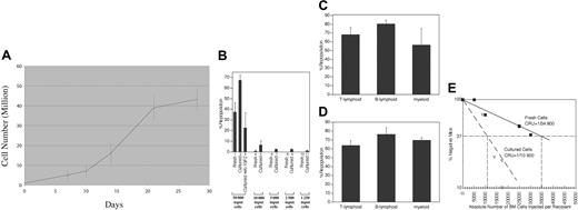
Previously, we identified an HSC supportive cell population, fetal liver CD3+Ter119- cells, and demonstrated that IGF-2 is a factor produced by these cells that binds to and stimulates LT-HSC proliferation in culture.10 Culture of fetal liver Lin-Sca-1+Kit+ cells or BM SP cells in serum-containing medium with 500 ng/mL IGF-2, 50 ng/mL SCF, and 100 ng/mL TPO stimulated a 2- to 3-fold expansion of LT-HSCs.10 To improve the efficiency of cultured HSC expansion we tested the effects of IGF-2 at different concentrations, together with different cytokine combinations and concentrations, serum contents, cell densities, and culture durations. In this way we developed a simple culture system that dramatically expands BM HSCs. Total mouse BM cells (Figure 1) or BM SP cells (Figure 2) were cultured in serum-free medium containing 10 ng/mL SCF, 20 ng/mL TPO, 20 ng/mL IGF-2, and 10 ng/mL FGF-1. All of these cytokines have been shown previously to bind to HSCs,10,24-28 but to our knowledge they have never been combined together for culturing hematopoietic cells. In Figure 1, we seeded 1 × 10⁶ total BM cells/mL medium. After 10 days of culture, the number of total cells increased about 7-fold to 7.2 ± 2.8 × 10⁶ (Figure 1A). The cultured cells contain mostly suspension cells with a minor adherent subpopulation.
We performed competitive repopulation assays to test whether ex vivo–expanded cells were capable of engraftment. Various numbers of fresh and cultured CD45.2 BM cells were mixed with 105 fresh CD45.1 BM competitors and injected into lethally irradiated CD45.1 recipients. A representative result from 3 independent experiments is shown in Figure 1B. When 3.5 × 10⁵ cultured cells (the progeny of 5 × 10⁴ initially plated CD45.2 cells) were transplanted, an average hematopoietic chimerism of 67.2% was observed 4 months after transplantation (lane 2). This is much higher than the 37.5% engraftment shown by the equivalent 5 × 10⁴ uncultured cells (lane 1). Importantly, this expansion of HSCs was dependent on the presence of IGF-2 in the culture medium, as the progeny of the same 5 × 10⁴ cells cultured in the absence of IGF-2 but otherwise in the same medium had a significantly lower engraftment level of 22.6% (lane 3, P < .05, Student t test).
To avoid donor HSC saturation, we also transplanted fewer cultured cells and compared their engraftment with equivalent numbers of freshly isolated cells. Thus 1 × 10⁴, 5000, 2500, or 1250 freshly isolated cells, after 10 days of culture, yielded 7 × 10⁴, 3.5 × 10⁴, 1.8 × 10⁴, and 9000 cells, respectively. While 1 × 10⁴, 5000, 2500, or 1250 freshly isolated BM cells showed an average of 0.6%, 0%, 0%, and 0% engraftment, respectively (lanes 4, 6, 8, 10), their cultured progeny repopulated 6.5%, 2.3%, 2.3%, and 1.5% of the recipients, respectively (lanes 5, 7, 9, 11). It is noteworthy that the progeny of 1250 input cells had a higher average level of engraftment than that resulting from 1 × 10⁴ input cells, suggesting a dramatic increase of stem cell activity during culture.
A novel culture system of total BM cells dramatically expands HSCs. (A) Total BM cells (106) were initiated in serum-free medium with SCF, TPO, IGF-2, and FGF-1 as described in “Materials and methods,” and total cell numbers were counted at days 7, 10, 14, 21, and 28. Results from 3 independent cultures were plotted. (B) Comparison of the long-term repopulation potential of 10-day cultured and freshly isolated BM cells. We mixed 5 × 10⁴, 1 × 10⁴, 5000, 2500, or 1250 freshly isolated CD45.2 BM cells or 3.5 × 10⁵, 7 × 10⁴, 3.5 × 10⁴, 1.75 × 10⁴, or 9000 10-day cultured BM cells (the product of 5 × 10⁴, 1 × 10⁴, 5000, 2500, or 1250 initially plated CD45.2 cells, respectively) with 105 CD45.1 competitor BM cells and transplanted them into lethally irradiated recipients (n = 6 mice). In 1 case IGF-2 was not added, but the remainder of the culture conditions was unchanged. Peripheral blood cells were analyzed for the presence of CD45.2+ cells at 4 months after transplantation. Three independent experiments were performed that gave similar results. (C) Multilineage contribution of 3.5 × 10⁵ cultured cells (derived from 5 × 10⁴ input cells) at 4 months after transplantation (n = 6). (D) Multilineage contribution of the 3.5 × 10⁵ cultured cells (equivalent to 5 × 10⁴ input cells) at 4 months after transplantation of mice receiving a secondary transplant (n = 4). (E) Limiting dilution analysis of the repopulating ability of total BM cells before and after culture. Irradiated CD45.1 congenic mice were injected with 105 CD45.1 BM competitor cells and the indicated numbers of freshly isolated CD45.2 BM cells (▪ and —) or their progeny after 10 days of culture in serum-free medium with SCF, TPO, IGF-2, and FGF-1 (▿ and —). Plotted is the percentage of recipient mice containing less than 1% CD45.2 lymphoid and myeloid subpopulations in nucleated peripheral blood cells 4 months after transplantation versus the number of injected cells. The curve was anchored by the 0 cells/100% negative mice point. Error bars indicate SEM.
A novel culture system of total BM cells dramatically expands HSCs. (A) Total BM cells (106) were initiated in serum-free medium with SCF, TPO, IGF-2, and FGF-1 as described in “Materials and methods,” and total cell numbers were counted at days 7, 10, 14, 21, and 28. Results from 3 independent cultures were plotted. (B) Comparison of the long-term repopulation potential of 10-day cultured and freshly isolated BM cells. We mixed 5 × 10⁴, 1 × 10⁴, 5000, 2500, or 1250 freshly isolated CD45.2 BM cells or 3.5 × 10⁵, 7 × 10⁴, 3.5 × 10⁴, 1.75 × 10⁴, or 9000 10-day cultured BM cells (the product of 5 × 10⁴, 1 × 10⁴, 5000, 2500, or 1250 initially plated CD45.2 cells, respectively) with 105 CD45.1 competitor BM cells and transplanted them into lethally irradiated recipients (n = 6 mice). In 1 case IGF-2 was not added, but the remainder of the culture conditions was unchanged. Peripheral blood cells were analyzed for the presence of CD45.2+ cells at 4 months after transplantation. Three independent experiments were performed that gave similar results. (C) Multilineage contribution of 3.5 × 10⁵ cultured cells (derived from 5 × 10⁴ input cells) at 4 months after transplantation (n = 6). (D) Multilineage contribution of the 3.5 × 10⁵ cultured cells (equivalent to 5 × 10⁴ input cells) at 4 months after transplantation of mice receiving a secondary transplant (n = 4). (E) Limiting dilution analysis of the repopulating ability of total BM cells before and after culture. Irradiated CD45.1 congenic mice were injected with 105 CD45.1 BM competitor cells and the indicated numbers of freshly isolated CD45.2 BM cells (▪ and —) or their progeny after 10 days of culture in serum-free medium with SCF, TPO, IGF-2, and FGF-1 (▿ and —). Plotted is the percentage of recipient mice containing less than 1% CD45.2 lymphoid and myeloid subpopulations in nucleated peripheral blood cells 4 months after transplantation versus the number of injected cells. The curve was anchored by the 0 cells/100% negative mice point. Error bars indicate SEM.
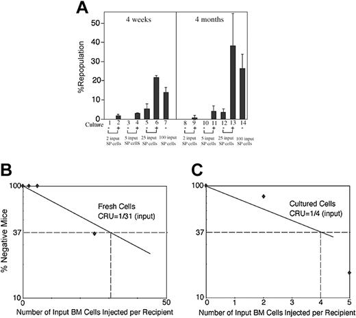
Culture dramatically increases in vivo repopulating stem-cell activity of BM SP cells. (A) Freshly isolated adult CD45.2 BM SP cells (2, 5, 25, or 100) or their progenies after 10 days of culture were transplanted (together with 1 × 10⁵ CD45.1 competitor BM cells per mouse, n = 5-8) into CD45.1 congenic mice. Peripheral blood engraftments at 4 weeks and 4 months after transplantation are shown. Error bars indicate SEM. (B) Limiting dilution analysis of the repopulating ability of BM SP cells before culture. Irradiated CD45.1 congenic mice were injected with 105 CD45.1 BM competitor cells and 2 (n = 7 mice), 5 (n = 6), 25 (n = 8), or 100 (n = 5) freshly isolated CD45.2 BM SP cells. Similar to Figure 1E, plotted is the percentage of recipient mice containing less than 1% CD45.2 lymphoid and myeloid subpopulations in nucleated peripheral blood cells 4 months after transplantation versus the number of injected cells. Input SP cells (100) resulted in 0% of negative mice, and this data point is not plotted. (C) Limiting dilution analysis of the repopulating ability of BM SP cells after culture. The same assay as used in panel B was carried out except the progenies of the input 2 (n = 5), 5 (n = 6), 25 (n = 4), or 100 SP cells (n = 5) after 10 days of culture were injected. The cultured progeny of 25 or 100 input SP cells resulted in 0% of negative mice, and the data points are not plotted.
Culture dramatically increases in vivo repopulating stem-cell activity of BM SP cells. (A) Freshly isolated adult CD45.2 BM SP cells (2, 5, 25, or 100) or their progenies after 10 days of culture were transplanted (together with 1 × 10⁵ CD45.1 competitor BM cells per mouse, n = 5-8) into CD45.1 congenic mice. Peripheral blood engraftments at 4 weeks and 4 months after transplantation are shown. Error bars indicate SEM. (B) Limiting dilution analysis of the repopulating ability of BM SP cells before culture. Irradiated CD45.1 congenic mice were injected with 105 CD45.1 BM competitor cells and 2 (n = 7 mice), 5 (n = 6), 25 (n = 8), or 100 (n = 5) freshly isolated CD45.2 BM SP cells. Similar to Figure 1E, plotted is the percentage of recipient mice containing less than 1% CD45.2 lymphoid and myeloid subpopulations in nucleated peripheral blood cells 4 months after transplantation versus the number of injected cells. Input SP cells (100) resulted in 0% of negative mice, and this data point is not plotted. (C) Limiting dilution analysis of the repopulating ability of BM SP cells after culture. The same assay as used in panel B was carried out except the progenies of the input 2 (n = 5), 5 (n = 6), 25 (n = 4), or 100 SP cells (n = 5) after 10 days of culture were injected. The cultured progeny of 25 or 100 input SP cells resulted in 0% of negative mice, and the data points are not plotted.
The progeny of 5 × 10⁴ cells, after culture, repopulated lymphoid and myeloid lineages 4 months after transplantation, with 68% of the T lineage, 80% of the B lineage, and 56% of the myeloid lineage chimeric at this time (Figure 1C). We pooled the BM cells of the mice that received primary transplants and transplanted them into irradiated secondary recipients. These cells repopulated 64% of the T lineage, 76% of the B lineage, and 70% of the myeloid lineage (Figure 1D). These data indicate a net expansion of LT-HSCs during the initial culture period.
The limiting dilution experiment18 in Figure 1E directly demonstrates that after a 10-day culture there was a more than 20-fold increase in numbers of LT-HSCs. The frequency of CRUs for freshly isolated BM cells is 1 per 34 800 (95% confidence interval for mean, 1/26 500 to 1/45 600, n = 36). That is, as calculated from Poisson statistics, injection of on average of 34 800 freshly isolated BM cells is sufficient to repopulate 63% (= 1-1/e) of mice receiving transplants. After culture in serum-free medium containing SCF, TPO, IGF-2, and FGF-1, the number of total cells was increased about 7-fold. Importantly, after culture the CRU frequency increased to 1/10 900 cultured cells (95% confidence interval for mean, 1/8 800 to 1/13 400, n = 38), 3.2-fold that of freshly isolated cells. Taken together with the fact that the total number of cells increased 6.98-fold, the total number of functional LT-HSCs increased 22.3-fold (= 6.98 × 3.2).
Ex vivo expansion of HSCs of cultured BM SP cells
To test whether these 4 cytokines can support expansion of highly enriched HSCs, we purified SP cells29 from adult mouse BM and cultured them in the same medium. As shown in Figure 2A, while 2 or 5 freshly isolated SP cells were incapable of repopulation (lanes 1, 3, 8, 10), the progeny of an equivalent number of SP cells after culture showed significant engraftments 4 weeks after transplantation (primarily measuring short-term [ST]–HSC activity) and 4 months after transplantation (primarily measuring LT-HSC activity; lanes 2, 4, 9, 11). (Because of the very small numbers of cells added to the cultures, we could not reliably count the cell number after the culture period; thus, we describe these experiments in terms of the number of SP cells initially added.) Freshly isolated SP cells (25) exhibited a small engraftment, with an average of 5.3% and 3.5% repopulation 4 weeks and 4 months after transplantation, respectively (lanes 5 and 12). In contrast, their cultured progeny exhibited engraftment of 21.6% and 38.2% at 4 weeks and 4 months, respectively (lanes 6, 13). These numbers were comparable to or greater than the average engraftment of 100 freshly isolated SP cells (14.0% and 26.2% at 4 weeks and 4 months, respectively; lanes 7, 14). Clearly both the ST- and LT-HSC activities of SP cells had increased significantly in culture.
With the dilution experiments (Figure 2B), the CRUs of freshly isolated BM SP cells is 1/31 (95% confidence interval for mean, 1/21 to 1/44, n = 26). That is, injection on average of 31 freshly isolated BM SP cells is sufficient to repopulate 63% of mice that received transplants mice. The data in Figure 2C show that, based on the number of cells initially added to the culture, the CRU of the cultured cells was 1/4 (Figure 2C; 95% confidence interval for mean, 1/3 to 1/6, n = 20). In other words, injection of the cultured progeny of only 4 initially isolated SP cells is sufficient to repopulate 63% of the mice. Thus, the data in Figure 2C show that the number of HSCs increases 7.75-fold (= 31/4) after in vitro culture.
Furthermore, this study suggests that the main effect of the 4 cytokines added is to act directly on HSCs to stimulate their proliferation. They may also stimulate an accessory or stromal cell to secrete other cytokines that, in turn, stimulate HSC proliferation.
Expression of marker surface proteins on LT-HSCs after culture
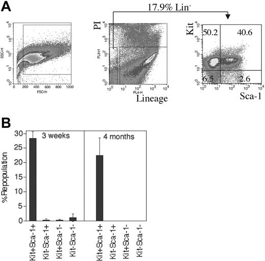
We next tested the status of the HSC markers Sca-1 and Kit in cultured BM HSCs. Figure 3A shows that after 10 days of culture 17.9% of total cells are Lin-. Of these Lin- cells the Kit+Sca-1+, Kit+Sca-1-, Kit-Sca-1+, and Kit-Sca-1- populations comprised 40.6%, 50.2%, 2.6%, and 6.5%, respectively. We isolated 1.3 × 10⁴ Sca-1+Kit+, Sca-1+Kit-, Sca-1-Kit+, and Sca-1-Kit- cells and measured their HSC activity by competitive repopulation. While Sca-1+Kit+ cells contained both ST- and LT-HSC activities, the 3 other fractions only contained minor ST-HSC and no LT-HSC activity (Figure 3B). This result establishes that, similar to freshly isolated HSCs, LT-HSC activity of cultured HSCs is in the Kit+Sca-1+ fraction.
All HSCs reside in the Kit- and Sca-1–positive fraction of cultured BM cells. (A) Ten-day cultured BM cells were stained with a cocktail of biotinylated lineage-specific antibodies, followed by streptavidin-APC, anti–Sca-1–FITC, and anti-Kit–PE. Forward scatter (FSC) and side scatter (SSC) on the left plot is used to gate on hematopoietic cells. In the middle and right plots, Lin- (negative APC-stained) and propidium iodide–negative (PI-) cells were gated to show surface expression of Sca-1 and Kit. Numbers in the graph are the percentages of each cell fraction. (B) Expanded HSCs in cultured BM cells are Sca-1+Kit+. After 10 days of culture of total BM cells, 1.3 × 10⁴ sorted CD45.2 Sca-1+Kit+, Sca-1+Kit-, Sca-1-Kit+, or Sca-1-Kit- cells were transplanted together with 2 × 10⁵ CD45.1 competitor cells into lethally irradiated CD45.1 mice (n = 4). Peripheral blood engraftments at 3 weeks and 4 months after transplantation are shown. Error bars indicate SEM.
All HSCs reside in the Kit- and Sca-1–positive fraction of cultured BM cells. (A) Ten-day cultured BM cells were stained with a cocktail of biotinylated lineage-specific antibodies, followed by streptavidin-APC, anti–Sca-1–FITC, and anti-Kit–PE. Forward scatter (FSC) and side scatter (SSC) on the left plot is used to gate on hematopoietic cells. In the middle and right plots, Lin- (negative APC-stained) and propidium iodide–negative (PI-) cells were gated to show surface expression of Sca-1 and Kit. Numbers in the graph are the percentages of each cell fraction. (B) Expanded HSCs in cultured BM cells are Sca-1+Kit+. After 10 days of culture of total BM cells, 1.3 × 10⁴ sorted CD45.2 Sca-1+Kit+, Sca-1+Kit-, Sca-1-Kit+, or Sca-1-Kit- cells were transplanted together with 2 × 10⁵ CD45.1 competitor cells into lethally irradiated CD45.1 mice (n = 4). Peripheral blood engraftments at 3 weeks and 4 months after transplantation are shown. Error bars indicate SEM.
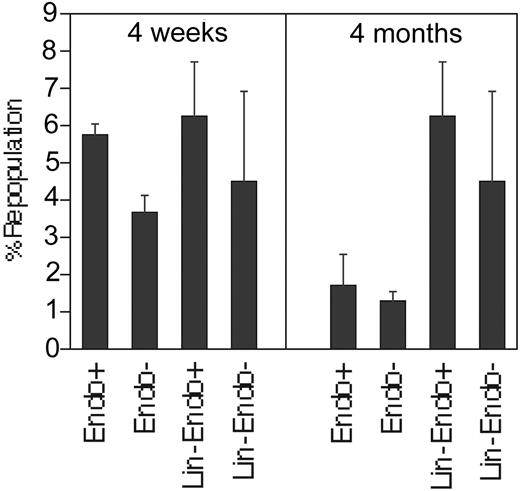
We also tested whether Endoglin, another surface protein found on freshly isolated HSCs, was also expressed on ex vivo–expanded HSCs. In cultured BM 26% of total cells and 20% of Lin- cells were Endoglin+ (Table 1). As shown by the competitive transplantation experiment in Figure 4, the 74% of the cultured cells that are Endoglin- have the similar repopulating ability as the 26% that are Endoglin+, both with respect to the ST-HSC and LT-HSC activities. Transplantation of similar numbers of Lin-Enodglin+ and Lin-Endoglin- cells, after culture, yielded similar reconstitution after 1 or 4 months. Thus, these cultured HSCs clearly are different from freshly isolated HSCs, where all of the LT-HSC activity resides in the Endoglin+ fraction.7
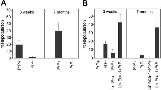
We recently showed that the prion protein PrP is a surface marker for LT-HSCs in freshly isolated BM (C.C.Z., A. Steele, S. Lindquist, and H.F.L., manuscript in preparation); thus, we determined whether following culture all HSCs express this protein. Whereas in freshly isolated BM PrP+ cells make up only 6% of Lin- cells (C.C.Z., A. Steele, S. Lindquist, and H.F.L., manuscript in preparation; and not shown), the PrP+ fraction increased substantially to 60% after 10 days of culture (Table 1). Figure 5A confirms our previous finding (C.C.Z., A. Steele, S. Lindquist, and H.F.L., manuscript in preparation) that, in freshly isolated BM, LT-HSC activity only resides in the PrP+ fraction. In contrast, when we sorted PrP+ and PrP- cells from 10-day cultured BM cells and transplanted 5000 of each, all ST-HSC and LT-HSC repopulating activity resided in the PrP- but not in the PrP+ fraction (Figure 5B).
We also competitively transplanted 3000 Lin-Sca-1+PrP+ and Lin-Sca-1+PrP- cells sorted from a 4-day culture; again, the PrP- but not PrP+ fraction contained all the repopulating LT potential of these cultured cells (Figure 5B). Therefore, the expression patterns of PrP on freshly isolated HSCs and cultured HSCs are different: PrP is expressed on all freshly isolated HSCs but not on ex vivo–expanded HSCs.
The results from Figures 4 and 5 suggest that surface proteins such as Endoglin and PrP that are characteristic of freshly isolated BM HSC markers are not necessarily found on cultured HSCs. To thoroughly investigate the presentation of surface proteins on cultured HSCs, we examined the expression on expanded cells of several additional HSC markers and cell surface proteins, including Tie-2,8 prominin-1 (or CD13330 ; see Fargeas et al31 for nomenclature suggestions), CD31,9 CD62L,32 CD34,6 IGF-2 receptors (which bind to IGF2-hFc),10 Mpl,5 Flk1,33 CD43,34 CD44,32 CD49D,35 CD49E,32 CD11A,32 CXCR4,36 CD24,37 and CD38.6 Expression of these surface proteins on total or Lin- BM cells, as determined by flow cytometry, is summarized in Table 1. The majority of 10-day cultured cells expressed Mpl, Flk1, CD43, CD44, CD49D, CD49E, CD11a, CXCR4, and CD24. By contrast, about half of the cultured cells were CD31+, CD62L+, or IGF2-hFc+, and a relatively small portion (0.3%-8%) of the cultured cells were Tie-2+, prominin-1+, CD34+, or CD38+.
Ex vivo–expanded BM HSCs are in both Endoglin-positive and -negative fractions. After 10 days of culture of total BM cells, 6000 sorted Endoglin+ and 2.4 × 10⁴ Endoglin- cells or 3000 Lin-Endoglin- and 3000 Lin-Endoglin+ cells were transplanted together with 2 × 10⁵ CD45.1 competitor cells into lethally irradiated CD45.1 mice (n = 4). Shown are peripheral blood engraftment at 4 weeks (left) and 4 months (right) after transplantation. Different numbers of sorted Endoglin+ Endoglin- cells were used because after culture there were 3 times more Endoglin- cells (76% of the total) than Endoglin+ cells (26%). Error bars indicate SEM.
Ex vivo–expanded BM HSCs are in both Endoglin-positive and -negative fractions. After 10 days of culture of total BM cells, 6000 sorted Endoglin+ and 2.4 × 10⁴ Endoglin- cells or 3000 Lin-Endoglin- and 3000 Lin-Endoglin+ cells were transplanted together with 2 × 10⁵ CD45.1 competitor cells into lethally irradiated CD45.1 mice (n = 4). Shown are peripheral blood engraftment at 4 weeks (left) and 4 months (right) after transplantation. Different numbers of sorted Endoglin+ Endoglin- cells were used because after culture there were 3 times more Endoglin- cells (76% of the total) than Endoglin+ cells (26%). Error bars indicate SEM.
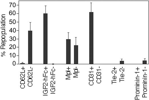
In the study in Figure 6, we sorted the positive and negative fractions of cultured cells after immunostaining with antibodies against CD62L, Mpl, CD31, Tie-2, or prominin-1, or after incubation with IGF2-hFc (a chimeric protein that binds to cells with receptors for IGF-210 ), followed by competitive transplantation. The LT-repopulating activity was found in the CD62L-, IGF2-hFc+, CD31+, Tie-2-, and prominin-1- fractions. Both Mpl+ and Mpl- cells contained LT-HSC activities (Figure 6). We also tested the engraftment of CD34+/- and CD38+/- cells after culture. More than 90% of cells are CD34- or CD38- (Table 1). While 105 CD34- or CD38- cells contained considerable HSC activity (data not shown), we could not obtain the same number of CD34+ or CD38+ cells so that we could test them by transplantation. We detected minor engraftment from 104 CD34+ cells and no engraftment from 104 CD38+ cells. These data suggested that repopulating cultured BM LT-HSCs are contained in the Sca-1+, Kit+, CD31+, IGF2-hFc+, PrP-, Tie-2-, prominin-1-, CD38-, and CD62L- population.
Discussion
We have developed a simple culture system that supports extensive expansion of LT-HSCs. Importantly, the expansion of LT-HSCs exceeded that of the more differentiated cells. Our most striking result is that the surface proteins on cultured HSCs differ significantly from those on freshly isolated HSCs. This suggests that HSCs possess a certain level of plasticity in surface protein expression that does not appear to alter significantly their functions as stem cells.
All ex vivo–expanded BM HSCs are PrP-. (A) Either 105 PrP- or 2 × 10⁴ PrP+ freshly isolated CD45.2 donor BM cells were mixed with 105 competitor CD45.1 cells and transplanted into lethally irradiated CD45.1 mice (n = 4). Donor CD45.2 contribution at 3 weeks (left) and 7 months (right) after transplantation are shown. (B) PrP+ and PrP- cells (5000) sorted from 10-day cultured total BM or 3000 Lin-Sca-1+PrP+ and Lin-Sca-1+PrP- cells sorted from 4-day cultured total BM cells were transplanted together with 2 × 10⁵ CD45.1 competitor cells into lethally irradiated CD45.1 mice (n = 4). Peripheral blood engraftments at 3 weeks (left) and 7 months (right) after transplantation are shown. Error bars indicate SEM.
All ex vivo–expanded BM HSCs are PrP-. (A) Either 105 PrP- or 2 × 10⁴ PrP+ freshly isolated CD45.2 donor BM cells were mixed with 105 competitor CD45.1 cells and transplanted into lethally irradiated CD45.1 mice (n = 4). Donor CD45.2 contribution at 3 weeks (left) and 7 months (right) after transplantation are shown. (B) PrP+ and PrP- cells (5000) sorted from 10-day cultured total BM or 3000 Lin-Sca-1+PrP+ and Lin-Sca-1+PrP- cells sorted from 4-day cultured total BM cells were transplanted together with 2 × 10⁵ CD45.1 competitor cells into lethally irradiated CD45.1 mice (n = 4). Peripheral blood engraftments at 3 weeks (left) and 7 months (right) after transplantation are shown. Error bars indicate SEM.
Ex vivo–expanded BM HSCs are CD62L-, IGF2-hFc+, CD31+, Tie-2-, and prominin-1-. After 10 days of culture of total BM cells, 4 × 10⁴ sorted CD62L+ and CD62L- cells, or IGF2-hFc+ and IGF2-hFc- cells, or Mpl+ and Mpl- cells, or CD31+ and CD31- cells, or 6000 sorted Tie-2+ and Tie-2- cells, or 6000 prominin-1+ and prominin-1- cells were transplanted, respectively, together with 105 CD45.1 competitor cells into lethally irradiated CD45.1 mice (n = 4). Peripheral blood engraftments at 4 months after transplantation are shown. Error bars indicate SEM.
Ex vivo–expanded BM HSCs are CD62L-, IGF2-hFc+, CD31+, Tie-2-, and prominin-1-. After 10 days of culture of total BM cells, 4 × 10⁴ sorted CD62L+ and CD62L- cells, or IGF2-hFc+ and IGF2-hFc- cells, or Mpl+ and Mpl- cells, or CD31+ and CD31- cells, or 6000 sorted Tie-2+ and Tie-2- cells, or 6000 prominin-1+ and prominin-1- cells were transplanted, respectively, together with 105 CD45.1 competitor cells into lethally irradiated CD45.1 mice (n = 4). Peripheral blood engraftments at 4 months after transplantation are shown. Error bars indicate SEM.
Numerous studies suggested that, while no single growth factor or cytokine can support expansion of HSCs ex vivo, certain combinations of 2 or more are able to stimulate HSC self-renewal.10,24-28 The key to our culture system is the combination of low levels of 4 cytokines (SCF, TPO, IGF-2, and FGF-1) in serum-free medium. The initial cell density and the culture time were also optimized for the increase of HSCs. Each of these 4 cytokines has been shown to be capable of directly binding to LT-HSCs, but each alone is unable to stimulate HSC expansion.10,24-28
In the initial phase of our culture the presence of low concentrations of these factors in the serum-free medium ensures that only HSCs, but not their differentiated progenies, are directly binding to and costimulated by multiple hormones that induce positive signals for survival and self-renewal. Since HSCs in both total BM and highly enriched SP populations can be expanded, it appears that HSCs are the primary target of these cytokines. Differentiated cells in the culture, either added initially or produced along culture, may also produce factors that stimulate (or inhibit) HSC self-renewal, prevent (or enhance) their death, or both. The direct utility of our cytokine combination for HSCs expansion is evident by our demonstration that both Kit (SCF receptor) and IGF-2 receptors are present on all cultured LT-HSCs, and that Mpl, the TPO receptor, is expressed on all freshly isolated and a large portion of cultured HSCs. In preliminary studies we also tested other growth factors, including FGF-2 and Wnt 3a. Neither of these supported additional HSC expansion when added to our standard medium containing SCF, TPO, IGF-2, and FGF-1 (data not shown).
The expression of surface proteins on ex vivo–expanded HSCs has not been systematically investigated previously. Our results provide evidence that the expression pattern of surface proteins on cultured HSCs is different from that on freshly isolated HSCs. One group of marker proteins, including Kit, Sca-1, CD31, and IGF-2 receptors, are present on freshly isolated HSCs and retained on ex vivo–expanded HSCs. A second group includes proteins such as Endoglin, Mpl, PrP, Tie-2, CD38, and prominin-1. These are present on all freshly isolated mouse HSCs but are not found on some or all of LT-HSCs after ex vivo expansion in our culture system. As expected, there is a large group of proteins, including CD62L, that is present neither on freshly isolated nor on ex vivo–expanded HSCs.
As noted in Figures 3B and 6, both Kit and IGF-2 receptors are likely important for maintenance or expansion of HSCs. Our culture medium contains their ligands SCF and IGF-2, and our preliminary studies indicated that the presence of each of them contributes to the approximate 8-fold expansion in HSC numbers we observe. We do not know whether these cytokines induce growth or survival signals for cultured HSCs.
After ex vivo culture the majority of Lin- cells are both Sca-1+ and Kit+. While we did show that LT-HSC activity resides in the Sca-1+Kit+ fraction after culture, neither Sca-1 nor Kit is a useful marker for purification of cultured HSCs unless other surface markers are also used. Because the majority of cultured BM cells express CD43, CD44, CD49D, CD49E, and CXCR4, we feel that these cell adhesion proteins or chemokine receptors, while likely expressed on the majority of cultured HSCs, are not useful as discriminating surface markers for HSC purification after culture.
We were surprised that PrP and Tie-2 proteins, present on the surface of all freshly isolated LT-HSCs, are not present on HSCs after their expansion in culture. Recently, we showed that PrP is not only expressed on all BM LT-HSCs, but it is also important for their self-renewal (C.C.Z., A. Steele, S. Lindquist, and H.F.L., manuscript in preparation). Like Tie-2,8 PrP may also maintain the quiescent state of BM HSCs. Because PrP, like Tie-2, is no longer expressed on cultured HSCs, we hypothesize that PrP and Tie-2 are sensors for signals generated by the BM microenvironment and play a role in the regulation of HSC quiescence. Once HSCs are removed from the BM, PrP and Tie-2 may become down-regulated and eventually lost from HSCs, possibly contributing to their ability to proliferate in culture. It will be interesting to transplant cultured HSCs, which are negative for both PrP and Tie-2, and determine whether PrP and Tie-2 reappear on HSCs in the recipient BM. If PrP or Tie-2 is functionally important for HSCs in BM, one might expect that their expression should be restored on the surface of such engrafted HSCs.
In contrast to PrP and Tie-2, which are completely lost from the surface of cultured HSCs, expression of Endoglin and Mpl are retained on some but not all cultured HSCs. Since the functions of these proteins on HSCs are not fully understood, further study is needed to offer an explanation for this result.
This culture system and our study of the phenotype of cultured HSCs may have broad applications. Our principle of using a combination of low levels of cytokines that directly stimulate HSCs in a serum-free medium may be applicable to expanding human HSCs for transplantation. This culture system may also be useful for introducing DNA into HSCs, which requires the culture of HSCs without losing their activity. In particular, the expanded HSC activity and the 10-day culture time are also ideal for drug selection of genetic-modified HSCs. Our phenotypic study of ex vivo–expanded HSCs will be valuable for stem cell purification and analysis, and it may be particularly useful for developing a screening system for the regulation of HSC activities, either by means of chemical genetics, cDNA libraries, or RNA interference (RNAi) libraries.
Prepublished online as Blood First Edition Paper, February 8, 2005; DOI 10.1182/blood-2004-11-4418.
Supported by Cambridge/Massachusetts Institute of Technology (MIT) Institute, the National Institutes of Health (grant R01 DK067356) (H.F.L.), and a postdoctoral fellowship from the Leukemia and Lymphoma Society.
The publication costs of this article were defrayed in part by page charge payment. Therefore, and solely to indicate this fact, this article is hereby marked “advertisement” in accordance with 18 U.S.C. section 1734.
We thank W. Tong for providing the antibody against mouse Mpl, and G. Paradis and M. Jennings in the MIT flow cytometry core facility for cell sorting. We thank J. Shuga for preparing this manuscript and M. Kaba for technical assistance.

