Activation of invariant natural killer T (iNKT) cells with CD1d-restricted T-cell receptor (TCR) ligands is a powerful means to modulate various immune responses. However, the iNKT-cell response is of limited duration and iNKT cells appear refractory to secondary stimulation. Here we show that the CD94/NKG2A inhibitory receptor plays a critical role in down-regulating iNKT-cell responses. Both TCR and NK-cell receptors expressed by iNKT cells were rapidly down-modulated by priming with α-galactosylceramide (α-GalCer) or its analog OCH [(2S,3S,4R)-1-O-(α-d-galactopyranosyl)-N-tetracosanoyl-2-amino-1,3,4-nonanetriol)]. TCR and CD28 were re-expressed more rapidly than the inhibitory NK-cell receptors CD94/NKG2A and Ly49, temporally rendering the primed iNKT cells hyperreactive to ligand restimulation. Of interest, α-GalCer was inferior to OCH in priming iNKT cells for subsequent restimulation because α-GalCer–induced interferon γ (IFN-γ) up-regulated Qa-1b expression and Qa-1b in turn inhibited iNKT-cell activity via its interaction with the inhibitory CD94/NKG2A receptor. Blockade of the CD94/NKG2–Qa-1b interaction markedly augmented recall and primary responses of iNKT cells. This is the first report to show the critical role for NK-cell receptors in controlling iNKT-cell responses and provides a novel strategy to augment the therapeutic effect of iNKT cells by priming with OCH or blocking of the CD94/NKG2A inhibitory pathway in clinical applications.
Introduction
Natural killer T (NKT) cells are a special T-cell population coexpressing the T-cell receptor (TCR) and NK-cell receptors such as NK1.1.1-4 Invariant NKT (iNKT) cells express a Vα14Jα18 chain (Vα24Jα15 in humans) and a semivariant TCR β-chain that is largely biased toward Vβ8.2 (Vβ11 in humans), Vβ2, and Vβ7.1-4 This TCR recognizes glycolipid antigens, such as α-galactosylceramide (α-GalCer), its analogs including OCH (a sphingosine-truncated analog of α-GalCer; (2S,3S,4R)-1-O-(α-D-galactopyranosyl)-N-tetracosanoyl-2-amino-1,3,4-nonanetriol), and isoglobotrihexosylceramide (iGb3), presented on the major histocompatibility complex (MHC) class I–like molecule CD1d.1-10 In addition, costimulatory signals, mediated through antigen-presenting cells that express CD80/86 interacting with CD28 expressed by iNKT cells, critically regulate iNKT-cell activation in a similar manner to conventional T cells.11,12 One of most striking characteristics of iNKT cells is their ability to promptly secrete various cytokines, including both interferon γ (IFN-γ) and interleukin 4 (IL-4), after their encounter with antigens (Ag's),1-4 which is reminiscent of effector/memory T cells. Accordingly, iNKT cells are thought to be potent immunoregulatory cells and their activation by ligands, such as α-GalCer and OCH, has been shown to be a powerful means to modulate various immune responses, including protective immunity, autoimmunity, and antitumor immunity.1-4,6,9,11,13,14
Despite an accumulation of studies concerning iNKT-cell activation of bystander immune cells, relatively little is known about the fate of Ag-primed iNKT cells themselves. Previous studies have shown that iNKT cells disappear quickly after their activation following TCR ligation or IL-12 stimulation.15-17 This phenomenon was initially attributed to increased activation-induced cell death (AICD) of iNKT cells, consistent with repopulation and homeostatic proliferation of peripheral iNKT cells after their rapid recruitment from the bone marrow.15 More recently, however, several studies have reported that down-regulation of TCR and NK1.1 cell surface expression on iNKT cells is the primary reason for the apparent disappearance of iNKT cells following α-GalCer treatment.18-20 Indeed a substantial iNKT-cell proliferation was observed in peripheral lymphoid organs following α-GalCer treatment.18-20 These findings suggested an interesting possibility that Ag-primed iNKT cells may develop an effector/memory subpopulation, which exerts even more potent function than unprimed iNKT cells. Here, we now demonstrate that priming of iNKT cells with their TCR ligands induces a dynamic modulation of TCR, NK-cell receptors (NK1.1, CD94/NKG2, NKG2D, and Ly49), and a costimulatory receptor (CD28). Differential kinetics of re-expression of these molecules on the cell surface temporally renders the primed iNKT cells hyperresponsive to secondary Ag stimulation. Of importance, the recall response of α-GalCer–primed iNKT cells is strictly regulated by an IFN-γ–dependent negative feedback mechanism where IFN-γ up-regulates Qa-1b expression and subsequent ligation of CD94/NKG2 inhibits iNKT-cell activity. Although Qa-1b can interact with NKG2A, NKG2C, and NKG2E, it has been previously reported that the majority of CD94/NKG2 expressed in mice is the inhibitory CD94/NKG2A receptor and that the Qa-1b–CD94/NKG2A interaction plays a critical role in negative regulation of NK-cell responses to self.21-23 We confirm that iNKT cells preferentially express CD94/NKG2A and hence blockade of the Qa-1b–NKG2A interaction markedly augmented recall responses of primed iNKT cells and primary responses of naive iNKT cells to α-GalCer. These findings are an important step to improve the efficacy of iNKT-cell–targeting therapeutics against tumor, infection, and autoimmune diseases since they demonstrate a means to modulate the adjuvant nature of iNKT cells by combined treatment with iNKT-cell glycolipid ligands and antagonistic monoclonal antibodies (mAbs).
Materials and methods
Mice
Wild-type (WT) C57BL/6 (B6) mice were obtained from Charles River Japan (Yokohama, Japan). IFN-γ–deficient (IFN-γ–/–) B6 mice were kindly provided by Y. Iwakura (University of Tokyo).24 All mice were maintained under specific pathogen-free conditions and used in accordance with the institutional guidelines of Juntendo University.
Reagents
A synthetic form of α-GalCer was obtained from Kirin Brewery (Gunma, Japan) and OCH was derived as described previously.6 In most experiments, mice were intraperitoneally injected with 2 μgof α-GalCer or OCH in 200 μL of phosphate-buffered saline (PBS) for priming and boosting. Dimethyl sulfoxide (DMSO; 0.1%) was used as the vehicle control. Phycoerythrin (PE)–conjugated tetrameric CD1d molecules loaded with α-GalCer (α-GalCer/CD1d) were prepared as described.17 The anti-NKG2A/C/E (NKG2) mAb, 20d5, and the anti-NKG2D mAb (CX5) were generated as described previously.21,25 Fab fragments of 20d5 and anti–Qa-1b mAb (BD Pharmingen, San Diego, CA) were prepared using the Fab preparation kit (Pierce, Rockford, IL) as described.26
Flow cytometric analysis
Mononuclear cells (MNCs) were prepared from spleen and liver as described.11 Cells were first preincubated with antimouse CD16/32 (2.4G2) mAb to avoid nonspecific binding of mAbs to FcγR. Surface and intracellular expression of molecules by iNKT cells were analyzed on electronically gated α-GalCer/CD1d tetramer+ cells on 4-color flow cytometry using a FACSCaliber (BD Bioscience, San Jose, CA). Intracellular staining was performed with a BD Cytofix/Cytoperm kit (BD Pharmingen) according to the manufacturer's instructions. Intracellular TCR in NKT cells was detected with a mixture of PE-conjugated antimouse Vβ2 TCR mAb (B20.6), anti-Vβ7 TCR mAb (TR310), and anti-Vβ8 TCR mAb (F23.1), or α-GalCer–loaded recombinant soluble dimeric mouse CD1d: immunoglobulin (CD1d:Ig; BD Pharmingen) and PE-conjugated anti–mouse IgG1 mAb (A85-1; BD Pharmingen). Surface and intracellular molecules were analyzed on electronically gated intracellular Vβ2/7/8+ cells 1 day after α-GalCer or OCH injection. Surface and intracellular molecules were stained with fluorescein isothiocyanate (FITC)– or allophycocyanin (APC)–conjugated NK1.1 mAb (PK136); FITC- or biotin-conjugated antimouse CD94 mAb (18d3); FITC- or biotin-conjugated antimouse NKG2A/C/E (NKG2) mAb (20d5); biotin-conjugated antimouse NKG2AB6 (16a11); FITC-conjugated antimouse Ly49 mAbs (anti-Ly49D mAb [4E5] or a mixture of anti-Ly49AB6 [A1], anti-Ly49C/I mAb [5E6], anti-Ly49G2 mAb [4D11], and antimouse Ly49I mAb [YL1-90]); biotin-conjugated anti-NKG2D mAb (CX5); APC- or biotin-conjugated anti-mouse CD28 mAb (37.51); biotin-conjugated antimouse Qa-1b mAb (6A8.6F10.1A6); FITC-, PE-, PE–cyanin 5 (PE-Cy5)–, APC-, or biotin-conjugated isotype-matched control mAbs (G155-178, MOPC-31C, R35-95, A95-1, R3-34, and Ha4/8); and Cy5-conjugated streptavidin. All of these reagents were purchased from BD Pharmingen, except for antimouse CD94 mAb, anti-NKG2AB6 mAb, anti-NKG2D mAb, and anti-CD28 mAb from eBioscience (San Diego, CA).
Cell preparation and in vitro stimulation
Freshly isolated splenic MNCs from vehicle-, α-GalCer–, or OCH-primed mice (5 × 10⁵) were cultured in RPMI 1640 medium supplemented with 10% heat-inactivated fetal calf serum (FCS), 2 mM L-glutamine, and 25 mM NaHCO3 in humidified 5% CO2 at 37°C in 96-well U-bottom plates (Corning, Corning, NY) as previously described.11 Cells were stimulated with 100 ng/mL α-GalCer, OCH, or vehicle (0.1% DMSO) in the presence or absence of 10-μg/mL Fab fragments of isotype-matched control mAbs, anti-NKG2 mAb, or antimouse Qa-1b mAb, or intact antimouse CD80 (16-10A1) and antimouse CD86 (PO 3.1) mAbs (eBioscience). After 24 to 48 hours, cell-free culture supernatants were harvested to determine IFN-γ and IL-4 levels by enzyme-linked immunosorbent assay (ELISA).
Coculture of iNKT cells and DCs
Freshly isolated hepatic MNCs were stained with PE-conjugated α-GalCer/CD1d tetramer, and positive cells were enriched by autoMACS using anti-PE microbeads (Miltenyi Biotec, Bergisch Gladbach, Germany) according to the manufacturer's instructions. Enriched iNKT cells were then sorted on a FACS Vantage (BD Bioscience) to obtain highly purified (98%-99%) iNKT cells. Splenic dendritic cells (DCs) were prepared according to the reported method.27,28 Purified iNKT cells (105) and DCs (5 × 10⁴) were cocultured as previously described11,29,30 with 100 ng/mL α-GalCer or vehicle (0.1% DMSO) in the presence or absence of 10-μg/mL Fab fragments of isotype-matched control mAbs, anti-NKG2A mAb, or anti–Qa-1b mAb. After 24 to 72 hours, cell-free culture supernatants were harvested to determine IFN-γ and IL-4 levels by ELISA.
ELISA
IFN-γ and IL-4 levels in the culture supernatants or the sera were determined by using mouse IFN-γ– or IL-4–specific ELISA kits (OptEIA; BD Bioscience Pharmingen) according to the manufacturer's instructions.
Cytotoxicity assay
Cytotoxic activity was tested against NK-cell–sensitive YAC-1 cells and NK-cell–resistant P815 cells by a standard 4-hour 51Cr-release assay as previously described.11 Effector cells (hepatic and splenic MNCs) were prepared from mice 24 hours after the last intraperitoneal injection of α-GalCer, OCH, or vehicle. Some mice were administered with 300 μg of isotype-matched control Ig or anti-NKG2 mAb intraperitoneally 2 days before the last α-GalCer injection. Specific cytotoxicity was calculated as previously described.11
Experimental lung metastases
B6 mice were intraperitoneally injected with OCH, α-GalCer, or vehicle, and then intravenously inoculated with B16 melanoma cells (1 × 10⁵, 3 × 10⁵, or 5 × 10⁵/200 μL) 2 hours later on day 0. B16 melanoma cells were prepared as previously described.11,16 Some mice were primed 2.5 days earlier with OCH or α-GalCer. Some mice were administered with 300 μg of isotype-matched control Ig or anti-NKG2 mAb intraperitoneally 2 days before the last α-GalCer injection. On day 14, the number of tumor colonies on the lungs was counted under a dissecting microscope (Olympus, Tokyo, Japan).
Statistical analysis
Data were analyzed by a 2-tailed Student t test. P values less than .05 were considered significant.
Results
Modulation of costimulatory and NK-cell receptors on iNKT cells upon priming with TCR ligands
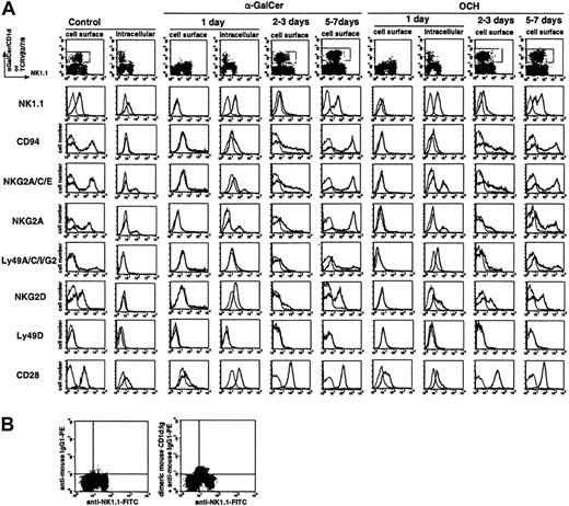
We analyzed the modulation of TCR, inhibitory NK-cell receptors (CD94/NKG2 and Ly49A/C/I/G2), activating NK-cell receptors (NK1.1, NKG2D, and Ly49D), and a costimulatory receptor (CD28) on iNKT cells after in vivo priming with synthetic TCR ligands, α-GalCer or OCH. Amongst liver iNKT cells from B6 WT mice, approximately 50% expressed CD94/NKG2 and NKG2D, less than 10% expressed Ly49A/C/I/G2, less than 2% expressed Ly49D, and all constitutively expressed CD28 (Figure 1A). Staining with an NKG2A-specific mAb showed that CD94/NKG2 expressed on iNKT cells was mainly composed of NKG2A (data not shown) but not NKG2C or NKG2E, as previously reported.22 Upon priming with α-GalCer or OCH, α-GalCer/CD1d tetramer+ iNKT cells seemingly began to disappear within 6 hours (data not shown) and almost completely disappeared at 16 to 24 hours, as previously reported.15-20 Consistent with recent reports,18-20 intracellular staining with anti-Vβ2/7/8 mAbs, detecting the predominant TCR β-chains expressed by iNKT cells, clearly showed the presence of liver iNKT cells expressing intracellular TCR at 24 hours after α-GalCer or OCH priming. NK-cell receptors and CD28 were also internalized, although some retention of cell surface CD28 was still detected (Figure 1A). Although staining intensity was relatively weak, intracellular staining with α-GalCer–loaded recombinant soluble dimeric mouse CD1d:Ig also demonstrated the internalized α-GalCer/CD1d–specific TCR coexpressed with intracellular NK1.1 one day after α-GalCer (Figure 1B) or OCH injection (data not shown). Similar results were obtained with spleen MNCs after in vivo priming and with liver MNCs after in vitro priming (data not shown).
After 2 to 3 days, the primed iNKT cells re-expressed TCR and CD28 on their surface. By contrast, a reduced level of surface NK-cell receptors (NK1.1, CD94/NKG2, Ly49, and NKG2D) was maintained for at least 3 to 4 days. After 5 to 7 days, some iNKT cells still expressed a low level of surface NK1.1, but another iNKT-cell population expressed relatively higher levels of NK1.1 compared with naive iNKT cells. After activation, the proportions of CD94/NKG2-, Ly49-, or NKG2D-expressing iNKT cells increased, and the expression levels of CD94/NKG2 and Ly49 were relatively higher than those found on naive iNKT cells. Again, CD94/NKG2 on these iNKT cells was mainly composed of NKG2A as estimated by staining with NKG2A-specific mAb. Consistent with previous reports,7,9,18-20 TCR ligand priming induced iNKT-cell expansion, although the expansion level was reduced following OCH priming (1.5-3 fold) compared with α-GalCer priming (5-8 fold) (data not shown). Similar results were obtained with spleen MNCs after in vivo priming and with liver MNCs after in vitro priming (data not shown).
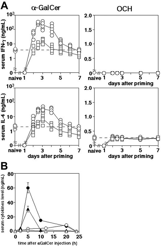
Consistent with a previous report that OCH selectively induced T-helper 2 (Th2) cytokine production by iNKT cells,6 a minor but significant serum IL-4 elevation was observed 3 to 5 hours after priming with OCH, but serum IFN-γ was not detected (Figure 2A). Moreover, we observed a similar modulation of iNKT-cell surface receptors by α-GalCer or OCH priming in IFN-γ–/– mice or in anti–IFN-γ mAb– and/or anti–IL-4 mAb–treated WT mice (data not shown). Taken together, these results indicated that priming of iNKT cells with TCR ligands resulted in a dramatic modulation of not only TCR and NK1.1 but also CD28 and inhibitory or activating NK-cell receptors on their surface, and this modulation was independent of IFN-γ or IL-4 production.
Modulation of NK1.1, CD94/NKG2, Ly49, NKG2D, and CD28 on α-GalCer– or OCH-activated liver iNKT cells. (A) Cell surface expression of the indicated molecules was analyzed on electronically gated α-GalCer/CD1d+ iNKT cells on the indicated days after intraperitoneal injection of α-GalCer or OCH. One day after α-GalCer or OCH injection, both cell surface and intracellular expression of the indicated molecules were analyzed in electronically gated intracellular Vβ2/7/8+ iNKT cells. The analysis gates are indicated by the gray line in dot plot panels. Bold lines indicate the staining with the respective mAb, and the thin lines indicate the staining with isotype-matched control Ig. Similar results were obtained from 3 independent experiments. (B) Existence of a cell population expressing intracellular α-GalCer/CD1d–specific TCR 1 day after α-GalCer injection. Liver MNCs were intracellularly stained with α-Gal-Cer–loaded recombinant soluble dimeric mouse CD1d:Ig and PE-conjugated anti–mouse IgG1 mAb, or PE-conjugated anti–mouse IgG1 mAb together with FITC-conjugated anti-NK1.1 mAb, 1 day after α-GalCer injection. Quadrant gates were set by staining with FITC-conjugated isotype-matched control and PE-conjugated anti–mouse IgG1 mAb.
Modulation of NK1.1, CD94/NKG2, Ly49, NKG2D, and CD28 on α-GalCer– or OCH-activated liver iNKT cells. (A) Cell surface expression of the indicated molecules was analyzed on electronically gated α-GalCer/CD1d+ iNKT cells on the indicated days after intraperitoneal injection of α-GalCer or OCH. One day after α-GalCer or OCH injection, both cell surface and intracellular expression of the indicated molecules were analyzed in electronically gated intracellular Vβ2/7/8+ iNKT cells. The analysis gates are indicated by the gray line in dot plot panels. Bold lines indicate the staining with the respective mAb, and the thin lines indicate the staining with isotype-matched control Ig. Similar results were obtained from 3 independent experiments. (B) Existence of a cell population expressing intracellular α-GalCer/CD1d–specific TCR 1 day after α-GalCer injection. Liver MNCs were intracellularly stained with α-Gal-Cer–loaded recombinant soluble dimeric mouse CD1d:Ig and PE-conjugated anti–mouse IgG1 mAb, or PE-conjugated anti–mouse IgG1 mAb together with FITC-conjugated anti-NK1.1 mAb, 1 day after α-GalCer injection. Quadrant gates were set by staining with FITC-conjugated isotype-matched control and PE-conjugated anti–mouse IgG1 mAb.
Augmented serum IFN-γ and IL-4 following α-GalCer treatment in OCH-primed mice. (A) Mice were primed with intraperitoneal injection of α-GalCer (□) or OCH (○) and then boosted with α-GalCer or OCH on the indicated day. Serum samples were obtained from 3 to 10 mice in each group 5 hours after the boost or the priming. Serum IFN-γ and IL-4 levels of primed naive mice were indicated on the y-axis (⋄), and the dotted horizontal line in each panel shows the mean level of the primary response. Serum IFN-γ or IL-4 in the vehicle-injected mice were not detectable (data not shown). (B) Kinetics of serum IFN-γ (circles) and IL-4 (triangles) levels following α-GalCer boost of vehicle-primed (open symbols) or OCH-primed (closed symbols) mice on day 2.5. Data are represented as the mean ± SD of 5 mice in each group. Similar results were obtained from 3 independent experiments.
Augmented serum IFN-γ and IL-4 following α-GalCer treatment in OCH-primed mice. (A) Mice were primed with intraperitoneal injection of α-GalCer (□) or OCH (○) and then boosted with α-GalCer or OCH on the indicated day. Serum samples were obtained from 3 to 10 mice in each group 5 hours after the boost or the priming. Serum IFN-γ and IL-4 levels of primed naive mice were indicated on the y-axis (⋄), and the dotted horizontal line in each panel shows the mean level of the primary response. Serum IFN-γ or IL-4 in the vehicle-injected mice were not detectable (data not shown). (B) Kinetics of serum IFN-γ (circles) and IL-4 (triangles) levels following α-GalCer boost of vehicle-primed (open symbols) or OCH-primed (closed symbols) mice on day 2.5. Data are represented as the mean ± SD of 5 mice in each group. Similar results were obtained from 3 independent experiments.
Augmented recall responses of OCH-primed iNKT cells to α-GalCer in vivo
Since the modulation of surface receptors on iNKT cells by Ag priming might modify iNKT-cell responses to subsequent Ag challenge, we next examined the responses of α-GalCer– or OCH-primed mice to secondary α-GalCer or OCH administration. We measured serum IFN-γ and IL-4 5 hours after α-GalCer or OCH injection to avoid the contribution of NK cells, since it has been previously reported that NK cells are activated after iNKT-cell activation and contribute significantly to IFN-γ production within 12 hours after α-GalCer injection.16,31 Of interest, serum IFN-γ and IL-4 levels were dramatically increased (10-30 fold) by secondary α-GalCer injection 2 to 4 days after OCH priming compared with primary α-GalCer injection (Figure 2A). By contrast, serum IFN-γ and IL-4 levels were only slightly increased by secondary α-GalCer injection in α-GalCer–primed mice compared with primary α-GalCer injection (Figure 2A). Very little serum IFN-γ and IL-4 were detected in mice injected with α-GalCer 1 day after α-GalCer or OCH priming, possibly due to the initial internalization of TCR in iNKT cells. OCH administration after α-GalCer or OCH priming did not augment serum IL-4 levels compared with primary administration of OCH, and serum IFN-γ was never detected (Figure 2A). We also examined the kinetics of serum IFN-γ and IL-4 induction by α-GalCer injection 2.5 days after OCH priming. Both IFN-γ and IL-4 levels were dramatically increased by the OCH priming and peaked at 5 hours after secondary α-GalCer injection (Figure 2B).
We next examined the α-GalCer–induced cytotoxic activity and antitumor effect in α-GalCer– or OCH-primed mice. OCH priming markedly augmented the α-GalCer–induced cytotoxic activities of liver and spleen MNCs against either NK-cell–sensitive YAC-1 or NK-cell–resistant P815 target cells compared with priming with the vehicle (Figure 3A). However, the iNKT-cell proportions in MNCs were similar among the groups when the mice were boosted by secondary α-GalCer injection (Table 1). In contrast, α-Gal-Cer priming did not significantly augment the secondary α-GalCer–induced cytotoxicity (Figure 3A). Moreover, α-Gal-Cer administration 2.5 days after OCH priming markedly augmented the antimetastatic effect against B16 melanoma compared with other prime/boost regimens (eg, α-GalCer/α-GalCer; Figure 3B). These results indicated that iNKT cells were hyperresponsive to secondary α-GalCer stimulation 2 to 3 days after OCH priming in vivo, resulting in a dramatic augmentation of effector functions, including IFN-γ and IL-4 production, cytotoxicity, and antitumor effect.
Induction of cytotoxic activity and antimetastatic effect by priming and boosting with α-GalCer and OCH. (A) Cytotoxic activity of liver and spleen MNCs was tested against NK-cell–sensitive YAC-1 cells or NK-cell–resistant P815 cells 24 hours after α-GalCer injection into mice primed 2.5 days earlier with vehicle (▵), αGalCer (□), or OCH (○). Control mice were primed and boosted with vehicle (▿). Proportion of iNKT cells (%) in respective MNC populations at the time of boosting injection is indicated in Table 1. Data are represented as the mean ± SD of triplicate samples. Similar results were obtained from 3 independent experiments. E/T indicates effector-to-target ratio. (B) Antimetastatic effect. Mice were primed and boosted with α-GalCer, OCH, or vehicle on days –3 and 0 as indicated. Then, the indicated number of B16 melanoma cells were intravenously inoculated into the mice 2 hours after the boost. On day 14, the number of tumor metastatic colonies in the lungs was counted. Data are represented as the mean ± SD of 5 mice in each group. Similar results were obtained from 3 independent experiments.
Induction of cytotoxic activity and antimetastatic effect by priming and boosting with α-GalCer and OCH. (A) Cytotoxic activity of liver and spleen MNCs was tested against NK-cell–sensitive YAC-1 cells or NK-cell–resistant P815 cells 24 hours after α-GalCer injection into mice primed 2.5 days earlier with vehicle (▵), αGalCer (□), or OCH (○). Control mice were primed and boosted with vehicle (▿). Proportion of iNKT cells (%) in respective MNC populations at the time of boosting injection is indicated in Table 1. Data are represented as the mean ± SD of triplicate samples. Similar results were obtained from 3 independent experiments. E/T indicates effector-to-target ratio. (B) Antimetastatic effect. Mice were primed and boosted with α-GalCer, OCH, or vehicle on days –3 and 0 as indicated. Then, the indicated number of B16 melanoma cells were intravenously inoculated into the mice 2 hours after the boost. On day 14, the number of tumor metastatic colonies in the lungs was counted. Data are represented as the mean ± SD of 5 mice in each group. Similar results were obtained from 3 independent experiments.
Suppression of α-GalCer–induced iNKT-cell activation by Qa-1b and CD94/NKG2A interaction
To evaluate the priming effects more precisely, spleen MNCs were periodically isolated from naive, α-GalCer–primed, or OCH-primed mice and then stimulated with α-GalCer or OCH in vitro (Figure 4A). At 24 hours after the in vitro stimulation with α-GalCer or OCH, spleen MNCs from naive mice did not produce either IFN-γ or IL-4 at detectable levels, but by contrast spleen MNCs from α-GalCer– or OCH-primed mice produced substantial amounts of IFN-γ and IL-4 (Figure 4A). Maximal cytokine secretion was obtained from spleen MNCs isolated 2.5 days after priming. Consistent with the in vivo data (Figure 2), OCH-primed spleen MNCs secreted greater amounts of cytokines compared with α-GalCer–primed spleen MNCs. At 48 to 72 hours after the in vitro stimulation, naive splenic MNCs produced substantial amounts of IFN-γ and IL-4 in response to α-GalCer, but the OCH-primed MNCs still produced increased levels of both IFN-γ and IL-4 in response to α-GalCer restimulation compared with naive or α-GalCer–primed MNCs (data not shown). These results indicated that OCH priming sensitized iNKT cells to secondary α-GalCer stimulation more effectively than α-GalCer priming.
We next explored the mechanism by which OCH or α-GalCer priming sensitized iNKT cells to α-GalCer restimulation. We first examined whether the CD94/NKG2 inhibitory receptor might regulate the hyperresponsiveness of iNKT cells, since CD94/NKG2 was down-modulated on the sensitized iNKT cells 2 to 3 days after α-GalCer or OCH priming (Figure 1). Blockade of the CD94/NKG2 and Qa-1b interaction by Fab fragments of anti-NKG2 mAb or anti–Qa-1b mAb markedly enhanced IFN-γ and IL-4 production by α-GalCer–primed MNCs in response to restimulation in vitro with α-GalCer (Figure 4B). Albeit to a lesser extent, IFN-γ and IL-4 production by OCH-primed or naive MNCs was also significantly enhanced by the blockade of Qa-1b or CD94/NKG2. Notably, while α-GalCer–primed MNCs produced lower levels of IFN-γ and IL-4 than OCH-primed MNCs in response to α-GalCer restimulation in vivo, this difference was abrogated by the blockade of Qa-1b or CD94/NKG2. The contribution of activating CD94/NKG2C/E NK-cell receptors may be negligible, since blockade of the Qa-1b–CD94/NKG2 interaction did not inhibit cytokine production by α-GalCer–activated iNKT cells in all cocultures, even if anti-NKG2 mAb or anti–Qa-1b mAb inhibited the function of activating CD94/NKG2C/E. Blockade of the CD80/CD86 interaction with CD28 abolished the cytokine production by naive or primed MNCs, irrespective of Qa-1b or CD94/NKG2 blockade. These results indicated that the Qa-1b and CD94/NKG2 interaction suppressed the TCR- and CD28-mediated activation of iNKT cells by α-GalCer, especially when the iNKT cells had first been primed with α-GalCer.
We also examined the impact of OCH or α-GalCer priming on antigen-presenting cells (APCs) by coculturing of purified iNKT cells and purified splenic DCs separately isolated 2.5 days after priming with α-GalCer, OCH, or vehicle (Figure 4C). Neither IFN-γ nor IL-4 was detected when iNKT cells were cocultured with any DCs in the absence of α-GalCer (data not shown). Notably, α-GalCer–primed DCs induced significantly lower levels of IFN-γ and IL-4 production by vehicle-primed iNKT cells compared with vehicle- or OCH-primed DCs. Of importance, this difference was abrogated by the blockade of CD94/NKG2. Moreover, OCH-primed iNKT cells produced significantly higher levels of IFN-γ and IL-4 compared with vehicle- or α-GalCer–primed iNKT cells. Again, the difference of cytokine production between OCH-primed and α-GalCer–primed iNKT cells was abrogated by CD94/NKG2 blockade. Significantly higher levels of IFN-γ and IL-4 were attained by both types of primed iNKT cells compared with vehicle-primed iNKT cells. A similar level (approximately 200 pg/mL) of IL-12 p40 was detected in the supernatants of all cocultures (data not shown), which suggested that DC function was not impaired by α-GalCer or OCH priming. These results suggested that the recall responses of iNKT cells in α-GalCer–primed mice were more strictly regulated by Qa-1b and CD94/NKG2-mediated suppression than in naive or OCH-primed mice. Consistent with this notion, Qa-1b expression on splenic MNCs was markedly up-regulated in α-GalCer–primed mice compared with naive or OCH-primed mice (Figure 5A).
NKG2 and CD28 regulate activation of naive or primed iNKT cells by α-GalCer. (A) Mice were primed with intraperitoneal injection of α-GalCer (□) or OCH (○). Splenic MNCs were prepared on the indicated days after priming and stimulated with α-GalCer or OCH in vitro for 24 hours. Data are represented as the mean ± SD of triplicate wells. Similar results were obtained from 3 independent experiments. (B) Mice were primed with intraperitoneal injection of α-GalCer, OCH, or vehicle on day –2.5. Then, splenic MNCs were prepared on day 0 and stimulated with α-GalCer in vitro for 24 or 48 hours in the presence or absence of the indicated mAbs. Data are represented as the mean ± SD of triplicate wells. Similar results were obtained from 3 independent experiments. (C) Liver iNKT cells and splenic DCs were isolated from naive mice or primed with α-GalCer or OCH 2.5 days before and then cocultured with α-GalCer for 48 hours in the presence or absence of anti-NKG2 mAb. Data are represented as the mean ± SD of triplicate wells. Similar results were obtained from 2 independent experiments.
NKG2 and CD28 regulate activation of naive or primed iNKT cells by α-GalCer. (A) Mice were primed with intraperitoneal injection of α-GalCer (□) or OCH (○). Splenic MNCs were prepared on the indicated days after priming and stimulated with α-GalCer or OCH in vitro for 24 hours. Data are represented as the mean ± SD of triplicate wells. Similar results were obtained from 3 independent experiments. (B) Mice were primed with intraperitoneal injection of α-GalCer, OCH, or vehicle on day –2.5. Then, splenic MNCs were prepared on day 0 and stimulated with α-GalCer in vitro for 24 or 48 hours in the presence or absence of the indicated mAbs. Data are represented as the mean ± SD of triplicate wells. Similar results were obtained from 3 independent experiments. (C) Liver iNKT cells and splenic DCs were isolated from naive mice or primed with α-GalCer or OCH 2.5 days before and then cocultured with α-GalCer for 48 hours in the presence or absence of anti-NKG2 mAb. Data are represented as the mean ± SD of triplicate wells. Similar results were obtained from 2 independent experiments.
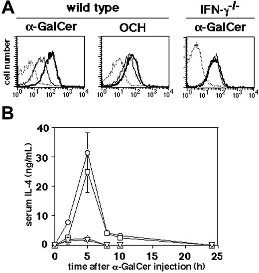
IFN-γ–induced Qa-1b inhibits reactivation of α-GalCer–primed iNKT cells by α-GalCer. (A) Qa-1b expression on splenic MNCs isolated from WT and IFN-γ–/– mice was analyzed 2.5 days after intraperitoneal administration of α-GalCer or OCH. Thin lines indicate the staining of MNCs from vehicle-treated mice with anti–Qa-1b mAb; bold lines, the staining of MNCs from α-GalCer– or OCH-treated mice with anti–Qa-1b mAb; and dotted lines, the staining with isotype-matched control Ig. Similar results were obtained from 3 independent experiments. (B) Kinetics of serum IL-4 induction after α-GalCer injection into vehicle-primed wild-type mice (▿), vehicle-primed IFN-γ–/– mice (▵), α-GalCer–primed IFN-γ–/– mice (□), or OCH-primed IFN-γ–/– mice (○). Priming was performed 2.5 days before. Serum IL-4 was not detectable in the vehicle-injected mice (data not shown). Data are represented as the mean ± SD of 5 mice in each group. Similar results were obtained from 3 independent experiments.
IFN-γ–induced Qa-1b inhibits reactivation of α-GalCer–primed iNKT cells by α-GalCer. (A) Qa-1b expression on splenic MNCs isolated from WT and IFN-γ–/– mice was analyzed 2.5 days after intraperitoneal administration of α-GalCer or OCH. Thin lines indicate the staining of MNCs from vehicle-treated mice with anti–Qa-1b mAb; bold lines, the staining of MNCs from α-GalCer– or OCH-treated mice with anti–Qa-1b mAb; and dotted lines, the staining with isotype-matched control Ig. Similar results were obtained from 3 independent experiments. (B) Kinetics of serum IL-4 induction after α-GalCer injection into vehicle-primed wild-type mice (▿), vehicle-primed IFN-γ–/– mice (▵), α-GalCer–primed IFN-γ–/– mice (□), or OCH-primed IFN-γ–/– mice (○). Priming was performed 2.5 days before. Serum IL-4 was not detectable in the vehicle-injected mice (data not shown). Data are represented as the mean ± SD of 5 mice in each group. Similar results were obtained from 3 independent experiments.
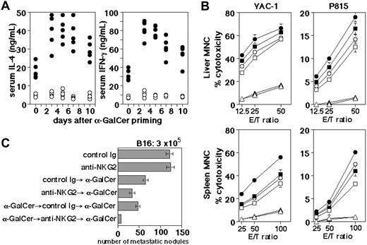
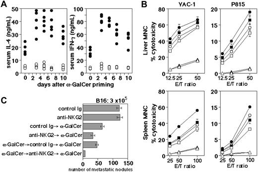
Blockade of NKG2 enhances activation of naive and α-GalCer–primed iNKT cells by α-GalCer in vivo. (A) Mice were primed with α-GalCer on day 0 and then boosted with α-GalCer on the indicated day. Anti-NKG2 mAb (•) or control Ig (○) was administered 2 days before the boost. Serum samples were obtained 5 hours after the boost. The mice indicated on day 0 were treated once with α-GalCer injection on day 0. (B) Cytotoxic activity of liver and spleen MNCs was tested against NK-sensitive YAC-1 cells and NK-resistant P815 cells 24 hours after the last α-GalCer injection. Mice were intraperitoneally injected with α-GalCer on day 0 (squares) or days –3 and 0 (circles), or injected with vehicle on days –3 and 0 (triangles), and intraperitoneally administered with anti-NKG2 mAb (closed symbols) or control Ig (open symbols) on day –2. Data are represented as the mean ± SD of triplicate samples. Similar results were obtained from 3 independent experiments. (C) Antimetastatic effect. Mice were intraperitoneally injected with α-GalCer on day 0 or days –3 and 0, and then intravenously inoculated with 3 × 10⁵ B16 melanoma cells 2 hours later. Anti-NKG2 mAb or control Ig was intraperitoneally administered on day –2. On day 14, the number of tumor colonies in the lungs was counted under a dissecting microscope. Data are represented as the mean ± SD of 5 to 8 mice in each group. Similar results were obtained from 3 independent experiments.
Blockade of NKG2 enhances activation of naive and α-GalCer–primed iNKT cells by α-GalCer in vivo. (A) Mice were primed with α-GalCer on day 0 and then boosted with α-GalCer on the indicated day. Anti-NKG2 mAb (•) or control Ig (○) was administered 2 days before the boost. Serum samples were obtained 5 hours after the boost. The mice indicated on day 0 were treated once with α-GalCer injection on day 0. (B) Cytotoxic activity of liver and spleen MNCs was tested against NK-sensitive YAC-1 cells and NK-resistant P815 cells 24 hours after the last α-GalCer injection. Mice were intraperitoneally injected with α-GalCer on day 0 (squares) or days –3 and 0 (circles), or injected with vehicle on days –3 and 0 (triangles), and intraperitoneally administered with anti-NKG2 mAb (closed symbols) or control Ig (open symbols) on day –2. Data are represented as the mean ± SD of triplicate samples. Similar results were obtained from 3 independent experiments. (C) Antimetastatic effect. Mice were intraperitoneally injected with α-GalCer on day 0 or days –3 and 0, and then intravenously inoculated with 3 × 10⁵ B16 melanoma cells 2 hours later. Anti-NKG2 mAb or control Ig was intraperitoneally administered on day –2. On day 14, the number of tumor colonies in the lungs was counted under a dissecting microscope. Data are represented as the mean ± SD of 5 to 8 mice in each group. Similar results were obtained from 3 independent experiments.
IFN-γ–induced Qa-1b expression negatively regulates recall NKT-cell responses in vivo
Although iNKT cells primed with α-GalCer strongly up-regulated the Qa-1b expression on splenic MNCs, those primed with OCH did so only weakly (Figure 5A). Notably, the α-GalCer–induced Qa-1b up-regulation was not observed in IFN-γ–/– mice (Figure 5A). Moreover, α-GalCer priming increased the secondary α-Gal-Cer–induced serum IL-4 to a level comparable with that induced by OCH priming in IFN-γ–/– mice (Figure 5B). These results indicated that IFN-γ induced by α-GalCer priming was responsible for Qa-1b up-regulation, which in turn resulted in CD94/NKG2-mediated suppression of recall iNKT-cell response in α-GalCer–primed mice.
We finally evaluated whether the blockade of CD94/NKG2 could augment the α-GalCer–induced iNKT-cell function in α-Gal-Cer–primed mice. The in vivo treatment with anti-NKG2 mAb alone did not induce cytokine production or cytotoxicity and did not deplete NK cells or iNKT cells (data not shown). The CD94/NKG2 blockade dramatically increased α-GalCer–induced serum IFN-γ and IL-4 levels, particularly in α-GalCer–primed mice (Figure 6A). It was notable that high levels of serum IFN-γ and IL-4 were inducible 10 days after α-GalCer priming if CD94/NKG2 was blocked at the time of boosting. Moreover, the CD94/NKG2 blockade also significantly augmented the α-GalCer–induced cytotoxicity of liver and spleen MNCs and the antimetastatic effect of treatment, particularly in α-GalCer–primed mice (Figure 6B-C). Even if anti-NKG2 mAb possibly inhibited the function of activating CD94/NKG2C/E, the contribution of activating CD94/NKG2C/E NK-cell receptors may be negligible, since blockade of the Qa-1b–CD94/NKG2 interaction did not inhibit cytokine production, cytotoxic activity, or the antimetastatic effect caused by the α-GalCer injection. These results indicated that the iNKT-cell activation by α-GalCer was limited by CD94/NKG2-mediated suppression and blockade of CD94/NKG2 could significantly improve the antitumor effect of a secondary α-GalCer treatment.
Discussion
In this study, we have shown that activation of iNKT cells is critically regulated by CD94/NKG2. In addition to TCR and CD28, naive iNKT cells express activating (NK1.1, NKG2D, and Ly49D) or inhibitory (CD94/NKG2 and Ly49A/C/I/G2) NK-cell receptors. All of these cell surface receptors were rapidly down-modulated upon priming of iNKT cells with their TCR ligands (α-GalCer or OCH). Two to 3 days after the priming, iNKT cells re-expressed TCR and CD28 on their surface, but CD94/NKG2 and Ly49 remained down-modulated. This pattern of expression enabled the primed iNKT cells to produce a larger amount of cytokines upon secondary stimulation with α-GalCer. Of interest, OCH was superior to α-GalCer in priming iNKT cells for a secondary response to α-GalCer, resulting in a markedly enhanced antimetastatic effect. We found that IFN-γ induced by α-GalCer priming up-regulated Qa-1b, which in turn suppressed the secondary iNKT-cell activation via CD94/NKG2. Thus, the blockade of CD94/NKG2 markedly enhanced the antimetastatic effect of α-GalCer after α-GalCer priming. These findings revealed a negative feedback regulation of iNKT-cell activation by IFN-γ–inducible Qa-1b and provided a novel strategy to improve the antimetastatic effect of α-GalCer by priming with OCH or by blocking CD94/NKG2-mediated suppression.
It was unexpected that OCH was far more effective than α-GalCer in priming the secondary iNKT-cell responses to α-Gal-Cer, since OCH and α-GalCer similarly modulated iNKT-cell surface receptors and OCH was rather inferior to α-GalCer in expanding iNKT cells upon priming (data not shown) as recently reported.6,9 We hypothesized that IFN-γ produced by iNKT cells upon priming with α-GalCer might be responsible for this difference, since OCH did not induce IFN-γ production in vivo. We found that α-GalCer priming up-regulated the expression of Qa-1b in an IFN-γ–dependent manner, which suppressed iNKT-cell activation in response to secondary α-GalCer stimulation in vitro and in vivo. Qa-1b is an MHC class Ib molecule broadly expressed on leukocytes and it predominantly presents a canonical signal peptide of classical MHC class Ia molecules, called Qa-1 determinant modifier (Qdm), in a transporter associated with antigen presentation (TAP)–dependent manner, thereby indirectly representing cellular MHC class Ia levels.32,33 The up-regulation of Qa-1b expression by IFN-γ might be due to increased transcription of Qa-1b and/or increased TAP-mediated loading of Qdm onto Qa-1b. The Qdm/Qa-1b complex is recognized by inhibitory CD94/NKG2A and activating CD94/NKG2C or E receptors.21-23 The CD94/NKG2 receptors expressed on naive and primed iNKT cells were predominantly CD94/NKG2A as estimated by staining with an NKG2A-specific mAb as previously reported.22 It has been shown that CD94/NKG2A expressed on NK cells and CD8+ T cells suppressed their activation.34 However, our present results are the first indication that iNKT-cell activation is critically regulated by CD94/NKG2A. Similarly, inhibitory Ly49 receptors, which recognize MHC class Ia molecules directly, have been reported to suppress α-GalCer–induced iNKT-cell activation.30,35 Since CD94/NKG2A is more frequently expressed on iNKT cells than Ly49, it may play a more dominant role in regulating iNKT-cell activation. The IFN-γ–mediated Qa-1b up-regulation may be a negative feedback mechanism to maintain self-tolerance of iNKT cells and avoid a pathogenic effect of iNKT cells.36-39 It will be interesting to explore the iNKT-cell functions in Qa-1b–deficient40 or CD94-deficient22 mice in future studies.
Consistent with recent reports,18-20 we observed a rapid down-modulation of TCR and NK1.1 on the surface of iNKT cells upon priming with their TCR ligands, which mostly accounted for the apparent disappearance of iNKT cells. However, intracellular staining 1 day after α-GalCer priming demonstrated a 20% to 30% reduction of iNKT-cell numbers compared with untreated mice (data not shown), and some annexin V–positive iNKT cells were detected in the liver and spleen promptly after α-GalCer injection as we reported previously.16 Therefore, some minor fraction of iNKT cells appeared to be susceptible to AICD upon α-GalCer priming. A significant increase of CD94/NKG2+ iNKT cells after α-GalCer priming suggested that these cells were more resistant to AICD than CD94/NKG2– iNKT cells. This preferential survival and/or expansion of CD94/NKG2+ iNKT cells might be at least partly responsible for the higher sensitivity of primed iNKT cells to Qa-1b and CD94/NKG2-mediated suppression.
We and others have recently shown that the expansion of iNKT cells is maximal 3 days after α-GalCer priming and then iNKT-cell numbers gradually return to normal levels by homeostatic mechanisms within 7 to 10 days.9,18-20 This is consistent with the kinetics of recall responses of α-GalCer– or OCH-primed mice to α-Gal-Cer, suggesting that the enhanced secondary responses were mainly due to expansion of iNKT cells after priming. However, the enhanced secondary responses were mostly maintained up to 10 days after α-GalCer priming if CD94/NKG2-mediated suppression was blocked at the secondary α-GalCer stimulation. This suggests that the primed iNKT cells with a high capacity to produce cytokines upon restimulation persist (typical of effector/memory T cells), although they are under a strict regulation by CD94/NKG2A-mediated suppression.
A recent study has shown that the recognition of α-GalCer analogues was influenced by the TCR Vβ repertoires of iNKT cells. OCH was preferentially recognized by Vβ8+ iNKT cells, which also have a higher avidity for α-GalCer than Vβ7+ iNKT cells.7,41 Thus, a preferential expansion of Vβ8+ iNKT cells after OCH or α-GalCer priming might also be responsible for the enhanced responses of primed iNKT cells to α-GalCer restimulation in vitro and in vivo.
We have previously shown that α-GalCer administration into naive mice induces sustained IFN-γ production and cytotoxic activity, which were mediated by NK cells secondarily activated by IFN-γ derived from iNKT cells and IL-12 derived from DCs.16,29,31 Thus, depletion of NK cells mostly abrogated the sustained α-GalCer response and consequently impaired the antimetastatic effect of α-GalCer. In contrast, α-GalCer administration into OCH-primed mice induced a greatly enhanced IFN-γ production at 5 hours but not at 16 to 20 hours, which was not reduced by NK-cell depletion (data not shown). This indicated that OCH priming mainly enhanced IFN-γ production by iNKT cells themselves, rather that secondary activated NK cells, upon the secondary α-GalCer stimulation. However, the markedly enhanced cytotoxic activity of liver and spleen MNCs 24 hours after α-GalCer boost in OCH-primed mice was mostly abrogated by NK-cell depletion (K.T., unpublished data, May 2004). In addition, the significantly augmented antimetastatic effect of α-GalCer in OCH-primed mice was significantly inhibited by NK-cell depletion (K.T., unpublished data, May 2004). These data suggested that IFN-γ–activated NK cells were mainly responsible for the antimetastatic effect of α-GalCer in the OCH-primed mice. In this context, the CD94/NKG2 blockade might augment the antimetastatic effect of α-GalCer by enhancing the activation of not only iNKT cells but also NK cells, since NK cells also express CD94/NKG2A inhibitory receptors. Thus, blockade of the CD94/NKG2A suppressive pathway may be effective at either the induction or effector phase of the α-GalCer–induced antitumor effect.
The most notable finding of this study is that the antimetastatic effect of α-GalCer was greatly improved by the OCH-priming or the CD94/NKG2A blockade. OCH was a weak inducer of iNKT-cell expansion and IL-4 production but did not induce IFN-γ production or antimetastatic activity by itself.6,7,9 However, OCH modulated iNKT-cell surface receptors as efficiently as α-GalCer. These OCH-primed iNKT cells produced a huge amount of IFN-γ upon secondary α-GalCer restimulation in vivo, resulting in a potent antimetastatic effect. The inability of OCH to induce IFN-γ was a beneficial property for priming secondary α-GalCer responses because IFN-γ down-regulated the secondary iNKT-cell responses by up-regulating Qa-1b and thus CD94/NKG2A-mediated suppression. Recent studies have demonstrated quantitative and qualitative differences in the in vivo response of iNKT cells to distinct α-GalCer analogues, including OCH and β-Gal-Cer.9 Like OCH, in our preliminary experiments, priming with β-GalCer a26,9 another weak iNKT-cell ligand inducing poor cytokine production, also potently enhanced iNKT-cell responses to α-GalCer restimulation (data not shown). α-GalCer and OCH have been shown to activate human Vα24 iNKT cells in a similar manner to murine Vα14 iNKT cells in vitro,3,4 and α-GalCer is now in early clinical trials in cancer patients.42,43 Therefore, priming with OCH may be a novel strategy to improve the therapeutic effect of α-GalCer in such patients. Further exploration of an α-GalCer analog with a better priming effect is also warranted. CD94/NKG2A blockade might be also applicable to improve the antitumor effect of α-GalCer in humans. In addition to an antitumor effect, α-GalCer has been shown to protect mice against infections and autoimmune diseases.1-4,13,14 Therefore, the priming with OCH and the blockade of CD94/NKG2A may also be applicable to improve the therapeutic effect of α-GalCer in these diseases. However, it has also been shown that α-GalCer occasionally exacerbated autoimmune diseases, depending on the model and/or administration protocol.44 Moreover, overactivation of iNKT cells can induce tissue pathologies.36-39 Therefore, further studies are needed to determine the optimal prime/boost protocol or blockade of NK-cell receptors in iNKT-cell–targeting therapy for the safe treatment of tumor, infections, and autoimmune diseases.
Prepublished online as Blood First Edition Paper, March 3, 2005; DOI 10.1182/blood-2004-11-4257.
Supported by research grants from the Human Frontier Science Program Organization; the Organization for Pharmaceutical Safety and Research; the Ministry of Education, Science, and Culture, Japan; the National Health and Medical Research Council of Australia; and the National Institutes of Health.
T.O. and K.T. contributed equally to this work.
The publication costs of this article were defrayed in part by page charge payment. Therefore, and solely to indicate this fact, this article is hereby marked “advertisement” in accordance with 18 U.S.C. section 1734.
We thank Lewis L. Lanier for reading the manuscript and helpful suggestions.


