Comment on Tassone et al, page 713
In this issue of Blood, Tassone and colleagues describe a novel in vivo model of human multiple myeloma (MM) that relies on the engraftment of the IL-6–dependent human myeloma cell line INA-6 in a human bone marrow milieu implanted in SCID-hu mice. Because of the presence of the human microenvironment, evaluation of anticancer therapeutics in this model is likely to yield information predictive of their activity in MM patients.
The limited efficacy of conventional chemotherapy in most malignant diseases and the significant progress in our understanding of the molecular mechanisms underlying tumor growth and interactions with the microenvironment have recently fueled the development of several alternative therapeutic strategies. Therefore there is a need for preclinical models that are suitable to screen a large array of anticancer therapeutics in an efficient way and uncover information predictive of their clinical activity. MM is no exception to this trend. Antiangiogenic compounds,1 proteasome inhibitors,2 and immunotherapeutic strategies3-5 have already been shown to be viable approaches for the treatment of this disease. These results suggest that a large range of therapeutic strategies will be developed in the coming years, emphasizing the need for a reliable animal model system that recapitulates the clinical features of MM.
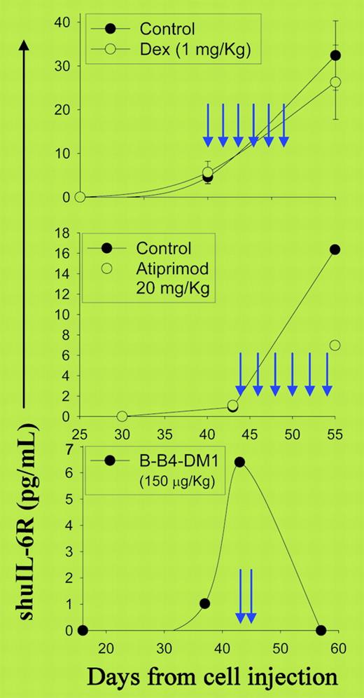
The animal model described by Tassone and colleagues in this issue of Blood appears to meet most of these criteria. These authors have successfully engrafted the interleukin 6 (IL-6)–dependent human MM cell line INA-6 transduced with green fluorescent protein (Gfp) gene into severe combined immunodeficiency (SCID) mice that previously received transplants of human fetal bone chips (SCID-hu mice). The chips provide the human microenvironment, which plays a major role in the outcome of therapies of hematologic malignancies, so the major limitations of conventional models of mice carrying subcutaneous or disseminated human tumors are eliminated. Furthermore, measurement of serum-soluble IL-6 receptor and fluorescent imaging of host animals provide reliable assays to monitor MM cell growth and assess the therapeutic potential of investigational drugs and experimental therapies (see figure).
Tassone et al are not the first to describe an in vivo model of MM, since Yaccoby et al6 have already demonstrated engraftment of primary MM cells in SCID-hu mice. So what is new about Tassone et al's model? Although Yaccoby et al's model accurately recapitulates the pathophysiology of MM, its application on a large scale is hindered by the limited number of MM cells harvested from individual bone marrow patient samples and the potential variability among MM cells obtained from different patients. In Tassone et al's model, the use of an established IL-6–dependent cell line provides an unlimited number of MM cellsFIG1 with homogeneous and reproducible characteristics available for engraftment in SCID-hu mice; therefore, no difficulties are envisioned in evaluating anticancer therapeutics using the number of mice required to perform experiments with the appropriate statistical power.
Effect of dexamethasone, atiprimod, and B-B4-DM1 treatment on serum soluble human IL-2 receptor (shuIL-2R) of SCID-hu mice engrafted with INA-6 cells. See the complete figure in the article beginning on page 713.
Effect of dexamethasone, atiprimod, and B-B4-DM1 treatment on serum soluble human IL-2 receptor (shuIL-2R) of SCID-hu mice engrafted with INA-6 cells. See the complete figure in the article beginning on page 713.
Does the model described by Tassone et al have limitations, and if so, what are they? The model relies on one single-cell line that reflects one molecular subtype of MM with unknown and unpredictable frequency, so we do not know if and to what extent the conclusions drawn from this cell line can be generalized. Furthermore, evaluation of immunotherapies is restricted to the tumor antigen(s) and HLA allospecificities expressed by the INA-6 cell line. Finally, the potential for changes associated with in vitro culture raises the question of how closely the INA-6 cell line resembles the in vivo MM cells. These limitations emphasize the need to develop mouse models with multiple MM cell lines that require the human bone milieu for their growth and survival. ▪


