Comment on Gampel et al, page 2624
VEGF stimulates neovascularization via activation of its key receptor VEGFR-2 signaling pathway in endothelial cells. In this issue of Blood, Gampel and colleagues report that a significant proportion of VEGFR-2 molecules are stored in an endosomal recycling compartment and that VEGF mobilizes these intracellular receptors to the cell surface, which might increase the sensitivity of endothelial cells to proangiogenic signals.
In addition to promoting angiogenesis, vascular endothelial growth factor (VEGF) as the key angiogenic factor has several unique and unusual features, including control of stem cell differentiation into endothelial lineages, establishment of the primitive vascular plexuses, regulation by hypoxia, and increase of vascular permeability, which collectively contribute to the onset, development, and progression of many severe human diseases such as cancer, metastasis, and ocular diseases.1-4 In fact, the first antiangiogenic drugs for the treatment of cancer and ocular disease are anti-VEGF reagents that are aimed to neutralize these unusual biologic functions. Deletion of only 1 allele of the VEGF gene in mice leads to the early embryonic lethality due to defective formation of hemangioblasts.1,4 These genetic studies demonstrate that not only can the functions of VEGF not be replaced by other gene products, but also that the optimal level of VEGF is crucial for vascular development in the embryo. The biologic functions of VEGF are mainly mediated by the VEGF receptor-2 (VEGFR-2; KDR for human and Flk-1 for mouse) receptor.5 Although much information becomes available for understanding the receptor signaling pathways, relatively little is known about their intracellular trafficking, which might play a pivotal role for regulation of VEGF functions.
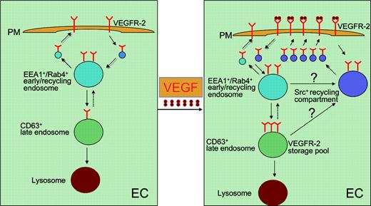
The initial surprising notion of the study by Gampel and colleagues is that 3 independent anti-VEGFR-2 antibodies give rise to a punctuated staining pattern rather than staining of the receptor at the cell surface in 2 endothelial cell lines. To reveal the identity of these intracellular VEGFR-2-positive structures, EEA1 (an early endosome-associated protein) and Rab4 (a marker of the “short-loop” recycling pathway) are colocalized with VEGFR-2+ vesicles. A significant number of VEGFR-2 molecules are also colocalized with CD63 (a marker for late endosomes). Quantification analysis demonstrates that roughly 40% of VEGFR-2 is stored in the endosomal compartment in endothelial cells (see figure).
Previous studies with other tyrosine kinase receptors (TKRs), including epidermal growth factor receptors (EGFRs) and platelet-derived growth factor receptors (PDGFRs), have demonstrated that upon ligand stimulation these receptors are down-regulated by endocytosis, followed by lyosomal degradation to down-regulate the persistent signaling.6,7 In contrast to these TKRs, stimulation of VEGFR-2 with VEGF does not lead to loss of the total intracellular pool of VEGFR-2, albeit redistribution of VEGFR-2 from the early endosomal compartments into the late endosomes. Intriguingly, most VEGFR-2 molecules are stored in the late endosomes without further degradation. One of the most important findings of this study is that stimulation of endothelial cells with VEGF leads to accumulation of VEGFR-2+ vesicles beneath the plasma membrane, particularly in the protrusion area. Using a novel biotinylation method, Gampel and colleagues have quantified the recycling event of VEGFR-2 and found that VEGF stimulation can dramatically increase the recycling of the intracellular VEGFR-2 to the cell surface. This finding is very surprising since VEGFR-2 is thought to be destroyed after signaling to desensitize the ligand. The increase of recycling of TKRs upon ligand stimulation seems to be a unique feature for the VEGFR-2 trafficking. Even more surprisingly, the intracellular recycling VEGFR-2+ vesicles also contain Src, which plays a crucial role in mediating VEGF-induced vascular permeability and endothelial motility. These findings suggest that Src as a downstream signaling component is also assembled in the same type of vesicles and is ready for signaling. Targeted recycling of VEGFR-2 might help us to understand the essential process of vascular sprouting and branch formation. Accumulation of VEGFR-2 molecules in a particular region of the endothelial cell plasma membrane might lead to formation of the protrusive filopodia at the sprout tip and guide movement of endothelial cells toward the VEGF gradient. This directed movement of endothelial cells is essential for vascular sprouting, branching, permeability, and the formation of a functional network.
Recycling and intracellular trafficking of VEGFR-2. In unstimulated endothelial cells (ECs), a significant proportion of VEGFR-2 is located in the EEA1+/Rab4+early/recycling endosomes. From this compartment, VEGFR-2 molecules could be recycled to the cell surface. Alternatively, a small portion of the receptor molecules could be transported to the CD63+late endosomes and eventually degraded in lyosomes. Upon VEGF stimulation, the VEGFR-2+vesicles stored in the EEA1+/Rab4+early/recycling endosomes are redistributed to the cell surface through the Src+recycling compartment and accumulated just beneath the plasma membrane (PM). VEGF stimulation also increases the distribution of VEGFR-2 in the late endosome compartment, where most of the receptor molecules are stored without further degradation.
Recycling and intracellular trafficking of VEGFR-2. In unstimulated endothelial cells (ECs), a significant proportion of VEGFR-2 is located in the EEA1+/Rab4+early/recycling endosomes. From this compartment, VEGFR-2 molecules could be recycled to the cell surface. Alternatively, a small portion of the receptor molecules could be transported to the CD63+late endosomes and eventually degraded in lyosomes. Upon VEGF stimulation, the VEGFR-2+vesicles stored in the EEA1+/Rab4+early/recycling endosomes are redistributed to the cell surface through the Src+recycling compartment and accumulated just beneath the plasma membrane (PM). VEGF stimulation also increases the distribution of VEGFR-2 in the late endosome compartment, where most of the receptor molecules are stored without further degradation.
Understanding of intracellular trafficking events of VEGFR-2 might help us to define novel therapeutic targets for intervention of the VEGF/VEGFR-2 signaling system. The present study has paved a new avenue for approaching this goal. However, at present there are several unanswered questions. Are the recycled VEGFR-2 molecules functional? Does VEGF-C, another ligand for VEGFR-2, also increase recycling of intracellular VEGFR-2 to the cell surface? What about VEGFR-1 and VEGFR-3? These are several important issues that warrant future studies. ▪


