Abstract
Acquired hemophilia A is a severe bleeding disorder caused by an autoantibody to factor VIII. Previous reports have focused on referral center patients and it is unclear whether these findings are generally applicable. To improve understanding of the disease, a 2-year observational study was established to identify and characterize the presenting features and outcome of all patients with acquired hemophilia A in the United Kingdom. This allowed a consecutive cohort of patients, unbiased by referral or reporting practice, to be studied. A total of 172 patients with a median age of 78 years were identified, an incidence of 1.48/million/y. The cohort was significantly older than previously reported series, but bleeding manifestations and underlying diseases were similar. Bleeding was the cause of death in 9% of the cohort and remained a risk until the inhibitor had been eradicated. There was no difference in inhibitor eradication or mortality between patients treated with steroids alone and a combination of steroids and cytotoxic agents. Relapse of the inhibitor was observed in 20% of the patients who had attained first complete remission. The data provide the most complete description of acquired hemophilia A available and are applicable to patients presenting to all centers.
Introduction
Acquired hemophilia A is a rare bleeding disorder caused by an autoantibody to factor VIII. The clinical characteristics of the disease have been previously described.1–3 The literature describing acquired hemophilia A is based on tertiary referral single-center cohorts,4–16 larger retrospective surveys of referral center patients,1,5,17–19 and patients reported because they have been treated for bleeding.20–23 These reports have been combined in a review and meta-analysis.24 This means that an accurate incidence of the disorder is unknown because not all of the patients would have been referred to the tertiary center and the referral population is not defined. Selection bias may also have been introduced because referral centers may manage younger, more complicated, and more severely affected patients than other hospitals and extrapolation of data from these patients may misrepresent the range of clinical presentation, natural history, and outcome of the condition.
Treatment of bleeding episodes in acquired hemophilia has been subjected to relatively large clinical trials and good hemostatic efficacy has been demonstrated for porcine factor VIII,21 recombinant factor VIIa,20 and factor 8 inhibitor bypassing activity (FEIBA).22,23 Data on inhibitor eradication, however, are based on relatively small, uncontrolled single-center cohorts4–16 and a meta-analysis.24 These reports are potentially unrepresentative because referral center patients may be a subgroup of patients. Furthermore, there may be positive reporting bias because authors are more likely to report and journals more likely to publish good outcomes. The one prospective randomized study in the field was unable to recruit sufficient patients to allow definite conclusions to be drawn.
Despite the shortcomings in the literature, many authorities recommend that patients with acquired hemophilia A should be immunosuppressed with steroids and a cytotoxic agent in an attempt to eradicate the inhibitor rapidly and decrease the length of time a patient is exposed to the risk of bleeding. Although this approach is logical, the extrapolation of uncontrolled studies on referral center patients to the whole patient group may not be correct because the risks of immunosuppression in elderly patients must be balanced against the potential benefits.
To address these issues, the UK Haemophilia Centre Doctors' Organisation undertook a 2-year national surveillance of acquired hemophilia A. The aim of the study was to identify all patients presenting with acquired hemophilia A in the United Kingdom between May 1, 2001, and April 30, 2003. This study allows a consecutive cohort of patients, which is unaffected by referral and reporting bias, to be studied and may lead to a better understanding of the presenting features and natural history of the disease and allow more informed treatment recommendations.
Patients, materials, and methods
Data acquisition
The study was approved by the Multicentre Research Ethics Committee for Wales. The ethics committee stated that written consent was not required by individual patients for the data to be reported. All National Health Service (NHS) hospitals in the United Kingdom were identified through the NHS website (http://www.nhs.uk). The hematology department covering each hospital was identified (some hematology departments cover more than 1 hospital) and a clinician within each hematology department with an interest in hemostasis and thrombosis was identified. This clinician was sent a simple questionnaire (form 1) asking how many patients with acquired hemophilia A had been diagnosed or treated by that hematology department in the previous 6 months. The patients' initials and referral details were requested to avoid double reporting. A reminder was sent after 2 months if no reply had been received. The clinician was telephoned personally if there was no response to the reminder. These questionnaires were sent to all United Kingdom hematology departments in November 2001, May 2002, November 2002, and May 2003. This process identified patients newly presenting with acquired hemophilia A in the United Kingdom between May 1, 2001 and April 30, 2003.
Within the United Kingdom all cases of acquired hemophilia A are diagnosed and treated within the NHS. It is not possible for a diagnosis of acquired hemophilia A to be made in the United Kingdom without a consultant hematologist being involved because United Kingdom hematologists have responsibility both for the laboratory and clinical services. Also, because the disease is a rare but significant event, the lead hematologist for thrombosis and hemostasis in each department would be aware of all cases. All reporting laboratories participate in the United Kingdom National External Quality Assurance (QA) and Accreditation Schemes and those diagnosing inhibitors participate in specific QA exercises involving acquired inhibitors and lupus anticoagulants. It is, therefore, unlikely that cases were not reported if a diagnosis had been made and it is also unlikely that cases of lupus anticoagulants would have been misdiagnosed as acquired hemophilia.
Clinical details of the patients identified were requested on a second questionnaire (form 2). Data requested included presenting age, factor VIII level and inhibitor titer at diagnosis, associated diagnoses, bleeding sites, type of hemostatic treatment given, type of immunosuppression given (steroids alone, steroids plus cytotoxics, intravenous immunoglobulin [IVIG], or other), date and cause of death if appropriate, and date inhibitor was eradicated. The data collected were deliberately kept to a minimum to increase response rates.
At the time of the study United Kingdom national guidelines relating to the diagnosis and management of acquired hemophilia were published.25 These recommended starting immunosuppression at diagnosis with either prednisolone or a combination of prednisolone and oral cyclophosphamide. If prednisolone-treated patients did not respond then addition of cyclophosphamide was recommended. The dose of prednisolone was recommended to be 1 mg/kg body weight and of cyclophosphamide 1 to 2 mg/kg body weight. The majority of patients were treated according to these guidelines.
The definition of complete remission (CR) used in the study was factor VIII normal, inhibitor undetectable, and immunosuppression stopped or reduced to doses used before acquired hemophilia developed without relapse. Some patients, for example, those with autoimmune disease, were taking low-dose steroids at the time of diagnosis of acquired hemophilia A and for this reason it was not possible to stop prednisolone completely, even when they had remitted from their acquired hemophilia. The study had no definition of partial remission because a decrease in inhibitor titer or increase in factor VIII level without CR does not remove the risk of severe bleeding.
In May 2004, a further follow-up questionnaire (form 3) was sent to the reporting clinician requesting details of patients' sex, whether they were alive or dead and, if appropriate, date and cause of death. In addition the questionnaire asked whether the patients had relapsed and requested details of the immunosuppressive regimen, and, in particular, for those treated with steroids and cytotoxics whether these drugs had been started together or sequentially. Details of treatment-related morbidity such as cytopenia, steroid-induced glucose intolerance, infection, and other morbidity were requested.
Statistics
To detect differences that may have introduced bias, the primary treatment comparison groups (steroids alone and steroids and cytotoxics started together) were compared with respect to the main risk factors. Continuous variables (age, factor VIII level at presentation, and inhibitor titer at presentation) were compared using the Mann-Whitney U test and categorical variables (underlying diagnosis, sex, and center type) were compared using Fisher exact test.
Multivariate analysis was used to investigate whether any of the presenting characteristics of age, sex, underlying diagnosis, factor VIII level, inhibitor titer, and center type (comprehensive care hemophilia center, hemophilia center, or other hospital) could be considered predictors of the outcomes of remission or survival. To take account of the fact that patients were observed for differing lengths of time, and therefore had differing “at-risk” periods, the multivariate technique used was the Cox proportional hazards regression analysis.
Similarly, when comparing the different treatment regimens, events such as remission and death were reported as the Kaplan-Meier estimate of median time after presentation that the event occurred, and differences between treatment groups were reported as the Peto log-rank test.
In situations where the rarity of the event precluded median estimation by the Kaplan-Meier method, such as time from remission to relapse, the median was reported directly from the data values. In these cases, the median observation times for the nonrelapsed group were also reported to give some indication of the relative at-risk periods. All analyses were performed using SPSS for Windows Release 11.5.0 (SPSS, Chicago, IL) and StatsDirect Statistical Software V2.4.3 (Altrincham, Cheshire, United Kingdom).
Results
The United Kingdom has 256 departments of hematology. Form one was returned by all departments for the first 6-month period and subsequently by 255 of the 256 centers. During the 2-year period, 172 patients with acquired hemophilia A were identified. Form 2, requesting clinical details, was returned for 156 (91%) of these patients. Form 3 was returned for 122 (71%) patients.
Presenting characteristics
Incidence, sex, age, and seasonal variation at diagnosis.
The population of the United Kingdom is 58 million according to the national census of 2001 (http://www.statistics.gov.uk/census2001/). The incidence rate of acquired hemophilia A in the United Kingdom is, therefore, 1.48/million/y. The median age of the 154 patients whose ages were known was 78 years (range, 2-98 years). The incidence of acquired hemophilia A increased with age (Table 1)Presentation in childhood was exceptional with only one case diagnosed in the whole 2-year period. This patient was a 2-year-old child with an underlying immunodeficiency syndrome. The patient cohort was significantly older than previously reported in a large retrospective study (P < .001).1
Of the 122 patients whose sex was reported, just under half (43%) were men although in the age group 21 to 40 years, all 4 patients were women, 3 with pregnancy-related disease and one with systemic lupus erythematosus (SLE). There was no seasonal variation in presentation. Similar numbers of patients presented in each month of the year for both years (P = .85, χ2 goodness-of-fit test).
Underlying diagnosis.
Data on associated diseases are available for 150 patients (87% of the whole cohort) and are shown in Table 1. Acquired hemophilia A was associated with pregnancy in 3 patients, representing 4.3% of the known women and 2% of the whole cohort. The pregnancy-related presentations occurred at day 1, week 8, and month 7 postpartum. This is an incidence within the United Kingdom of 1 case/350 000 births.
Age was known in 148 of the 150 patients in whom an underlying diagnosis was recorded, and in these 148 patients the likelihood of having an underlying diagnosis was inversely related to age. An underlying diagnosis was found in all of the 5 patients under age 40 years, 55% of those between 40 and 59 years, 42% of those between 60 and 79 years, and 23% of those aged 80 or over (P < .01, χ2 test for trend).
Bleeding
Bleeding symptoms and hemostatic treatment.
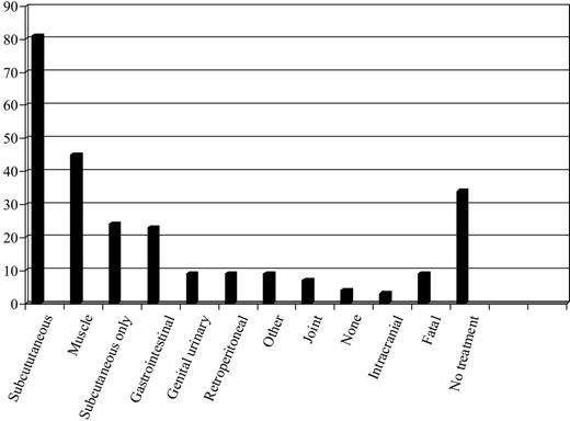
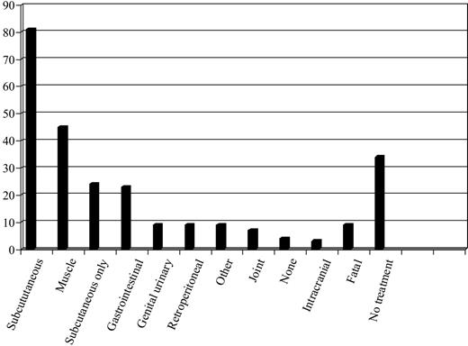
The presence or absence of bleeding symptoms was known for 149 patients. The categories of bleeding are shown in Figure 1 and are similar to previously reported cohorts.1,21 Hemostatic therapy was not required at all for 51 (34%) of these patients excluding one patient who died of bleeding before hemostatic treatment could be given. Two of these patients were asymptomatic and presented following routine preoperative coagulation screens, 22 patients had subcutaneous bleeds only, 9 had gastrointestinal bleeding, 6 hematuria, 11 soft tissue hematoma, one had a subconjunctival hemorrhage, and in one no data were available.
Sites of bleeding in patients with acquired hemophilia. The percentage of cohort with each bleeding subtype is shown. Many patents had more than one type of bleeding. No hemostatic treatment was required in 34% of the patients and in 8% bleeding was the primary cause of death.
Sites of bleeding in patients with acquired hemophilia. The percentage of cohort with each bleeding subtype is shown. Many patents had more than one type of bleeding. No hemostatic treatment was required in 34% of the patients and in 8% bleeding was the primary cause of death.
The number and percentage of patients receiving each hemostatic treatment are shown in Table 2 Some patients received more than one treatment modality. Data were not collected on the hemostatic response to treatment. Furthermore, when it has been reported that patients received more than one modality of hemostatic treatment, it is not known whether treatments were given for the same episode together or sequentially or for different bleeding episodes.
Fatal bleeding.
Of the 143 patients of known bleeding status, bleeding was the cause of death in 13 (9.1%) at a median of 19 days (range, 1-146 days). As shown in Table 3, early deaths (within the first week) were generally caused by gastrointestinal and lung bleeding, whereas later deaths were predominantly secondary to soft tissue bleeds such as intracranial and retroperitoneal hemorrhage.
Factor VIII level and inhibitor titer at presentation were not useful for predicting the severity of bleeding events. The median presenting factor VIII in patients who had a fatal bleed was 4 IU/dL (range, < 1-12 IU/dL), whereas for the group that required no hemostatic therapy the median factor VIII was 3 IU/dL (range, < 1-25 IU/dL). The inhibitor titer at presentation was similar in the patients who had fatal bleeds and the patients who required no hemostatic therapy with a median of 7.2 BU/mL (range, 1.4-219 BU/mL) and 7 BU/mL (range, 0.8-717 BU/mL), respectively. Furthermore, in the 46 patients who presented with a factor VIII level of 1 IU/dL or less, 14 (30%) did not require hemostatic treatment, a proportion similar to the whole cohort.
Presenting characteristics as predictors of outcome
The Cox proportional hazards regression analysis was applied to all patients whose presentation characteristics were known to investigate whether these characteristics were associated with time to attaining first remission (105 in analysis) or time to death (113 in analysis). The presenting characteristics entered into the Cox model were age, underlying diagnosis, factor VIII level, and inhibitor titer at diagnosis, sex, and center type.
Age was associated with survival and also with achieving a CR. The direction of the association was that older patients were more likely to have died during the follow-up period (P < .001) but also achieved remission more quickly (P < .042). The other variables investigated were not significantly associated (5% significance level) with outcome.
Comparability of primary treatment groups
As shown in Table 4, immunosuppression was given to 143 (95%) of the 151 patients on whom the data were reported. Patients were treated at the discretion of the local clinician according to United Kingdom national guidelines.25 Steroids were almost invariably prescribed as prednisolone 1 mg/kg. Those treated with steroids and a cytotoxic agent received prednisolone 1 mg/kg usually combined with oral cyclophosphamide 1 to 2 mg/kg daily, although 3 patients received oral azathioprine 100 to 150 mg daily.
The most informative comparison is between those treated with steroids alone (n = 40) and those treated with steroids and cytotoxic agents initiated together at presentation (n = 48). For the purposes of this study, these were designated the primary treatment groups. Allocation of patients to treatment groups was not randomized and it is possible, therefore, that treatment choices were influenced by presenting characteristics that had previously been reported to affect outcome. This was investigated by analyzing whether any presenting features were disproportionately represented in the either of the primary treatment groups.
The patients treated with steroids alone had a lower median inhibitor titer (8 BU/mL) than those treated with steroids and cytotoxics (18 BU/mL), P < .01. The median factor VIII level was 5 IU/dL in the steroid group compared to 2 IU/dL in the steroid and cytotoxic group (not significant at the 5% level). However, neither inhibitor titer nor factor VIII level had been found significantly associated with outcome by the Cox regression analysis.
Age, sex, and underlying diagnosis did not differ significantly between the steroid and the steroid and cytotoxic groups. Patients who were not treated at a hemophilia or a comprehensive care hemophilia center were more likely to have been treated with steroids alone (P < .05).
The 8 patients who did not receive any immunosuppressive treatment had a median age of 81 years (range, 67-95 years). Six of these patients were known to have died, all within 22 days of diagnosis of either malignancy or medical problems related to old age except for one patient who died on day 1 of gastrointestinal bleeding. It is likely that the decision not to immunosuppress was predominantly based on the underlying diagnosis in these cases.
Inhibitor eradication
Of the 144 patients with remission follow-up information, 102 (71%) achieved CR. The accumulated observation period for the 93 patients for whom remission dates were known was 37 years. The Kaplan-Meier estimate of median time to remission for all patients irrespective or treatment group or order of starting treatment was 57 days (95% CI, 46-74).
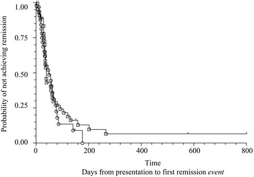
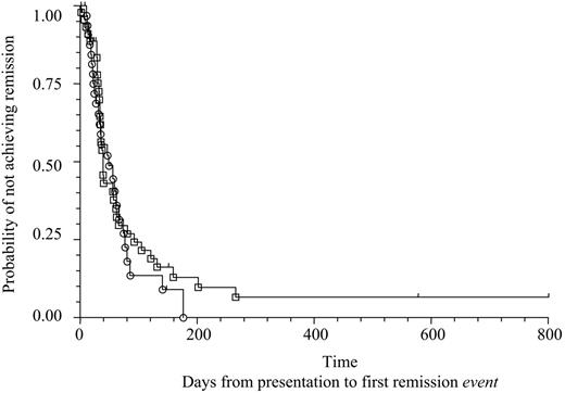
Comparison of time to CR in the primary treatment groups is shown in the Kaplan-Meier plot in Figure 2. The Kaplan-Meier estimate of median time to CR in those treated with steroids alone was 49 days (95% CI, 31-62) compared to 39 days (95% C, 34-57) in the group in whom steroids and cytotoxics were started together on day 1 (Peto log-rank test P = .51).
Complete remission dependent on treatment group. Kaplan-Meier plot of probability that patients will not have achieved remission at a given time dependent on treatment group. ○, remission event in steroids alone group; □, remission event in steroids and cytotoxics started together at presentation group. There is no evidence of a difference between treatment groups in terms of time to first remission (Peto log-rank test, P = .51).
Complete remission dependent on treatment group. Kaplan-Meier plot of probability that patients will not have achieved remission at a given time dependent on treatment group. ○, remission event in steroids alone group; □, remission event in steroids and cytotoxics started together at presentation group. There is no evidence of a difference between treatment groups in terms of time to first remission (Peto log-rank test, P = .51).
Although reporting the overall percentages of patients who attained CR while under study observation does not take into account the variable time they were followed, other studies in the field have reported results in this way and the data are included to allow comparison (Table 5)There was no evidence that the addition of IVIG to either treatment regimen improved outcome. In the group of patients treated with steroids alone, those who received IVIG (n = 15) achieved CR after a median (95% CI) of 59 days (27-66), whereas those who did not receive IVIG (n = 19) achieved CR after 36 days (24-62). In the group of patients treated with steroids and a cytotoxic agent those who received IVIG (n = 16) achieved CR after 40 days (30-57), whereas those who did not receive IVIG (n = 29) achieved CR after 37 days (34-61).
Contrary to previous reports that suggested that patients with rheumatoid arthritis are resistant to treatment with steroids alone, we found that of the 11 patients with rheumatoid arthritis 4 were treated with steroids alone and all achieved CR after 13, 21, 22, and 80 days, respectively. Three of these patients were further observed for 2, 20, and 21 months, respectively, and during these periods none had a relapse.
Inhibitor relapse
Of the 102 patients who were known to have achieved CR, data on relapse are available for 90, of whom 18 (20%) had a relapse. The median time to relapse for the 11 patients for whom these data were available was 7.5 months after stopping immunosuppression (range, 1 week to 14 months). The median observation time after remission in the 68 patients who did not have a relapse and for whom these data were available was 13 months (range, 0-37 months).
A second CR was induced in 10 (56%) patients and in a further 4 (22%) the inhibitor was eradicated, factor VIII normalized but immunosuppression could not be stopped without relapse. In 4 (22%) patients a second remission could not be achieved.
Survival
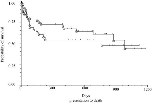
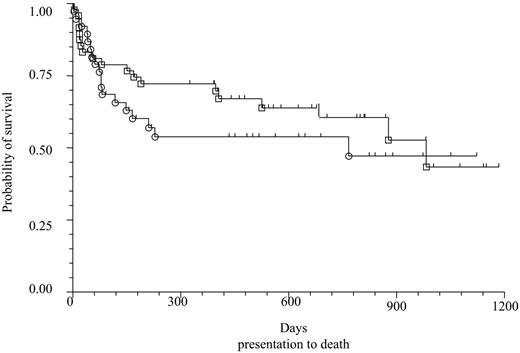
A comparison of survival in patients treated with steroids alone compared to those treated with steroids and cytotoxics started together is shown by the Kaplan-Meier plot in Figure 3. The Kaplan-Meier estimate of median survival time between presentation and death in those treated with steroids alone was 767 days (95% CI, 148-1122) compared to 975 days (95% CI, 526-1176) in patients who received steroids and cytotoxics together (Peto long-rank test P = .33).
Survival from all cause mortality dependent on treatment group. The figure shows a Kaplan-Meier plot of the probability of surviving after presentation dependent on treatment group. ○, a death in steroids alone group; □, a death in steroids and cytotoxics started together at presentation group. There is no evidence of a difference between treatment groups in terms of length of survival after presentation (Peto log-rank test, P = .33).
Survival from all cause mortality dependent on treatment group. The figure shows a Kaplan-Meier plot of the probability of surviving after presentation dependent on treatment group. ○, a death in steroids alone group; □, a death in steroids and cytotoxics started together at presentation group. There is no evidence of a difference between treatment groups in terms of length of survival after presentation (Peto log-rank test, P = .33).
A summary of survival for the whole cohort is shown in Table 5. To be consistent with other study reports, this table reports percent of patients who died, but similar to remission, these percentages do not take into account the variable time patients were observed.
Morbidity unrelated to bleeding episodes
Data were received on 112 patients relating to non–bleeding-related morbidity. No morbidity was reported in 55 (49%) patients. Sepsis was reported in 37 (33%) patients and contributed to death in 12 (11%) cases. Neutropenia was reported in 13 (12%) patients and 2 patients had thrombocytopenia. Twelve of the cytopenic patients had been treated with steroids and a cytotoxic agent and one had received cyclophosphamide alone. Six of these patients were reported to have had infections and 2 required growth factor support. None of the patients in whom sepsis contributed to death were reported to have neutropenia.
A raised blood sugar level was reported in 8 patients and one had worsening control of previously diagnosed non–insulin-dependent diabetes mellitus. Other major morbidity reported was steroid-induced psychosis (n = 2), proximal myopathy (n = 2), syndrome of inappropriate antidiuretic hormone (n = 2), congestive cardiac failure secondary to steroids (n = 2), gastrointestinal bleeding induced by steroids (n = 2), and osteoporosis secondary to steroids (n = 1).
Discussion
This study reports data on a population of unselected, consecutive patients with acquired hemophilia A, thus reducing the potential for referral and reporting bias and provides the most complete description of the disease to date.
Previously published data are almost exclusively of referral center patients, either as retrospective surveys1,5,17–19 or single-center cohorts.4–16 This is likely to have led to the reporting of more severely affected and younger patients. This view is supported by the finding that the patients reported in this series1,20,21 are significantly older than previously reported, have a lower incidence of pregnancy-related acquired hemophilia, and a higher incidence of malignancy.1 Furthermore, although this study confirmed that the pattern of bleeding in patients with acquired hemophilia differs from patients with congenital hemophilia and was very similar to that described in previous studies,1,3,24 the variation in bleeding symptoms is more marked in this study with 33% of patients requiring no hemostatic treatment and 8% suffering fatal bleeds. Together these findings show that the cohort reported here is different from other studies and the most likely explanation for this is that the cohort is a more complete representation of patients with acquired hemophilia A compared with tertiary referral center patients.
The current study has identified almost all patients who presented with acquired hemophilia in the United Kingdom over a 2-year period allowing for an accurate incidence of 1 in 1.48 million/y to be calculated. This result equates closely with the only other reported incidence in a defined population of 1.34/million/yr.7 This study also confirms that acquired hemophilia becomes more likely with increasing age. The disorder is extremely rare in childhood with only one patient under the age of 30 years presenting over the 2-year period. This is in contrast to a previous report in which 8% of patients were below the age of 20 years and 3.7% younger than 11 years,1 but is comparable to the very rare reports of childhood acquired hemophilia elsewhere in the literature.26 Similarly, pregnancy-related acquired hemophilia is a rare presentation with only 3 patients (1:350 000 births) reported in a 2-year period. This is a similar rate to that noted in an Italian registry, a country with a similar population to the United Kingdom, where 25 patients were seen in 10 years.18 In the United Kingdom cohort the likelihood of an underlying diagnosis decreases with age. This suggests that either old age alone is a risk factor for acquired hemophilia, older patients were investigated less intensively than the younger patients, or both.
The severity of bleeding did not correlate with the factor VIII level or the inhibitor titer and was not useful in predicting those patients who would have fatal bleeding or those with sufficiently mild bleeding not to have required hemostatic treatment. Although patients often present with severe or life-threatening bleeding, this study demonstrates that fatal bleeding can occur up to 5 months after presentation if the inhibitor is not eradicated. These data do not support the view that management decisions should be based on the presenting factor VIII level or inhibitor titer.27
Although spontaneous remission may occur,28 immunosuppression to eradicate the inhibitor is recommended for all patients with acquired hemophilia A to reduce the length of time the patient is at risk of severe bleeding. The best regimen to achieve this, however, is not known. Standard therapy in most centers is either to use steroids alone or in combination with a cytotoxic agent, most commonly cyclophosphamide. More recently, reports of treatment with rituximab have been published.29,30 The studies reporting immunosuppressive regimens have almost invariably been single-center cohorts that have not included controls.4–16 These studies will tend to preferentially report good outcomes as opposed to average or poor outcomes and journals will similarly be more likely to publish good outcome studies. One randomized study published to date found no difference between nonremitters randomized to cyclophosphamide and those that continued with steroids after an initial 3-week course of steroids.31
The study presented here is the largest cohort reported and shows that both the proportion of patients who achieve CR and the median time to CR was the same for steroids alone and steroids plus cytotoxics. The patients were assigned to an immunosuppressive regimen according to the preference of their clinician. Analysis of treatment groups showed that patients in the steroid and cytotoxic combined group had a higher inhibitor titer at presentation than the patients in the steroids alone group but this did not relate to time taken to reach remission or survival time. A higher inhibitor titer at presentation may have led clinicians to prescribe a more intensive immunosuppressive regimen that potentially improved outcome such that a difference between the treatment arms was not observed. There were 16 centers that contributed 3 or more patients and of these 11 centers prescribed a single regimen to all patients irrespective of inhibitor titer. Five centers prescribed variable regimens, in 3 centers the choice of regimen was not associated with inhibitor level, whereas in 2 centers there was a trend to use combined steroids and cytotoxic agents in patients with higher inhibitor titers. This potentially biasing effect is an unavoidable consequence of the nonrandomized study design used but in the absence of adequately powered randomized studies remains the best available data.
Age was the only factor shown to be associated with survival, but the 2 treatment groups did not differ with regard to age. The comparison of the treatments groups therefore gives useful information in the absence of adequately powered prospective randomized studies.
A meta-analysis of 249 patients from 20 papers (range, 5-34 patients per report), none of which were controlled studies, found that 70% of patients achieved remission with steroids and 89% with the combination of steroid and cyclophosphamide but showed no advantage in survival. The authors point out that the cohort studies included are prone to the reporting of good outcomes and it is possible that the apparent advantage of the combination of steroid and cyclophosphamide in inhibitor eradication may have resulted from this.24
The 2 studies to date that directly compare treatment groups (Green et al31 and this study) do not provide evidence that cyclophosphamide and steroids are superior to steroids alone despite a combination of cohort studies suggesting otherwise. Although prospective randomized trials are the optimum method to assess treatment regimens, in a rare disease such as acquired hemophilia A it is very difficult to perform these trials. An adequately powered study would need to randomize hundreds of patients. It is unlikely that this sort of study is feasible within the current regulatory system and reports of cohorts of patients are likely to be the only data available for the foreseeable future.
It has been reported that adding IVIG to the immunosuppressive regimen may improve CR rates and survival.32 Our data do not support this. A similar conclusion was reached in a review of the literature and an open-label, uncontrolled study of steroids and IVIG reported a similar remission rate (57%)8 to this study.
One important finding presented here is that about 20% of patients had a relapse after immunosuppression had been stopped. The relapses recorded here occurred between 1 week and 14 months with a median of 7.5 months. This means that long-term follow-up of patients with acquired hemophilia should be recommended and patients should be counseled to report signs of bleeding or bruising so that relapse can be detected early to minimize the length of time patients are at risk of bleeding.
Although eradication of the inhibitor is important to decrease the risk of severe bleeding, immunosuppression of an elderly group of patients is likely to be associated with significant morbidity. This is confirmed in the cohort presented here and nonbleed morbidity was reported in about half the patients. Sepsis resulting from immunosuppression in at-risk patients was the most commonly reported adverse event and contributed to death in 12 patients.
Our data demonstrate no difference in mortality between the treatment arms. This supports the conclusion of the meta-analysis, which also found no difference in mortality when steroids were compared to cyclophosphamide. The discrepancy in the meta-analysis between inhibitor eradication and survival was attributed to a higher mortality in the steroid and cyclophosphamide group.24 This was not seen in our study but highlights the need to report both efficacy and toxicity of treatment regimens and to consider their potential side effects in an elderly population.
In conclusion, our data show that acquired hemophilia A presents with an incidence of 1.48/million/y. In this nonrandomized study, treatment with steroids alone or steroids plus cytotoxic agents was indistinguishable and at present it is reasonable for clinicians to use either regimen depending on the patient's individual circumstances and local preferences. Many patients do not require hemostatic treatment but if the inhibitor is not eradicated fatal bleeding remains a risk. No clinical or laboratory features of the disease were found to identify the high-risk patients and so all patients should be immunosuppressed as soon as the diagnosis is made.
Although prospective randomized studies are the gold standard for assessing treatment regimens, the rarity of acquired hemophilia and the number of patients required for such studies means that international collaboration is required and is unlikely to occur. In this context our study, which reports on a comparison of treatment regimens in an unselected but nonrandomized cohort of patients, provides the best data on response to immunosuppression currently available. On the basis of these data and a review of the literature, new guidelines for the treatment of acquired hemophilia have been compiled as part of general guidelines on the management of inhibitors.33
Authorship
Contribution: P.C. designed and initiated the study, performed data collection, performed data interpretation, and coordinated writing the manuscript; S.H. was involved in data interpretation, undertook statistical analysis, and contributed to manuscript preparation; T.B. was involved in study design, entered more than 5% of the study patients, and contributed to data analysis and writing the manuscript; G.D. entered more than 5% of the study patients, and contributed to data analysis and writing the manuscript; J.H. entered more than 5% of the study patients, and contributed to data analysis and writing the manuscript; M.M. entered more than 5% of the study patients, and contributed to data analysis and writing the manuscript; D.K. was involved in study design, entered more than 5% of the study patients, and contributed to data analysis and writing the manuscript; R.L. was involved in study design, and contributed to data analysis and writing the manuscript; S.B. was involved in study design, entered more than 5% of the study patients, and contributed to data analysis and writing the manuscript; and C.H. was involved in study design, entered more than 5% of the study patients, and contributed to data analysis and writing the manuscript.
Conflict-of-interest disclosure: The authors declare no competing financial interests.
A complete list of the participating members of the UK Haemophilia Centre Doctors' Organisation appears as a data supplement to the online version of this article (Document S1, available on the Blood website; see the Supplemental Document link at the top of the online article).
Correspondence: Peter W. Collins, Department of Haematology, University Hospital of Wales, and Medical School Cardiff University, Heath Park, Cardiff CF14 4XN, United Kingdom; e-mail: peter.collins@cardiffandvale.wales.nhs.uk.
The online version of this article contains a data supplement.
An Inside Blood analysis of this article appears at the front of this issue.
The publication costs of this article were defrayed in part by page charge payment. Therefore, and solely to indicate this fact, this article is hereby marked advertisement in accordance with 18 USC section 1734.
All United Kingdom hematologists who contributed data are acknowledged and thanked for their contribution. B. Jenkins and Elinor Collins helped in organizing and performing the data collection.

