Abstract
Peroxiredoxin 2 (Prx2), a thiol-dependent peroxidase, is the third most abundant protein in the erythrocyte, and its absence in knock-out mice gives rise to hemolytic anemia. We have found that in human erythrocytes, Prx2 was extremely sensitive to oxidation by H2O2, as dimerization was observed after exposure of 5 × 10⁶ cells/mL to 0.5 μM H2O2. In contrast to Prx2 in Jurkat T lymphocytes, Prx2 was resistant to overoxidation (oxidation of the cysteine thiol to a sulfinic/sulfonic acid) in erythrocytes. Reduction of dimerized Prx2 in the erythrocyte occurred very slowly, with reversal occurring gradually over a 20-minute period. Very low thioredoxin reductase activity was detected in hemolysates. We postulate that this limits the rate of Prx2 regeneration, and this inefficiency in recycling prevents the overoxidation of Prx2. We also found that Prx2 was oxidized by endogenously generated H2O2, which was mainly derived from hemoglobin autoxidation. Our results demonstrate that in the erythrocyte Prx2 is extremely efficient at scavenging H2O2 noncatalytically. Although it does not act as a classical antioxidant enzyme, its high concentration and substrate sensitivity enable it to handle low H2O2 concentrations efficiently. These unique redox properties may account for its nonredundant role in erythrocyte defense against oxidative stress.
Introduction
The peroxiredoxins (Prxs) constitute a family of homodimeric peroxidases that reduce H2O2 and alkyl hydroperoxides to water and alcohol, respectively. They rely on a conserved cysteine residue to catalyze peroxide reduction. There are 6 known mammalian isoforms (Prx1-6), classified as typical 2-Cys, atypical 2-Cys, or 1-Cys Prxs based on the mechanism and number of cysteines involved during catalysis.1 When peroxiredoxin 2 (Prx2; a typical 2-Cys Prx) reacts with peroxide, the peroxidatic cysteine at the active site on one subunit is oxidized to a sulfenic acid. A second conserved cysteine at the C-terminal end of the other subunit (the resolving cysteine) then reacts with the sulfenic acid to form a disulfide bridge. Reduction of the disulfide by thioredoxin (Trx) regenerates Prx2 and completes the cycle. Trx is in turn regenerated by thioredoxin reductase (TrxR), with reducing equivalents derived from NADPH.2 An intriguing feature of mammalian 2-Cys Prxs is that in the presence of high levels of peroxide, the peroxidatic Cys becomes overoxidized to the sulfinic (SO2H) or sulfonic (SO3H) acid form.3 This abolishes the enzyme's peroxidase activity, although overoxidized Prx can be slowly reverted to the reduced state by sulfiredoxin.4 It has been suggested that overoxidation allows intracellular accumulation of H2O2, which can then function as a signal transducer for various pathways.5,6
Compared with other somatic cells, erythrocytes are exposed to oxidative stress from a wide variety of sources. They contain high levels of O2 and hemoglobin (Hb), which continually autoxidizes to produce O2− and H2O2. They also have membranes rich in polyunsaturated fatty acids, and the heme group of Hb is able to initiate a wide array of free radical reactions.7,8 In addition, as anucleated cells, erythrocytes are unable to synthesize new proteins to replace damaged ones during their 120-day lifespan. Therefore, erythrocytes are well equipped with many antioxidant proteins.
For a long time, it was considered that catalase and glutathione peroxidase (GPx) constitute the erythrocyte's defense against H2O2, and there has been continuous debate about which of these is the more significant.9–14 Until recently, little attention has been given to the antioxidant role of Prxs in erythrocytes. Erythrocyte Prx2 is the third most abundant erythrocyte protein,15 and has previously been called calpromotin for its ability to regulate calcium-activated potassium efflux.16–18 However, whether this property is linked to peroxide metabolism is not understood. Recently, Johnson et al19 found that modeling peroxide metabolism in GPx-deficient erythrocytes required the inclusion of Prx2 to fit their data. This suggests that the significance of Prx2 in peroxide consumption may have been overlooked thus far.
Erythrocytes also possess Prx1 and Prx6, although in lesser amounts than Prx2. Prx2 expression is induced at the early stages of erythroid differentiation, before Hb accumulation occurs.20 It has been purified from erythrocytes for use in early structural studies.15 Prx2 is able to protect Hb from exogenous oxidation,21 and is thought to remove hydroperoxides at the erythrocyte membrane.22 It is notable that mice lacking the Prdx2 gene develop hemolytic anemia, with their Hb showing signs of oxidation and precipitation as Heinz bodies.23 Intriguingly, these mice possess fully functional catalase and GPx. This indicates that Prx2 serves a nonredundant function in protecting erythrocytes against oxidative damage.
Since the discovery of the enzymatic activity of the Prx family, there has been intense focus on how they contribute to antioxidant defense and redox signaling. Various cell lines and organisms have been studied, but little has been done with erythrocytes. It is not known whether erythrocyte Prxs play a significant role in peroxide removal, and whether they undergo typical Trx-dependent redox cycling with a propensity to overoxidize. We have investigated Prx2 in human erythrocytes and have found that its behavior is very different from that reported for other cells.
We show that erythrocyte Prx2 is remarkably sensitive to reversible oxidation by H2O2 concentrations in the low micromolar range. However, recycling of the oxidized dimer occurs very slowly. Unexpectedly, Prx2 was found to be resistant to overoxidation in the erythrocyte. Thus, although overall consumption is low, Prx2 is able to act as an efficient scavenger of low concentrations of H2O2 without undergoing inactivation due to overoxidation.
Materials and methods
Materials
N-ethyl maleimide (NEM), reduced glutathione (GSH), xylenol orange, 1-chloro-2,4-dinitrobenzene (DNCB), sorbitol, dimethylsulfoxide (DMSO), Tween-20, and ethyleneglycoltetraacetic acid (EGTA) were from Sigma (St Louis, MO). Sodium azide was from Fisons (Loughborough, Leics, United Kingdom). Ferrous ammonium sulfate was obtained from J. T. Baker (Phillipsburg, NJ), and glycerol was from Merck (Darmstadt, Germany). Glucose, ethylenediaminetetraacetic acid (EDTA), N-2-hydroxyethylpiperazine-N′-2-ethanesulfonic acid (HEPES), bromophenol blue, sodium dodecyl sulfate (SDS), and H2O2 (30% wt/vol) were supplied by BDH (Poole, England). Tris(hydroxymethyl)aminomethane (Tris), CHAPS, and Complete protease cocktail tablets were purchased from Roche (Mannheim, Germany).
Rabbit polyclonal antibodies to Prx2 and overoxidized Prx (which detects Prx-SO2H and Prx-SO3H) were obtained from LabFrontier (Seoul, Korea), and goat antirabbit horseradish peroxidase was purchased from Sigma. Polyvinylidene difluoride membrane and enhanced chemiluminescence reagents were supplied by Amersham Biosciences (Buckinghamshire, United Kingdom).
Preparation of erythrocytes and treatment with hydrogen peroxide
Blood was drawn from healthy human volunteers into heparinized tubes and used according to the policy of the New Zealand Blood Transfusion Service for donated blood. Informed consent was provided in accordance with the Declaration of Helsinki. After plasma removal, cells were washed 3 times with at least 1.5 volumes of ice-cold phosphate-buffered saline (PBS; 10 mM phosphate in 137 mM NaCl and 2.7 mM KCl, pH 7.4), with successive removals of visible buffy coat and/or the top layer. The approximate number of cells per milliliter of packed erythrocytes was determined using a hemocytometer. The concentration of H2O2 in stock solutions was quantified by measuring A240 (ϵ = 43.6 M−1 cm−1). For treatment with H2O2, 5 × 10⁶ cells were suspended in 1 mL PBS containing 5 mM glucose unless otherwise stated, and incubated at 37°C. Where required, cells were pretreated with 1 mM sodium azide for 5 minutes, or 30 μM DNCB dissolved in dimethyl sulfoxide (DMSO) for 10 minutes prior to H2O2 addition. In experiments involving DNCB, controls were treated with the equivalent volume of DMSO. For inhibition of Hb autoxidation, cells were bubbled with carbon monoxide (CO) until a color change to pink was observed. Formation of carboxyhemoglobin was verified by spectral analysis.24
Trapping Prx2 in its native redox state
After each treatment, erythrocytes were pelleted, the supernatant was removed, and cells were resuspended in 1 mL PBS containing 100 mM N-ethyl maleimide (NEM). After 15-minute incubation at room temperature, cells were pelleted and lysed in 100 μL nonreducing lysis buffer (65.8 mM Tris-HCl, pH 6.8, 10.5% glycerol [vol/vol], 2.1% SDS [wt/vol], 0.053% bromophenol blue [wt/vol]) containing 100 mM NEM.
Immunoblot analysis
Proteins (approximately 20 μg per lane for erythrocyte samples) were resolved by nonreducing 12% SDS–polyacrylamide gel electrophoresis (PAGE) and transferred electrophoretically to a polyvinylidene difluoride membrane. Blocking was performed for 2 hours at room temperature in 5% (wt/vol) nonfat dried milk, 15 mM sodium azide, and 2% H2O2 (to inhibit the pseudoperoxidase activity of Hb) in Tris-buffered saline (TBS)/0.05% Tween 20. Incubation with antibodies to Prx2 or Prx-SO2H was performed overnight at 4°C in 5% (wt/vol) nonfat dried milk in TBS/0.05% Tween 20. Bands were detected with goat antirabbit horseradish peroxidase using enhanced chemiluminescence reagents, and visualized with the ChemiDoc XRS gel documentation system (Bio-Rad, Segrate, Italy). Quantification of band intensities was performed with Quantity One analysis software (Bio-Rad, Hercules, CA).
All immunoblots were representative of experiments performed with erythrocytes from at least 2 volunteers.
Treatment of Jurkat cells
Jurkat T lymphocytes (American Type Cell Culture, Rockville, MD) were maintained in RPMI-1640 supplemented with 10% (vol/vol) heat-inactivated fetal bovine serum, 2 mM glutamine, 100 U/mL penicillin, and 100 μg/mL streptomycin at 37°C in a humidified atmosphere with 5% CO2. For treatment with H2O2, 1 × 10⁶ cells were resuspended in 1 mL fresh media and incubated at 37°C. After treatment, the cells were pelleted, washed with PBS, and lysed in buffer (40 mM HEPES, 50 mM NaCl, 1 mM EDTA, 1 mM EGTA, pH 7.4) with protease inhibitors and 1% CHAPS to a density of 5 × 10⁷ cells/mL. The lysate was incubated for 15 minutes on ice, and insoluble material was removed by centrifugation. Protein quantities were measured with the Bio-Rad Protein Assay (Hercules, CA), using bovine serum albumin as the standard.
Measurement of H2O2 consumption
H2O2 consumption was measured using the ferrous oxidation of xylenol orange assay,25 adapted for low peroxide concentrations. The assay reagent consisted of 400 μM xylenol orange, 1 mM ferrous ammonium sulfate, 400 mM sorbitol, and 75 mM sulfuric acid. After treatment of erythrocyte suspensions with H2O2 for the appropriate durations, erythrocytes were pelleted and 700 μL supernatant was added to 250 μL assay reagent. The mixture was vortexed immediately, left at room temperature for 35 to 40 minutes, and the absorbance at 560 nm recorded.
Thioredoxin reductase activity assays and glutathione measurement
Activity was measured in Jurkat cells by monitoring NADPH-dependent reduction of DTNB by 100 μg lysate protein (derived from approximately 1 × 10⁶ cells).26 The difference in rates of TNB formation before and after NADPH addition was used to calculate enzyme activity, and a molar extinction coefficient of TNB of 14 150 at 412 nm was used.27
To measure TrxR activity in erythrocytes, the assay was modified to overcome interference by the high Hb absorbance at 412 nm. Ten milliliters of 5 × 10⁶ cells/mL suspensions were pretreated with 60 μM DNCB or the equivalent volume of DMSO, and incubated for 30 minutes at 37°C with slow rotation. Cells were pelleted and lysed in 55 μL 5 mM phosphate buffer (pH 7.4) containing 5 mM DTNB. The hemolysate was incubated at 37°C for 1 minute, after which NADPH was added to one sample to a final concentration of 200 μM. An equivalent volume of buffer was added to the control. After further incubation at 37°C for 7 to 10 minutes, the lysate was passed through a Micro Bio-Spin 30 column (Bio-Rad, Hercules, CA). The column was washed with 40 μL 5 mM phosphate buffer (pH 7.4) to elute residual Hb retained on the column, and TNB was eluted with 800 μL 5 mM phosphate buffer (pH 7.4). The eluate was measured for absorption at 412 nm. (Assays of known TNB standards confirmed that TNB is completely recovered by this method.) Activity was calculated from the difference in A412 between hemolysates incubated with and without NADPH. The degree of inhibition by DNCB was calculated from the difference in absorbance increases between DNCB-treated cells and control cells upon NADPH addition.
TrxR activity in Jurkat cells was also analyzed by the modified assay. Cells (1 × 10⁶) suspended in 1 mL cell culture medium were pretreated with 60 μM DNCB or the equivalent volume of DMSO. After 10-minute incubation at 37°C, cells were washed with 1 mL PBS and lysed as for treatments with H2O2. Lysates were vacuum-concentrated at ambient temperature until dry, then reconstituted in 60 μL DTNB buffer (5 mM phosphate buffer [pH 7.4] containing 5 mM DTNB). After incubation for 1 minute at 37°C, NADPH was added to a final concentration of 200 μM and incubated for 7 to 8 minutes. Samples were processed and TNB formation measured as with erythrocytes, except with omission of the wash step.
To measure the effect of DNCB on glutathione (GSH) content, 100 mL erythrocyte suspensions (5 × 10⁶ cells/mL) were incubated with DNCB under the same conditions as used for examining Prx2 oxidation state. GSH levels were measured by the method according to Beutler,28 which monitors DTNB reduction.
Results
The native redox state of erythrocyte Prx2
Erythrocytes were lysed in sample buffer, run on SDS-PAGE, and immunoblotted with antibodies to Prx2. The monomer was observed under reducing conditions, while under nonreducing conditions Prx2 was almost completely dimerized (Figure 1A). From this procedure, it was not apparent whether Prx2 is oxidized following cell lysis, or whether it is already present in its oxidized form in intact cells. To address this issue, erythrocytes were incubated with 100 mM NEM (a thiol-blocking agent) prior to lysis to prevent dimerization, then subjected to immunoblotting under nonreducing conditions. Most of the Prx2 was recovered as the monomer (Figure 1B), and the presence of NEM in preincubation and lysis buffers resulted in optimal recovery. This suggests that the dimer observed under nonreducing conditions was an artifact of Prx2 oxidation upon cell lysis. With different erythrocyte samples from many donors, the reduced form consistently comprised the major redox state, thus confirming that Prx2 exists mostly in the reduced state under normal conditions. Although the small proportion of dimers may be an indication of constitutive oxidation, it could still represent artifactual oxidation during sample analysis. The latter is supported by the observation that addition of catalase (5 μg/mL) to the incubation buffer could further improve capture of reduced Prx2 (not shown). This facile oxidation during preparation of erythrocyte lysates meant that in subsequent experiments, there was some variability in the amount of dimer in control samples.
The redox state of Prx2 in erythrocytes. (A) Cells were lysed in SDS-sample buffer, analyzed under reducing (R) or nonreducing (NR) conditions, and immunoblotted with antibodies against Prx2. Molecular weight (MW) markers are indicated on the left. (B) Cells were preincubated with or without 100 mM NEM for 15 minutes, then lysed in nonreducing SDS-sample buffer in the absence or presence of 100 mM NEM. Samples were then immunoblotted for the presence of Prx2. D indicates dimeric Prx2; M, monomeric Prx2.
The redox state of Prx2 in erythrocytes. (A) Cells were lysed in SDS-sample buffer, analyzed under reducing (R) or nonreducing (NR) conditions, and immunoblotted with antibodies against Prx2. Molecular weight (MW) markers are indicated on the left. (B) Cells were preincubated with or without 100 mM NEM for 15 minutes, then lysed in nonreducing SDS-sample buffer in the absence or presence of 100 mM NEM. Samples were then immunoblotted for the presence of Prx2. D indicates dimeric Prx2; M, monomeric Prx2.
Sensitivity of erythrocyte Prx2 to oxidation by H2O2
Having established a suitable protocol to monitor the redox transitions of Prx2, we investigated the sensitivity of erythrocyte Prx2 to H2O2-induced oxidation. Cells (5 × 10⁶/mL) were treated at a range of concentrations for 10 minutes and immunoblotted under nonreducing conditions. We found that a significant proportion of the protein was oxidized at 0.5 μM H2O2, as reflected by a transition from monomer to dimer, and oxidation was complete with 5 μM (Figure 2A-B). With all H2O2 treatments, reducing gels gave a single monomer band (not shown), and spectral analysis of Hb showed that it was not oxidized at this range of H2O2 concentrations (data not shown).
The effect of H2O2 on the redox state of Prx2 in cells. (A) Erythrocytes were treated at the indicated concentrations for 10 minutes and immunoblotted. Dimeric Prx2 ran as a doublet. (B) Quantification of band intensities as measured by chemiluminescent densitometry. Data are means ± SD of 3 experiments. (C) Erythrocyte suspensions of 5 × 10⁷ cells/mL were treated with the indicated concentrations of H2O2 for 10 minutes and immunoblotted. (D) Erythrocytes (5 × 10⁷ cells/mL) were pretreated with 10 mM azide for 5 minutes, then with the indicated concentrations of H2O2 for 10 minutes and immunoblotted. (E) Jurkat cells were treated at the indicated concentrations of H2O2 for 10 minutes and immunoblotted. Protein (15 μg) was loaded per lane. D indicates dimeric Prx2; M, monomeric Prx2.
The effect of H2O2 on the redox state of Prx2 in cells. (A) Erythrocytes were treated at the indicated concentrations for 10 minutes and immunoblotted. Dimeric Prx2 ran as a doublet. (B) Quantification of band intensities as measured by chemiluminescent densitometry. Data are means ± SD of 3 experiments. (C) Erythrocyte suspensions of 5 × 10⁷ cells/mL were treated with the indicated concentrations of H2O2 for 10 minutes and immunoblotted. (D) Erythrocytes (5 × 10⁷ cells/mL) were pretreated with 10 mM azide for 5 minutes, then with the indicated concentrations of H2O2 for 10 minutes and immunoblotted. (E) Jurkat cells were treated at the indicated concentrations of H2O2 for 10 minutes and immunoblotted. Protein (15 μg) was loaded per lane. D indicates dimeric Prx2; M, monomeric Prx2.
Increasing the cell density to 5 × 10⁷ cells/mL resulted in at least 10-fold more H2O2 being required to yield equivalent dimerization (Figure 2C). For example, 5 μM H2O2 at this cell density and 0.5 μM H2O2 at 5 × 10⁶ cells/mL induced comparable dimerization additional to controls. At higher (∼ 50-200 μM H2O2) treatments, oxidation was incomplete and stabilized at approximately 80% dimerization. This could be due to more efficient H2O2 consumption by catalase at higher concentrations of H2O2. To examine this, erythrocyte suspensions of 5 × 10⁷ cells/mL were pretreated with 10 mM azide to inhibit catalase, then subject to the same treatment with H2O2 (Figure 2D). Dimerization was slightly greater than without azide at low H2O2 concentrations (compare 5 μM in Figure 2C-D), and proceeded to completion above 10 μM H2O2. Thus, catalase activity was protective particularly at higher levels of H2O2.
In contrast to the erythrocyte, no increase in oxidized Prx2 was observed in Jurkat cells treated with H2O2 (Figure 2E). Instead, the small amount of dimer present in control cells was progressively lost with increasing H2O2. This pattern of oxidation is in accordance with previous findings that the amount of reversibly oxidized Prx2 decreased upon treatment of Jurkat cells with 200 μM H2O2.29
Sensitivity of erythrocyte Prx2 to overoxidation by H2O2
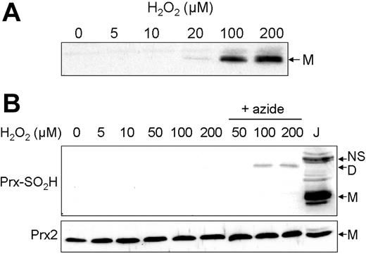
Prx2 overoxidation has been observed when various cell types, including Jurkat cells, are treated with H2O2.3,29,30 We observed this typical behavior when Jurkat cells were treated with a range of H2O2 concentrations, and immunoblots performed with antibodies to overoxidized Prx. Overoxidized bands were evident at 100 μM H2O2 and above (Figure 3A). Overoxidized Prx ran only as a monomer, and became evident concurrently with the switch to the monomeric form shown in Figure 2E.
The effect of H2O2 on overoxidation of Jurkat and erythrocyte Prx2. (A) Jurkat cells were treated with the indicated H2O2 concentrations and immunoblotted with antibodies against overoxidized Prx (Prx-SO2H). Protein (25 μg) was loaded per lane. (B) Erythrocytes (5 × 10⁶/mL) were pretreated with 1 mM azide for 5 minutes where specified, then treated with the indicated H2O2 concentrations for 10 minutes. Immunoblotting was then performed under nonreducing conditions against Prx-SO2H (top panel), and under reducing conditions against Prx2 to serve as a loading control (bottom panel). J indicates 40 μg extract from Jurkat cells treated with 200 μM H2O2 for 10 minutes as in panel A as a positive control. The nonspecific band was consistently present in untreated Jurkat extracts. NS indicates nonspecific band; D, overoxidized Prx dimer; and M, overoxidized Prx monomer.
The effect of H2O2 on overoxidation of Jurkat and erythrocyte Prx2. (A) Jurkat cells were treated with the indicated H2O2 concentrations and immunoblotted with antibodies against overoxidized Prx (Prx-SO2H). Protein (25 μg) was loaded per lane. (B) Erythrocytes (5 × 10⁶/mL) were pretreated with 1 mM azide for 5 minutes where specified, then treated with the indicated H2O2 concentrations for 10 minutes. Immunoblotting was then performed under nonreducing conditions against Prx-SO2H (top panel), and under reducing conditions against Prx2 to serve as a loading control (bottom panel). J indicates 40 μg extract from Jurkat cells treated with 200 μM H2O2 for 10 minutes as in panel A as a positive control. The nonspecific band was consistently present in untreated Jurkat extracts. NS indicates nonspecific band; D, overoxidized Prx dimer; and M, overoxidized Prx monomer.
The situation was completely different in the erythrocyte. When erythrocytes were probed with antibodies to overoxidized Prx, no overoxidation was detected either under basal conditions, or with H2O2 treatments up to 200 μM (Figure 3B, lanes 1-6). Inhibition of catalase prior to H2O2 treatment resulted in some overoxidation occurring at 100 μM H2O2 and above (Figure 3B, lanes 7-9). Even then, the overoxidized band ran in the dimeric position, suggesting overoxidation on only one peroxidatic cysteine.
Regeneration of reduced Prx2
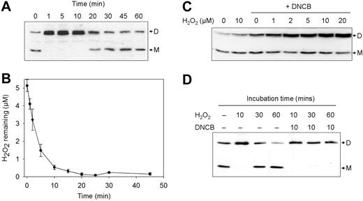
The accumulation of Prx2 dimer in erythrocytes at low micromolar concentrations of H2O2 indicates that, unlike in Jurkat cells, it is not rapidly recycled. To examine how efficient the erythrocyte is at recycling the dimer, erythrocytes were treated with 5 μM H2O2 and incubated for varying durations. Dimerization occurred rapidly, and was complete within 1 minute (Figure 4A). Total oxidation persisted for approximately 20 minutes, and then the monomer reappeared, reflecting reduction of the disulfide. Analysis of H2O2 in the medium showed that almost all H2O2 was consumed by the erythrocytes within 10 minutes, with none detectable at 20 minutes (Figure 4B). The Prx2 dimer remained while there was still H2O2 present, and reformation of the monomer required at least 10 minutes after complete consumption had occurred. The slow rate of reversal of the redox state suggests that regeneration is not limited by the persistence of H2O2 in the reaction mix.
The redox state of Prx2 in 5 μM H2O2-treated erythrocytes over time. (A) A representative immunoblot of the redox transitions of Prx2 following a bolus treatment with 5 μM H2O2. (B) Extracellular H2O2 levels over time after a bolus treatment of 5 μM H2O2. Data are the means ± SD from 3 experiments. (C) Jurkat cells were pretreated with 30 μM DNCB for 10 minutes prior to the indicated concentrations of H2O2, then immunoblotted (30 μg protein per lane). (D) The inhibition of erythrocyte Prx2 regeneration by treatment with 30 μM DNCB prior to 5 μM H2O2. D indicates dimeric Prx2; M, monomeric Prx2.
The redox state of Prx2 in 5 μM H2O2-treated erythrocytes over time. (A) A representative immunoblot of the redox transitions of Prx2 following a bolus treatment with 5 μM H2O2. (B) Extracellular H2O2 levels over time after a bolus treatment of 5 μM H2O2. Data are the means ± SD from 3 experiments. (C) Jurkat cells were pretreated with 30 μM DNCB for 10 minutes prior to the indicated concentrations of H2O2, then immunoblotted (30 μg protein per lane). (D) The inhibition of erythrocyte Prx2 regeneration by treatment with 30 μM DNCB prior to 5 μM H2O2. D indicates dimeric Prx2; M, monomeric Prx2.
As Trx and TrxR are understood to be responsible for the regeneration of reduced Prx2,2 we examined how inhibiting TrxR with DNCB affected the regeneration of reduced Prx2 after H2O2 exposure. Jurkat cells were pretreated with 30 μM DNCB, which inhibited TrxR activity by 75% (data not shown). Subsequent exposure of these cells to H2O2 led to accumulation of the oxidized dimer (Figure 4C, lanes 3-8), which is consistent with previous findings.29 Indeed, treatment with DNCB alone induced dimer accumulation greater than that achieved by 10 μM H2O2 alone (Figure 4C, lane 3 vs lane 2). This indicated that continual turnover was occurring as a result of reaction with endogenous H2O2, and that as long as TrxR is active, the cells can cope with both endogenous and low exogenous H2O2 (Figure 4C, lanes 1 and 2, respectively). Thus, the lack of dimer accumulation in Jurkat cells under normal conditions can be explained by rapid reduction by the Trx system. In erythrocytes, pretreatment with 30 μM DNCB followed by 5 μM H2O2 also resulted in persistence of the dimer for at least 60 minutes (Figure 4D). This suggests that the regeneration pathway for Prx2 in the erythrocyte also depends on the Trx system, but is very inefficient.
Thioredoxin reductase activity in the erythrocyte
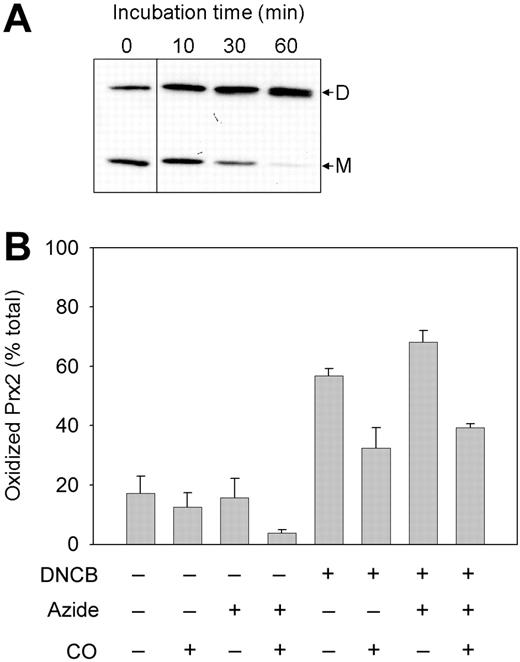
The slow rate of reduction of oxidized Prx2 in erythrocytes indicated that there was a limiting factor in the regeneration process. The abundance of Trx in the erythrocyte has been well established immunologically.31 In comparison, it is equivocal whether the cells contain TrxR. Immunologic evidence obtained in one study32 has been questioned in other more extensive negative studies.33 However, others have found indirect evidence for TrxR activity.34 GSH has been proposed as an alternative to TrxR for regenerating Trx.33 This is unlikely because while a 30-μM treatment with DNCB (a known depletor of GSH) was sufficient to completely inhibit Prx2 recycling (Figure 5A), the concentration was too low to affect GSH levels in these cells (97% of control levels; data not shown).
The inhibition of Prx2 regeneration in erythrocytes treated with DNCB. (A) Erythrocytes were incubated with 30 μM DNCB for the indicated times and immunoblotted. The portions left and right of the dividing line are derived from nonadjacent lanes on the same blot. D indicates dimeric Prx2; M, monomeric Prx2. (B) Erythrocytes were pretreated with CO or 1 mM azide as indicated, then with 30 μM DNCB for 30 minutes. Data are means ± SE from 3 experiments.
The inhibition of Prx2 regeneration in erythrocytes treated with DNCB. (A) Erythrocytes were incubated with 30 μM DNCB for the indicated times and immunoblotted. The portions left and right of the dividing line are derived from nonadjacent lanes on the same blot. D indicates dimeric Prx2; M, monomeric Prx2. (B) Erythrocytes were pretreated with CO or 1 mM azide as indicated, then with 30 μM DNCB for 30 minutes. Data are means ± SE from 3 experiments.
We performed TrxR activity assays on erythrocyte extracts to re-evaluate its presence. A common assay relies on monitoring DTNB reduction at 412 nm.26 However, as the heme component of Hb also absorbs strongly at 412 nm, this assay could not be used for detecting low activity in erythrocytes. Therefore, modifications were made to remove Hb by size exclusion chromatography before measuring TNB absorbance. Activity in erythrocytes was consistently detected, but on a per-cell basis was only 2% of that in Jurkat cells assayed by the same protocol (Table 1). Therefore, even allowing for their smaller volume, erythrocytes showed much lower TrxR activity. Pretreatment of cells with 60 μM DNCB decreased activity by approximately 75% in Jurkat cells and approximately 55% in erythrocytes (Table 1), which is consistent with most of the absorbance increase observed being attributable to TrxR.
Endogenously generated H2O2 in the erythrocyte
When erythrocytes were treated with DNCB alone, oxidized Prx2 progressively accumulated, with complete oxidation by 60 minutes (Figure 5A). Endogenously generated H2O2 was the most likely cause of this, which highlights the redox sensitivity of the enzyme. Hb continually undergoes slow autoxidation to form methemoglobin and superoxide, which dismutates to H2O2.35,36 To determine the degree to which Hb autoxidation contributes to endogenous Prx2 oxidation, erythrocyte suspensions were bubbled with CO to convert oxyhemoglobin to carboxyhemoglobin (which does not autoxidize to generate H2O2), and exposed to DNCB for 30 minutes (Figure 5B). Treatment with DNCB alone resulted in an accumulation of oxidized Prx2 (Figure 5B column 5). However, inhibition of Hb autoxidation inhibited the increase in Prx2 oxidation by approximately 65% (Figure 5B column 6). Similarly, while treatment with azide increased the Prx2 oxidation seen with DNCB (Figure 5B column 7), prior inhibition of Hb autoxidation lowered oxidation levels by about the same degree (Figure 5B column 8). This suggests that Hb autoxidation contributes much of the endogenously produced H2O2 in the erythrocyte.
Discussion
We found that Prx2 exists mostly in the reduced state under normal conditions, which is expected given the reducing environment of the red cell milieu. Although the small proportion of dimers observed in control samples may be an indication of constitutive oxidation, it could still represent artifactual oxidation during sample analysis. Prx2 was easily oxidized during cell preparation and sample analysis, and thiol blocking was required to reduce artifactual oxidation once cellular integrity was lost. Our data suggest that very small amounts of H2O2 present in the buffers used may contribute to Prx2 oxidation even before cell lysis. The high sensitivity of Prx2 to H2O2 is in agreement with other experiments in which we have shown that purified Prx2 reacts faster with H2O2 than it does with thiol-reactive reagents such as NEM and iodoacetamide (A.V.P., F.M.L., M.B.H. and C.C.W., manuscript in preparation).
We have shown that the erythrocyte Prx2 is remarkably sensitive to oxidation when exposed to exogenous H2O2. Treatment with even submicromolar concentrations resulted in its accumulation as a disulfide-linked homodimer. Prx2 oxidation was seen in cells containing active catalase and with glucose present to ensure a full reducing capacity via NADPH generated from the pentose phosphate pathway. Catalase inhibition increased the extent of Prx2 oxidation when higher concentrations of H2O2 were added, but had a relatively minor effect at low concentrations. Thus, Prx2 was able to compete effectively with catalase at scavenging small amounts of H2O2. The ability of erythrocyte Prx2 to sense micromolar concentrations of H2O2 was surprising, given the prevailing view that the high amounts of catalase and GPx are responsible for the elimination of H2O2 from erythrocytes. In fact, these results support the notion that the Prx rate constant is much higher than the proposed value of approximately 105 M−1 s−1.37 We have shown in competition experiments between Prx2 and catalase that both enzymes react at similar rates (A.V.P., F.M.L., M.B.H., and C.C.W., manuscript in preparation), which would be more consistent with the rate constant of 4 × 10⁷ M−1 s−1 recently determined for a bacterial Prx.38 Our results are also direct evidence in support of the modeling prediction by Johnson et al19 that peroxide metabolism in the erythrocyte is not exclusive to catalase and GPx, but requires Prx activity.
While treatment of erythrocytes with H2O2 resulted in dimerization, we found that treatment of Jurkat cells with H2O2 resulted in progressive loss of the dimer. The behavior of Prx2 in Jurkat cells is consistent with the view that the dimer is rapidly reduced and the oxidation step of the Prx cycle is rate limiting.37 Also in contrast to Jurkat cells, Prx2 was resistant to overoxidation in the erythrocyte. It is unlikely that this was due to high levels of erythrocyte sulfiredoxin. When hemolysate supplemented with Mg2+ and ATP4 was coincubated with Jurkat lysates containing overoxidized Prx, immunoblotting with antibodies to overoxidized Prx showed that levels of overoxidation were not decreased (F.M.L., unpublished observations, September 8, 2006).
While oxidized Prx2 is rapidly reduced in Jurkat cells, Prx2 oxidation was reversible in the erythrocyte, but only slowly after all the H2O2 had been consumed. Inhibition by DNCB suggested that TrxR was involved in regeneration, and the slow reversibility could be attributed to its very low concentration. Consistent with this, direct activity assays showed low but consistently detectable activity in erythrocytes, which was about 2% of that seen in an equivalent number of Jurkat cells. Our assay involved removal of Hb prior to measurement of absorbance increase, which eliminated problems with artifactual absorbance contributed by Hb. Furthermore, by assaying whole-cell lysates, measurements were representative of activity levels in the presence of the erythrocyte's endogenous levels of Trx and NADPH.
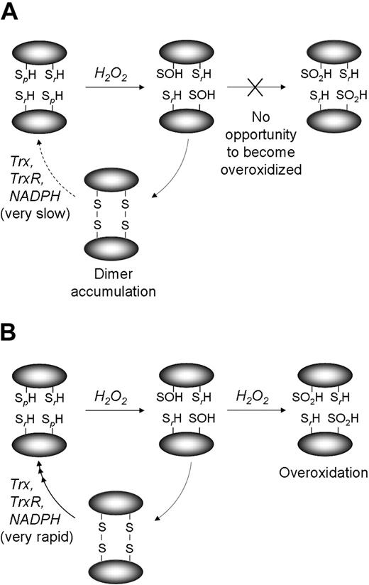
Thus the redox behavior of Prx2 in erythrocytes differs in 2 major respects from that in Jurkat cells, which appear typical of other cell types.30,39 It accumulates as a dimer and does not become overoxidized. We propose that this difference is due to the different levels of TrxR activity that change the rate-determining step in Prx2 cycling (Figure 6A). Prx2 is rapidly oxidized by very low concentrations of H2O2, and in the erythrocyte the low TrxR level results in slow recycling and accumulation of dimer. In this situation, regeneration of Trx (and therefore Prx2) by TrxR represents the rate-limiting step. Conversely, the high TrxR activity in Jurkat cells means that oxidized Prx2 does not accumulate over basal levels, and its catalytic rate is mostly determined by availability of H2O2. The different rate-determining steps therefore result in different levels of dimer accumulation. At each reaction cycle, a small proportion of the intermediate sulfenic acid form of Prx2 is overoxidized to a sulfinic or sulfonic acid before it can form an interchain disulfide with the resolving cysteine (Figure 6B). Thus rapid turnover, as occurs in Jurkat cells, allows the overoxidized form to accumulate. However, in the erythrocyte, slow turnover and continual persistence of the dimer limit the opportunity for the peroxidatic cysteines to become overoxidized.
Proposed scheme of the redox transitions of Prx2. Schematic representation of the proposed redox behavior of Prx2 when erythrocytes (A) and Jurkat cells (B) are treated with high concentrations of H2O2 (∼ 100-200 μM). SpH indicates peroxidatic cysteine, Cys50; SrH, resolving cysteine, Cys171.
Proposed scheme of the redox transitions of Prx2. Schematic representation of the proposed redox behavior of Prx2 when erythrocytes (A) and Jurkat cells (B) are treated with high concentrations of H2O2 (∼ 100-200 μM). SpH indicates peroxidatic cysteine, Cys50; SrH, resolving cysteine, Cys171.
Prx2 appears to be a sensitive scavenger of H2O2 in the erythrocyte, provided it can react in a noncatalytic manner. However, with excess H2O2, Prx2 becomes fully oxidized and the erythrocyte requires catalase and/or GPx activity to remove it. The slow rate of regeneration in the erythrocyte implies that once oxidized, Prx2 remains inactivated for a long period. However, Prx2 is one of the most abundant proteins in the erythrocyte (∼ 250 μM in the cytosol, equivalent to ∼ 15 million copies per cell). This enables it to handle up to an equivalent concentration of H2O2 without the need for recycling. Cells generate H2O2 in a variety of metabolic processes, producing concentrations that are orders of magnitude less than 250 μM. This large excess of Prx2 over its substrate suggests that Prx2 does not function in the erythrocyte as a classical erythrocyte antioxidant enzyme, but as a very effective H2O2 scavenging protein.
Our finding that DNCB treatment of erythrocytes resulted in gradual Prx2 dimerization indicates that Prx2 undergoes oxidation due to endogenous H2O2 generation. The fact that Prx2 remained a monomer when TrxR was not inhibited indicates that the regeneration rate, although low, was sufficient to maintain the reduced state. Hb autoxidation to methemoglobin occurs continuously and is thought to be a significant source of H2O2 in the erythrocyte. This was clearly demonstrated in erythrocytes where inhibition of Hb autoxidation and subsequent incubation with DNCB resulted in Prx2 oxidation. Various methods have been used in the past to measure rates of Hb autoxidation in erythrocytes, such as measuring catalase inactivation in the presence of aminotriazole.40 Our method of tracking Prx2 oxidation following CO treatment of erythrocytes may provide an alternative, more direct approach. Moreover, the lack of a sensitive yet specific probe for quantifying very low intracellular H2O2 generation has been noted.41 In this regard, the ubiquity and high reactivity of Prx2 with H2O2 could be exploited by tracking Prx2 dimerization in other cell types upon inhibition of TrxR activity.
Overoxidation of Prxs results in ablation of their peroxidase activity. The rationale for Prx2 overoxidation in other cells has been explained by the “floodgate” model of catalysis.42 This model hypothesizes that normal Prx activity keeps basal H2O2 low, and overoxidation enables H2O2 accumulation for participation as a secondary messenger in receptor-mediated signal transduction.5,6 However, it is unlikely that this mechanism applies to erythrocytes, where the absence of a nucleus precludes transcriptional regulation. In addition, unlike other cell types erythrocyte catalase is cytosolic and not compartmentalized in peroxisomes. Thus, even if Prx2 is overoxidized and inactivated, catalase should act in concert with GPx to effectively scavenge high H2O2 concentrations. Our findings with Prx2 raise the possibility that oxidation to the disulfide could serve as a regulatory switch for other Prx2-attributed roles, such as Ca2+-activated K+ transport17 or chaperone activity.43 Plishker et al18 have found that stimulation of K+ transport and inhibition of calpain in erythrocytes induced membrane association of high-molecular-weight oligomers of Prx2, and that these oligomers consisted of disulfide-linked dimers. Surface accumulation of oligomerized Prx2 has also been postulated to serve as a means of labeling oxidatively stressed erythrocytes.44
The results from this paper support the notion that Prx2 can protect the erythrocyte from low-level H2O2, such as that generated endogenously by Hb autoxidation. It is interesting that while mice lacking Prx2 present with hemolytic anemia and damaged erythrocytes containing Heinz bodies, mice deficient in catalase or GPx show no signs of Hb oxidation or hemolysis.13,14 This suggests a specific role for Prx2 in protecting Hb against endogenous H2O2. The role of Prx2 in keeping endogenous H2O2 low is supported by further studies on Prx2 knock-out mice, where constitutive production of endogenous reactive oxygen species was detected in spleen cells45 and thymocytes.46 In view of the redox sensitivity of Prx2, and its nonredundant role in protecting erythrocytes, we are investigating the mechanism by which it protects Hb from oxidation and denaturation.
Authorship
Contribution: F.M.L. performed research, analyzed data, and wrote paper, M.B.H. and C.C.W. designed research and contributed to interpretation and writing; A.V.P. contributed to data interpretation.
Conflict-of-interest disclosure: The authors declare no competing financial interests.
Correspondence: Christine C. Winterbourn, Free Radical Research Group, Department of Pathology, Christchurch School of Medicine & Health Sciences, University of Otago, Christchurch, New Zealand; e-mail: christine.winterbourn@chmeds.ac.nz.
The publication costs of this article were defrayed in part by page charge payment. Therefore, and solely to indicate this fact, this article is hereby marked “advertisement” in accordance with 18 USC section 1734.
Acknowledgments
This work was supported by a University of Otago Special Health Research Scholarship (F.M.L.) and the Health Research Council of New Zealand.
We thank Tessa Mocatta for performing venipunctures.


