Histone H2AX is required to maintain genomic stability in cells and to suppress malignant transformation of lymphocytes in mice. H2ax−/−p53−/− mice succumb predominantly to immature αβ T-cell lymphomas with translocations, deletions, and genomic amplifications that do not involve T-cell receptor (TCR). In addition, H2ax−/−p53−/− mice also develop at lower frequencies B and T lymphomas with antigen receptor locus translocations. V(D)J recombination is initiated through the programmed induction of DNA double-strand breaks (DSBs) by the RAG1/RAG2 endonuclease. Because promiscuous RAG1/RAG2 cutting outside of antigen receptor loci can promote genomic instability, H2ax−/−p53−/− T-lineage lymphomas might arise, at least in part, through erroneous V(D)J recombination. Here, we show that H2ax−/−p53−/−Rag2−/− mice exhibit a similar genetic predisposition as do H2ax−/−p53−/− mice to thymic lymphoma with translocations, deletions, and amplifications. We also found that H2ax−/−p53−/−Rag2−/− mice often develop thymic lymphomas with loss or deletion of the p53+ locus. Our data show that aberrant V(D)J recombination is not required for rapid onset of H2ax/p53-deficient thymic lymphomas with genomic instability and that H2ax deficiency predisposes p53−/−Rag2−/− thymocytes to transformation associated with p53 inactivation. Thus, H2AX is essential for suppressing the transformation of developing thymocytes arising from the aberrant repair of spontaneous DSBs.
Introduction
Chromosomal double-strand breaks (DSBs) are among the most hazardous cellular lesions and among the most difficult to repair because liberated DNA ends can separate irreversibly or join promiscuously. Unfortunately, such unwanted lesions also are common. They can arise in S phase through DNA replication errors and in any phase of the cell cycle from exogenous factors such as ionizing radiation and endogenous factors such as oxidative stress.1 When unrepaired or mis-repaired, DSBs can result in genomic instability and malignant transformation.1 Many human tumors contain chromosomal translocations and deletions that activate oncogenes or inactivate tumor suppressor genes.2 Such DNA lesions are probably causative for driving malignant transformation in a wide variety of human cell types. Consistent with this notion, mutations in genes encoding proteins involved in either of the 2 main DSB repair pathways, nonhomologous end-joining (NHEJ) and homologous recombination (HR), are associated with an increased predisposition to cancer in humans, whereas in mice, gene-targeted inactivation of NHEJ and HR factors can result in an increased predisposition to tumors, which are often immature αβ T-cell lymphomas.2,3
In humans and mice, mature αβ T lymphocytes develop through a highly regulated differentiation program that includes the assembly of T-cell receptor (TCR) variable region exons from germ line variable (V), diversity (D), and joining (J) gene segments and periods of rapid cellular proliferation.4 V(D)J recombination is initiated by the lymphocyte-specific RAG1 and RAG2 (RAG) proteins, which introduce DSBs between gene segments and their flanking recombination signal (RS) sequences, and completed by the NHEJ proteins, which repair RAG-generated DSBs.5 TCRβ variable region exons are assembled in the G1 phase of cycling CD4−/CD8− (double negative, DN) thymocytes.4 The assembly of in-frame VDJβ rearrangements generates TCRβ chains that, when expressed on the cell surface with pre-Tα chains, rescue DN thymocytes from apoptosis and signal rapid cellular proliferation, expansion, and differentiation to the CD4+/CD8+ (double positive, DP) cell stage.4 TCRα variable region exons are assembled in DP thymocytes. In-frame VJα rearrangements generate TCRα chains that, when expressed on the cell surface with TCRβ chains to form the αβ TCR, rescue DP cells from apoptosis and promote differentiation to CD4+ or CD8+ thymocytes.4 Developing αβ T cells may also generate spontaneous replication-associated DSBs that can occur in phase during the TCRβ-independent proliferation of DN cells and the TCRβ-dependent proliferation of thymocytes during DN to DP expansion. Thus, we hypothesize that the aberrant repair of either programmed or spontaneous DSBs can lead to thymic lymphomas associated with translocations, deletions, and genomic amplifications.
Histone H2AX is rapidly phosphorylated in chromatin around both programmed and spontaneous chromosomal DSBs.6,–8 H2AX is not required absolutely for either general DSB repair or V(D)J recombination; however, H2ax−/− cells exhibit defective chromosomal DSB repair, increased levels of spontaneous genomic instability, and a significant percentage of nonmalignant αβ T cells from H2ax−/− mice contain potential TCR translocations.9,10 Despite the dramatic instability of H2ax−/− cells, H2ax−/− mice exhibit only a slight increase in predisposition to thymic lymphoma, where the one tumor examined contained a potential TCR translocation.11 On induction of DSBs, p53 activates cell-cycle checkpoints to facilitate normal repair or apoptosis if the DNA damage is too severe to repair.12 Although p53 deficiency does not lead to an increased level of genomic instability in H2ax−/− cells,11,13,14 H2ax−/−p53−/− mice rapidly succumb to TCRαβ−/CD4+/CD8+ or TCRαβ−/CD4−/CD8+ thymic lymphomas with translocations, deletions, and gene amplifications that mostly do not involve TCR loci (only one clonal TCRα/δ translocation was found in 17 tumors analyzed) and, which, therefore have been proposed to arise through the mis-repair of spontaneous DSBs.11,14 In contrast, p53−/− mice succumb to TCRαβ−/CD4+/CD8+ or TCRαβ−/CD4−/CD8+ thymic lymphomas that usually are aneuploid with occasional nonclonal translocations, deletions, and gene amplifications.11,14,15 Therefore, p53 most likely suppresses the transformation of H2ax−/− cells by preventing continued proliferation of cells with DNA breaks and inducing apoptosis of cells with persistent DSBs, translocations, or both that activate oncogenes.13
In addition to thymic lymphomas, H2ax−/−p53−/− mice develop at a lower frequency pro-B lymphomas with immunoglobulin heavy chain (IgH) translocations in which the IgH locus is joined to DNA sequences near the c-myc locus,11,14 presumably by fusion of RAG-initiated DSBs at the IgH locus to general DSBs near c-myc. Similar translocations in NHEJ/p53-deficient pro-B lymphomas were shown to be RAG dependent.16 In vitro and in cell lines, the RAG proteins can introduce DSBs at both nonstandard DNA structures and fortuitous RS sequences that lie outside of antigen receptor loci.17,18 In addition, intrachromosomal deletions in human T-lineage lymphomas often contain fortuitous RS sequences near the breakpoints,19,–21 and such deletions in mouse thymic lymphomas are RAG dependent.22,23 Accordingly, a significant fraction of the genomic instability of H2ax−/−p53−/− thymic lymphomas could, theoretically, arise through the aberrant repair of promiscuous RAG-generated DSBs induced at genomic locations outside of TCR loci. Such a phenomenon also might account for the rapid development of lymphoma versus nonlymphoid tumors in H2ax−/−p53−/− mice. To investigate this issue, we generated H2ax−/−p53−/−Rag2−/− mice and characterized their genetic predisposition to cancer.
Methods
Generation of mice
Cohort mice were generated through breeding 129Sv H2ax−/−11 , 129Sv p53−/−,24 and 129Sv Rag2−/−25 mice. We initially bred H2ax−/−p53−/− mice with Rag2−/− mice to generate H2ax−/−p53−/−Rag2−/− mice. Because of sterility of H2ax−/− males and the inability of H2ax−/−p53−/− mice to breed, we interbred H2ax+/−p53+/−Rag2+/− mice to obtain H2ax+/−p53+/−Rag2−/− males and H2ax−/−p53−/−Rag2−/− females, which were interbred to generate the cohort H2ax−/−p53−/−Rag2−/− mice.
Characterization of tumors
Lymphomas were characterized by flow cytometry using anti-Thy1.2, anti-CD4, anti-CD8, and anti-TCRβ antibodies (Pharmingen, San Diego, CA). Sarcomas were characterized by H&E staining. Thymic lymphomas were grown in DMEM supplemented with 15% FBS, ConA, and IL-2 as described to prepare metaphase spreads for Spectral Karyotyping (SKY) as described.11 Tumor cells were incubated in Laird lysis buffer, and genomic DNA was isolated as described.26 Comparative genomic hybridization (CGH) was conducted by hybridization of HindIII- and XbaI-digested, biotinylated tumor and control DNAs to the Affymetrix (Santa Clara, CA) Gene Chip MOE 430_2 arrays as described.27 Southern blot analysis was conducted on EcoRI-digested genomic DNA as described26 using either a 400-kb (kilobase) AvaI fragment that hybridizes just 3′ of the p53 first exon or a polymerase chain reaction (PCR) product that hybridizes to the last exon of p53. The PCR product was generated using primers 5′-GTTGATATCAGCATAAGCTGTCTGGG-3′ and 5′-GGGGGTGGGTGAGAGGGTGTTAGGCT-3′. Protein was isolated as described28 and subjected to Western blot analysis using either an anti-p53 antibody or an anti-histone H4 antibody as described.11,28
Results
H2ax−/−p53−/−Rag2−/− mice rapidly succumb to TCRαβ−/CD4+/CD8+ thymic lymphomas
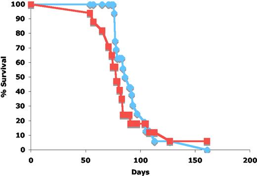
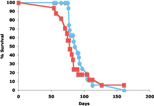
We previously found that 12 (70%) of 17 cohort H2ax−/−p53−/− mice succumbed to TCRαβ−/CD4+/CD8+ thymic lymphomas at a median day of death of 79 days (Figure 1; Table 1).11,14 To evaluate whether aberrant RAG activity or TCRβ-mediated survival, differentiation, or proliferation or both are required for the malignant transformation of H2ax−/−p53−/− thymocytes, we generated a cohort of 16 H2ax−/−p53−/−Rag2−/− mice and characterized their genetic predisposition to cancer. Similar to H2ax−/−p53−/− mice, we found that 14 (88%) of 16 cohort H2ax−/−p53−/−Rag2−/− mice succumbed to thymic lymphomas with a median day of death of 88 days (Figure 1; Table 1). Flow cytometry analysis of 11 tumors showed that 10 were TCRαβ−/CD4+/CD8+ and the other was TCRαβ−/CD4−/CD8− (data not shown), similar to the thymic lymphomas that arose in H2ax−/−p53−/− mice.11,14 Four of these 16 H2ax−/−p53−/−Rag2−/− mice also developed sarcomas by the time they succumbed to thymic lymphomas (Table 1). Of the remaining 2 cohort H2ax−/−p53−/−Rag2−/− mice, one succumbed to a sarcoma at 113 days of age, and the other mouse died from tooth malocclusion at 105 days of age (Table 1). Neither of these mice showed any visible signs of a thymic lymphoma at the time of their death. Notably, although 2 of 17 H2ax−/−p53−/− mice succumbed to B-lineage lymphomas (Table 1),11 none of the 16 cohort H2ax−/−p53−/−Rag2−/− mice developed B-lineage lymphomas (Table 1). Thus, H2ax−/−p53−/−Rag2−/− mice exhibit a similar genetic predisposition to TCRαβ−/CD4+/CD8+ thymic lymphomas as do H2ax−/−p53−/− mice, showing that neither aberrant V(D)J recombination nor TCRβ-mediated signals are required for the transformation of H2ax−/−p53−/− thymocytes.
Effect of Rag2 deficiency on survival of H2ax−/−p53−/− mice. Kapplan-Meier curve representing the percentage of survival of H2ax−/−p53−/−Rag2−/− (blue diamonds; n = 16) and H2ax−/−p53−/−Rag2+/+ (red squares; n = 17) cohort mice versus age in days. The H2ax−/−p53−/−Rag2+/+ cohort was previously characterized.11
Effect of Rag2 deficiency on survival of H2ax−/−p53−/− mice. Kapplan-Meier curve representing the percentage of survival of H2ax−/−p53−/−Rag2−/− (blue diamonds; n = 16) and H2ax−/−p53−/−Rag2+/+ (red squares; n = 17) cohort mice versus age in days. The H2ax−/−p53−/−Rag2+/+ cohort was previously characterized.11
H2ax−/−p53−/−Rag2−/− thymic lymphomas contain clonal and nonclonal translocations
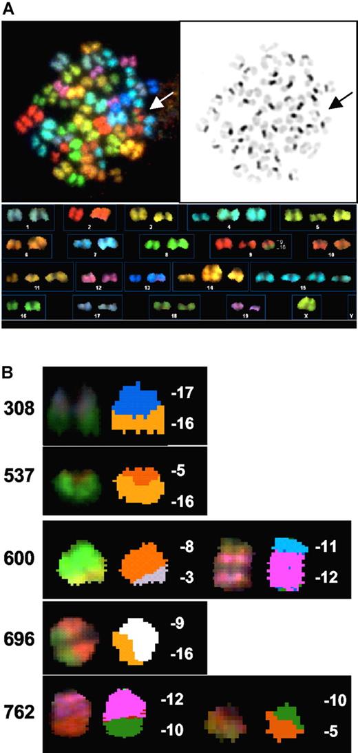
To evaluate whether a significant percentage of the translocations in H2ax−/−p53−/− thymic lymphomas could arise through the aberrant repair of RAG-generated DSBs at general genomic locations, we conducted SKY on metaphases prepared from early passage primary tumor cells. SKY permits the simultaneous classification of all mouse chromosomes using a single probe mixture.29 We analyzed 5 H2ax−/−p53−/−Rag2−/− thymic lymphomas by SKY and found that each tumor contained at least one clonal translocation (Figure 2; Table 2), whereas 4 of the 5 H2ax−/−p53−/−Rag2−/− tumors contained a number of additional nonclonal translocations (Figure 1; Table 2). Moreover, in contrast to the nearly tetraploid thymic lymphomas of p53−/− mice30 and mice with inactivation of p53 and Rag1 or Rag2,15 the H2ax−/−p53−/−Rag2−/− thymic lymphomas were close to diploid, similar to the thymic lymphomas of H2ax−/−p53−/− mice.11,14 Thus, H2ax−/−p53−/−Rag2−/− thymic lymphomas harbor clonal and nonclonal translocations similar to H2ax−/−p53−/− thymic lymphomas. These results show that TCRβ-mediated proliferation, aberrant V(D)J recombination, or both are not required for the generation of oncogenic translocations in H2ax−/−p53−/− thymic tumors.
H2ax−/−p53−/−Rag2−/− thymic lymphomas harbor clonal translocations. (A) Shown is a spectral, DAPI, and karyotype image of a representative metaphase from H2ax−/−p53−/−Rag2−/− thymic lymphoma 696. (B) Shown are spectral images of the clonal translocations of H2ax−/−p53−/−Rag2−/− thymic lymphomas 308, 537, 600, 696, and 762.
H2ax−/−p53−/−Rag2−/− thymic lymphomas harbor clonal translocations. (A) Shown is a spectral, DAPI, and karyotype image of a representative metaphase from H2ax−/−p53−/−Rag2−/− thymic lymphoma 696. (B) Shown are spectral images of the clonal translocations of H2ax−/−p53−/−Rag2−/− thymic lymphomas 308, 537, 600, 696, and 762.
H2ax−/−p53−/−Rag2−/− thymic lymphomas harbor genomic amplifications and deletions
To evaluate whether a significant percentage of the chromosomal amplifications and deletions in H2ax−/−p53−/− thymic lymphomas also could arise through the aberrant repair of RAG-generated DSBs at general genomic locations, we conducted CGH on genomic DNA prepared from early passage primary tumor cells. CGH permits the simultaneous identification of allelic gains and losses within a population of cells.31 We performed CGH on the genomic DNA isolated from 5 H2ax−/−p53−/−Rag2−/− thymic lymphomas generated in this study and from 5 H2ax−/−p53−/− thymic lymphomas generated in our previous study.11 We found that all 10 tumors contained gains and losses of multiple subchromosomal regions on most chromosomes (data not shown) and shared recurrent gains and losses of several subchromosomal regions (Tables 3,4). Thus, H2ax−/−p53−/−Rag2−/− thymic lymphomas harbor chromosomal amplifications and deletions similar to H2ax−/−p53−/− thymic lymphomas. These results show that TCRβ-mediated proliferation, aberrant V(D)J recombination, or both are not required for the generation of genomic amplification or deletions in H2ax−/−p53−/− thymic tumors.
H2ax−/−p53−/−Rag2−/− mice develop thymic lymphomas with loss of the p53+ allele
Compared with p53+/+ mice, p53−/− mice display an enhanced cancer predisposition with 25% of p53−/− mice succumbing by 18 months to thymic lymphomas with loss of heterozygosity (LOH) of the p53+ allele,32 most likely generated through interchromosomal mitotic recombination.33 In contrast, radiation (DSB)–induced mouse lymphomas develop in association with internal p53 deletions.34 During our breeding to generate H2ax−/−p53−/−Rag2−/− mice, we found that H2ax−/−p53−/−Rag2−/− mice died at an earlier median age than did H2ax−/−p53+/+Rag2−/− mice (161 days versus 232 days; Figure S1, available on the Blood website; see the Supplemental Materials link at the top of the online article), with one-third of H2ax−/−p53−/−Rag2−/− mice succumbing to TCRαβ−/CD4+/CD8+ thymic lymphomas (data not shown).
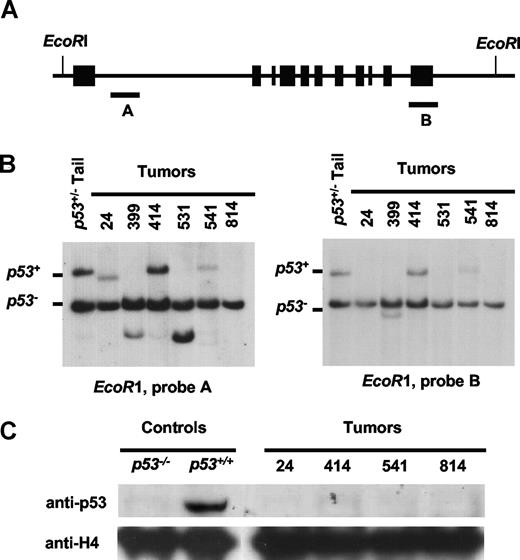
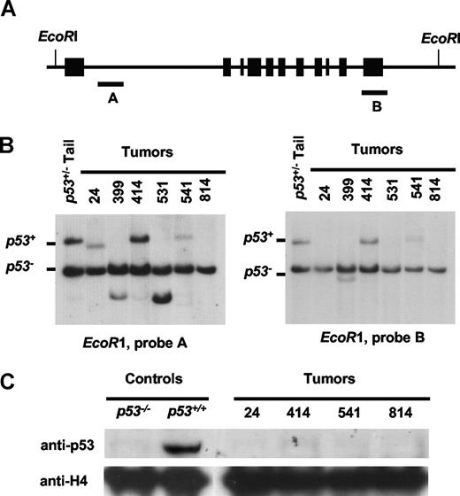
To determine whether H2ax−/−p53−/−Rag2−/− thymic lymphomas harbor p53+ LOH or internal p53+ deletions, we analyzed the genomic structure of the p53 locus in 6 of these tumors. We conducted Southern blot analysis with a probe that hybridizes just 3′ of the p53 first exon or a probe that hybridizes to the last exon of p53 on EcoRI-digested genomic DNA isolated from 6 H2ax−/−p53−/−Rag2−/− tumors. Both probes hybridize to the same 20-kb EcoRI fragment on the p53+ allele and different 8-kb EcoRI fragments on the p53− allele (Figure 3A). As expected, we observed both p53+ and p53− bands in control DNA (Figure 3B). However, we found loss of the 20-kb fragment corresponding to the p53+ allele in genomic DNA isolated from tumors 541 and 814 (Figure 3B), indicating that LOH of p53 occurred in 2 of 6 H2ax−/−p53−/−Rag2−/− thymic lymphomas. Strikingly, we found loss of the 20-kb p53+ band in DNA isolated from tumors 24, 399, and 531, with the concomitant appearance of novel-sized fragments that hybridized with only the p53 first exon probe in tumors 24 and 531 or both the p53 first and last exon probes in tumor 399 (Figure 3B), showing that either an internal or 3′ deletion of the p53 locus occurred in 3 of 6 H2ax−/−p53−/−Rag2−/− thymic lymphomas. Western blot analysis with an anti-p53 antibody on protein isolated from H2ax−/−p53−/−Rag2−/− thymic lymphomas 24, 414, 541, and 814 revealed that none expressed p53 protein (Figure 3C), confirming that p53 was inactivated by LOH or deletion in tumors 24, 541, and 814. The inactivation of p53 in tumor 414 may have occurred through a small deletion, point mutation, or epigenetic silencing.
H2ax−/−p53−/−Rag2−/− mice develop thymic lymphomas with p53 inactivation. (A) Schematic of the p53+ allele. The relative locations of where the 11 exons of p53 are located within the 20-kb EcoRI genomic fragment. The location of the 400-bp AvaI (A) and the p53 last exon (B) probes used for Southern blotting are indicated by black rectangles. (B) Southern blot analysis of EcoRI-digested genomic DNA isolated from 6 H2ax−/−p53−/−Rag2−/− thymic lymphomas and a p53−/− mouse-tail probed with either probe A or B. The fragments corresponding to the p53+ alleles are indicated. (C) Western blot analysis of protein isolated from 4 H2ax−/−p53−/−Rag2−/− thymic lymphomas and p53−/− or p53−/− control cells using either an anti-p53 antibody or an anti-H4 histone antibody as a loading control.
H2ax−/−p53−/−Rag2−/− mice develop thymic lymphomas with p53 inactivation. (A) Schematic of the p53+ allele. The relative locations of where the 11 exons of p53 are located within the 20-kb EcoRI genomic fragment. The location of the 400-bp AvaI (A) and the p53 last exon (B) probes used for Southern blotting are indicated by black rectangles. (B) Southern blot analysis of EcoRI-digested genomic DNA isolated from 6 H2ax−/−p53−/−Rag2−/− thymic lymphomas and a p53−/− mouse-tail probed with either probe A or B. The fragments corresponding to the p53+ alleles are indicated. (C) Western blot analysis of protein isolated from 4 H2ax−/−p53−/−Rag2−/− thymic lymphomas and p53−/− or p53−/− control cells using either an anti-p53 antibody or an anti-H4 histone antibody as a loading control.
LOH and deletion of genetic loci can arise in association with chromosomal translocations through the loss of genomic material around the translocation breakpoint. In mice, p53 resides on chromosome 11. We conducted SKY on metaphases prepared from primary cells of tumors 24 and 814. Tumor 24 contained 3 to 5 copies of chromosome 11 per metaphase and tumor 814 contained 3 copies of chromosome 11 per metaphase, but neither harbored detectable translocations involving chromosome 11 (data not shown). Thus, the LOH of p53 in tumor 814 and the structural alteration of p53 in tumor 24 were probably the result of internal deletions on chromosome 11 rather than either loss of entire chromosome 11 derivatives or translocations involving chromosome 11. These findings indicate that inactivation of H2AX predisposes p53−/−Rag2−/− thymocytes to transformation associated with p53+ deletions.
Discussion
Aberrant V(D)J recombination is not required for rapid development of H2ax/p53-deficient thymic lymphomas with genomic instability
Here, we generated a cohort of H2ax−/−p53−/−Rag2−/− mice and characterized their genetic predisposition to cancer to evaluate whether aberrant V(D)J recombination, or TCRβ-mediated survival, differentiation, or proliferation, or both are required for the malignant transformation of H2ax−/−p53−/− thymocytes. We found that H2ax−/−p53−/−Rag2−/− mice exhibit a similar genetic predisposition to TCRαβ−/CD4+/CD8+ thymic lymphomas as do H2ax−/−p53−/− mice, showing that neither promiscuous RAG activity nor TCRβ-mediated signals are required for the rapid development of thymic lymphomas in H2ax−/−p53−/− mice. Moreover, we found that H2ax−/−p53−/−Rag2−/− and H2ax−/−p53−/− thymic lymphomas each harbor translocations, deletions, and genomic amplifications, showing that neither TCRβ-mediated proliferation nor aberrant V(D)J recombination are required for the generation of a similar pattern of genomic instability in H2ax−/−p53−/− thymic lymphomas. In contrast, although Ku80−/−p53−/−, Xrcc4−/−p53−/−, and Dnapkcs−/−p53−/− mice each invariably succumb to pro-B lymphomas with clonal IgH/c-myc translocations, Ku80−/−p53−/−Rag2−/− mice develop thymic lymphomas that lack clonal translocations, Xrcc4−/−p53−/−Rag2−/− mice die at a later age without any sign of lymphoma, and Dnapkcs−/−p53−/−Rag2−/− mice rapidly succumb to pro-B lymphomas with clonal translocations that do not involve Ig loci.16,30,35 Thus, although we cannot rule out that some level of genomic instability in H2ax−/−p53−/− thymic lymphomas arises through the aberrant repair of promiscuous RAG-generated DSBs, our data firmly show that aberrant V(D)J recombination is not required to generate the pattern of genomic instability observed in H2ax−/−p53−/− thymic lymphomas.
Notably, none of the 16 cohort H2ax−/−p53−/−Rag2−/− mice developed B-lineage lymphomas, despite the observation that 5 of 35 H2ax−/−p53−/− cohort mice did.11,14 Four of these H2ax−/−p53−/− mice developed pro-B lymphomas with clonal IgH/c-myc translocations that probably formed by fusion of RAG-generated DSBs at the IgH locus to DSBs near c-myc,11,14 whereas one H2ax−/−p53−/− mouse developed a more mature B-lineage lymphoma with a clonal IgH/c-myc translocation that probably formed by fusion of AID-initiated IgH locus DSBs induced during IgH class switch recombination (CSR) to DSBs near c-myc11 . Because CSR occurs only in mature B cells and Rag2−/− mice lack mature lymphocytes, we expected that H2ax−/−p53−/−Rag2−/− mice would not develop B-lineage lymphomas with AID-initiated IgH/c-myc translocations. In addition, because IgH loci are not cleaved in pro-B cells of Rag2−/− mice, we anticipated that H2ax−/−p53−/−Rag2−/− mice would not develop pro-B lymphomas with RAG-initiated IgH/c-myc translocations. However, a much larger cohort of H2ax−/−p53−/−Rag2−/− mice, cohorts of Rag2+/+ and Rag2−/− mice, or both with conditional inactivation of H2ax and p53 in pro-B cells would need to be characterized to determine whether the mis-repair of RAG-generated DSBs is required for the malignant transformation of H2ax−/−p53−/− pro-B cells or whether other clonal translocations can be causative for pro-B lymphomas in this genetic background, as is the case for Dnapkcs−/−p53−/−Rag2−/− mice.16
H2AX suppresses thymic lymphomas with LOH and deletion of the p53 locus
We also found that H2ax−/−p53−/−Rag2−/− mice succumb to thymic lymphoma more rapidly than do H2ax−/−p53+/+Rag2−/− mice, with approximately half of these H2ax−/−p53−/−Rag2−/− tumors containing internal or 3′ deletion of the p53+ allele. H2ax−/− cells exhibit a substantially increased level of chromosome breaks and chromatid breaks compared with H2ax+/+ cells.9,10,13 Thus, H2AX may function in p53−/−Rag2−/− thymocytes to prevent spontaneous deletions at random locations throughout the genome that could, by chance, inactivate p53 or other tumor suppressor genes and, thereby, contribute to malignant transformation. Alternatively, the spontaneous deletion and inactivation of the p53+ allele may contribute to the malignant transformation of H2ax−/−p53−/−Rag2−/− thymocytes by allowing continued proliferation of H2ax-deficient cells with DNA breaks or preventing apoptosis of H2ax-deficient cells with genomic instability that activates oncogenes.11,13
Germ line H2ax/p53/Rag2-deficient mice exhibit a predisposition to thymic lymphomas
Our observation that H2ax−/−p53−/−Rag2−/− mice succumb predominantly to TCRαβ−/CD4+/CD8+ thymic lymphomas indicates that H2AX and p53 cooperate to suppress transformation of DP thymocytes or cells of an earlier developmental stage that continue to differentiate after their malignant conversion. Rag1−/− and Rag2−/− mice exhibit a complete block in thymocyte development at the DN stage because of an inability to assemble TCRβ chains that signal survival, expansion, and differentiation.25,36,37 This developmental block is not rescued in Rag2−/−p53−/− mice because the lack of p53-mediated apoptosis in DN cells cannot substitute for TCRβ-mediated survival and proliferation38 ; however, a low number of CD4+/CD8+ thymocytes can develop in Rag2−/−p53−/−38 and H2ax−/−p53−/−Rag2−/− mice (C.H.B. and F.W.A., unpublished observations, January 2005). Because H2ax−/−p53−/−Rag2−/− mice succumb to thymic lymphomas by a similar median age as do H2ax−/−p53−/− mice, it seems likely that these tumors arise from genetic or epigenetic changes that occur through the accumulation of genomic instability events in DN thymocytes, earlier stage cells, or both, perhaps even in stem cells, rather than in the rare DP cells that can develop. Our observation that H2ax−/−p53−/−Rag2−/− and H2ax−/−p53−/− thymic lymphomas often harbor amplification or deletion or both of the same genomic regions, suggests these sequences could either be highly prone to spontaneous DNA breakage and occur before cellular transformation or contain oncogenes and tumor suppressor genes, the activation or inactivation of which contributes to the transformation of developing thymocytes. Of note, the commonly deleted regions on chromosomes 4 and 11 contain, respectively, absent in melanoma 1-like and granulin, which are each homologues of potential human tumor suppressor genes,39,40 suggesting that inactivation of these genes may contribute to transformation of H2ax−/−p53−/−Rag2−/− and H2ax−/−p53−/− thymocytes. Finally, because H2ax−/−p53−/−Rag2−/− and H2ax−/−p53−/− mice invariably succumb to clonal thymic lymphomas with genomic instability, rather than other cancers, thymocytes may exhibit higher levels of spontaneous DNA damage, possess a lower intrinsic threshold of malignant transformation, or reside in a niche that supports the rapid expansion of cancerous cells, compared with cells of other tissues.
An Inside Blood analysis of this article appears at the front of this issue.
The online version of this article contains a data supplement.
The publication costs of this article were defrayed in part by page charge payment. Therefore, and solely to indicate this fact, this article is hereby marked “advertisement” in accordance with 18 USC section 1734.
Acknowledgments
We thank Donna Neuberg for statistical advice, Ed Fox for CGH analysis, and Shan Zha for critical discussion of the manuscript.
This work was supported by the National Cancer Institute (grant CA 125195, C.H.B.) and the National Institutes of Health (grant CA 109901, F.W.A.).
C.H.B. was a Lymphoma Research Foundation Fellow and is currently a Pew Scholar in the Biomedical Sciences. S.R. was supported by a Genentech/IDEC Fellowship from the American Cancer Society and is currently supported by the Dana-Farber Cancer Institute Postdoctoral Training Program in Cancer Immunology (DFCI/NCI CA70083). V.S. is supported by a Cancer Research Institute training grant awarded to the University of Pennsylvania Medical Center. F.W.A. is an investigator of the Howard Hughes Medical Institute.
National Institutes of Health
Authorship
Contribution: C.H.B. and F.W.A. designed the research; C.H.B., S.R., M.M., V.S., and M.G. performed research and collected data; C.H.B., S.R., and F.W.A. analyzed and interpreted data; and C.H.B. and F.W.A. drafted the manuscript.
Conflict-of-interest disclosure: The authors declare no competing financial interests.
Correspondence: Frederick W. Alt, The Children's Hospital, Karp Research Building, Rm 9216, 1 Blackfan Circle, Boston, MA 02115; e-mail: alt@enders.tch.harvard.edu.

