Abstract
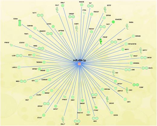
Primary Myelofibrosis (PMF) belongs to the Philadelphia negative Myeloproliferative Neoplasms (MPNs) and is characterized by hematopoietic stem-cell derived clonal myeloproliferation, involving especially megakaryocyte (MK) lineage, bone marrow fibrosis and extramedullary hematopoiesis. Recent studies have suggested that alterations in miRNAs expression could play a critical role in MPN's pathogenesis. In order to shed some light on this issue, we have previously performed the integrative analysis (IA) of gene and miRNA expression profiles of PMF CD34+ hematopoietic stem/progenitor cells (HSPCs) isolated from 42 PMF patients compared with 31 healthy donors (R. Norfo et al., Blood, 2014). IA identified miR-494-3p as one of the most upregulated miRNAs in PMF CD34+ cells associated to the highest number of downregulated predicted targets (eighty-six, Fig. 1).
In order to study the role of miR-494-3p in hematopoietic commitment and differentiation, and to elucidate its possible involvement in PMF pathogenesis, we performed miRNA overexpression experiments in cord blood (CB) CD34+ cells through miRNA mimic electroporation. The data showed that miR-494-3p promotes MK differentiation of HSPCs. Indeed, the fraction of cells expressing MK surface antigen CD41 was steadily increased in miR-494-3p overexpressing samples compared with controls (75.4±0.3% vs 53.2±3.5% at day 8, 82.6±1.3% vs 60.4±4.3% at day 10 of culture, p<0.05), as well as the percentage of cells expressing the late MK antigen CD42b was similarly increased. Furthermore, the percentage of MK colonies was increased in collagen-based clonogenic assay upon miR-494-3p overexpression compared to control (44.8±4.1% vs 24.1±2.1%, p<0.05). Next, to better characterize the molecular mechanisms underlying megakaryocytopoiesis stimulation by miR-494-3p, we profiled CB CD34+ cells overexpressing this miRNA using the Affymetrix HG-U219 microarray platform. Gene Expression profile analysis allowed the identification of 134 differentially expressed genes between cells overexpressing miR-494-3p and controls. In particular, we highlighted the presence of 13 genes downregulated both upon miRNA overexpression and in PMF CD34+ cells. Among them, Suppressor of Cytokine Signaling 6 (SOCS6) turned out to be the miR-494-3p predicted target associated to the most favorable prediction scores according to TargetScan, microRNA.org and miRDB prediction algorithms. Furthermore, 3' UTR luciferase reporter assays, performed in K562 cell line, proved the predicted interaction between miR-494-3p and SOCS6 3'UTR. Subsequently, we studied the role of SOCS6 in HSPCs differentiation by inhibiting its expression in CB CD34+ cells through small interfering RNAs. SOCS6 silencing stimulated megakaryocytopoiesis in CB CD34+ cells, as demonstrated by the expansion of CD41+ and CD42b+ cell fractions in SOCS6 silenced samples compared with controls (52.8±7.0% vs 37.7±4.5% at day 8, 66.9±7.2% vs 50.7±7.2% at day 10 for CD41+ cells, p<0.05). Moreover, MK colonies were increased upon SOCS6 silencing in collagen-based clonogenic assays (62.4±7.7% vs 51.3±6.5%, p<0.05) and morphological analysis further supported these results. Finally, in order to study the possible contribution of miR-494-3p overexpression to PMF pathogenesis, we performed inhibition experiments in PMF CD34+ cells by means of miR-494-3p antagomiR. As a result, miR-494-3p silencing led to SOCS6 upregulation and impaired MK differentiation in PMF HSPCs as demonstrated by the decrease in CD41+ cell fraction in silenced samples compared with controls (28.6±7.1% vs 39.2±7.7% at day 12 of culture, p<0.05) and by the reduction of MK colonies in collagen-based clonogenic assay (44.4±3.6% vs 54.7±2.5%, p<0.05). The values are reported as mean±S.E.M from 3 independent experiments.
Taken together, our results showed that miR-494-3p overexpression promotes megakaryocytopoiesis in HSPCs. Moreover, we demonstrated for the first time that SOCS6 is a direct target of miR-494-3p. Since SOCS6 downregulation promotes MK differentiation of HSPCs, SOCS6 could be considered, at least in part, responsible for the biological effects observed after miR-494-3p overexpression. As miR-494-3p and SOCS6 showed the same expression trend in PMF CD34+ cells, our results could suggest that miR-494-3p/SOCS6 axis is involved in the induction of MK hyperplasia typically observed in PMF patients.
Vannucchi:Novartis: Consultancy, Research Funding, Speakers Bureau; Baxalta: Speakers Bureau; Shire: Speakers Bureau.
Author notes
Asterisk with author names denotes non-ASH members.


