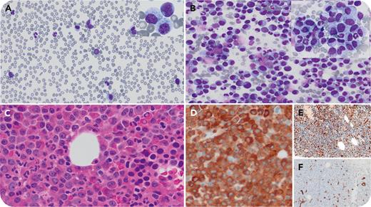
We illustrate a peculiar morphology of dysplastic myelocyte arrest in acute myeloid leukemia, myelodysplasia related (AML-MR), with comutation of stromal antigen 2 (STAG2) and serine and arginine rich splicing factor 2 (SRSF2) and overexpression of wild-type p53. An 81-year-old man with no history of hematological disorder was investigated for acute leukemia. Peripheral blood (PB) showed the following: leukocytes, 26.3 × 10⁹/L; neutrophils, 9.36 × 10⁹/L; myelocytes, 7.7 × 10⁹/L; blasts, 5.68 × 10⁹/L; hemoglobin, 105 g/L; and platelets, 35 × 109/L. PB film illustrated ∼30% blasts plus dysplastic myelocytes/granulocytes (panel A, May-Grünwald Giemsa stain, 40× lens objective; blast inset, 100× lens objective). PB flow cytometry confirmed blasts were positive with CD34, CD117, CD13, CD33, human leukocyte antigen-DR, and myeloperoxidase (MPO). Marrow was hypercellular with dysplastic myelocytes (>80%) (aspirate panel B, May-Grünwald Giemsa stain, 40× lens objective; inset, 100× lens objective; biopsy panel C, hematoxylin and eosin stain, 40× lens objective), positive for MPO (panel D, 40× lens objective), 80% p53 (panel E, 40× lens objective), and 5% to 10% CD34+ myeloblasts (panel F, 40× lens objective). Cytogenetics showed 46,XY[20]. Myeloid panel demonstrated STAG2:c.1005dupT, p.Met336Tyrfs∗3 (variant allele frequency [VAF], 86.8%), SRSF2:c.284C>A, p.Pro95His (VAF, 51.0%), ASXL1:c.1934dupG, p.Gly646Trpfs∗12 (VAF, 40.9%), 2 RUNX1 mutations, 2 TET2 mutations, and 2 NRAS mutations and absence of TP53 mutations. With PB blasts >20%, a diagnosis of AML-MR was rendered. The patient’s leukocytes tripled in 2 weeks. Death occurred 2 months later.
Wild-type p53 overexpression has been associated with AML with normal karyotype and poorer prognosis. The morphology of florid granulocytic hyperplasia with myelocyte arrest is in keeping with a mouse model of comutation of STAG2 with SRSF2, which shows increased granulocyte/monocyte fraction.
For additional images, visit the ASH Image Bank, a reference and teaching tool that is continually updated with new atlas and case study images. For more information, visit https://imagebank.hematology.org.


