Abstract
We examined the IgM VH gene subgroup use-distribution in serial blood samples of 37 human immunodeficiency virus (HIV)-infected patients and a group of HIV-seronegative healthy adults. The IgM VH gene repertoires of healthy adults were relatively similar to one another and were stable over time. In contrast, individuals infected with HIV had IgM VH gene repertoires that were significantly more heterogeneous and unstable. Persons at early stages of HIV infection generally had abnormal expression levels of Ig VH3 genes and frequently displayed marked fluctuations in the relative expression levels of this VHgene subgroup over time. In contrast, persons with established acquired immunodeficiency syndrome (AIDS) had a significantly lower incidence of abnormalities in Ig VH3 expression levels, although continued to display abnormalities and instability in the expression levels of the smaller Ig VH gene subgroups. Moreover, the skewing and/or fluctuations in the expressed-IgM VHgene repertoire appeared greatest for persons at earlier stages of HIV infection. These studies show that persons infected with HIV have aberrant and unstable expression of immunoglobulin genes suggestive of a high degree humoral immune dysregulation and ongoing humoral immune responses to HIV-associated antigens and superantigens.
© 1998 by The American Society of Hematology.
PATIENTS INFECTED WITH human immunodeficiency virus (HIV) are at increased risk for B-cell lymphoproliferative disease and B-cell lymphoma.1,2 Various mechanisms are hypothesized to account for this. These include direct infection of B cells with viruses, such as Epstein-Barr virus (EBV); chronic antigenic stimulation due to HIV or opportunistic infections; immune dysregulation secondary to attrition of CD4 T cells or other immune-accessory cells; or antigen-induced B-cell activation and depletion.3-6
Recent studies have provided evidence that antigen may play a role in acquired immunodeficiency syndrome (AIDS)-associated B-cell lymphomagenesis. Certain Ig heavy chain variable region genes (VH genes) that have a higher-relative intrinsic capacity to incur nonconservative mutations in the complementarity-determining regions (CDR) appear to be expressed most frequently by AIDS-associated B-cell lymphomas.7 Furthermore, the Ig VH genes expressed by such lymphomas have somatic mutations similar to those expressed by B cells engaged in secondary immune responses to antigen.7-9 This is striking, considering the profound immune deficiency of the patients who develop such lymphomas, limiting their capacity to mount such secondary immune responses. As such, the B cells that subsequently develop into B-cell lymphoma may have originated at stages of the HIV infection that are earlier than that of end-stage AIDS when most lymphomas become clinically apparent.
Previous studies have noted alterations in the B-cell compartment in patients infected with HIV, which may precede the development of B-cell lymphoma. Indeed, persons infected with HIV have been found to have a skewed Ig VH repertoire depleted of IgM+, IgM/IgD+, and IgG+ B cells that express Ig encoded by VH genes of the VH3 subgroup.10,11 This subgroup is one of seven human Ig VH gene subgroups, each subgroup containing VHgenes that share more than 80% nucleic acid sequence homology.12 In part because this VH gene subgroup has the largest number of functional VH genes in the human haploid genome,13,14 VH3 genes ordinarily are used by approximately 45% to 60% of the blood B cells of normal adults.15 16 As such, depletion of B cells expressing Ig VH3 genes reflects a major alteration in the B-cell compartment.
The relative depletion of B cells that express Ig VH3 could be secondary to loss of B cells that express Ig VH3 genes and/or a relative expansion of B cells that express Ig VH genes of other VH gene subgroups. However, in view of recent findings that B cells are generated continuously throughout life,17 it appears unlikely that there would be a fixed depletion of B cells expressing Ig VH3 genes unless HIV-infected patients also acquire defects in B-cell lymphopoiesis. However, detailed analyses of the relative expression of all the VH gene subgroups in patients infected with HIV are lacking. Also lacking are data on whether such clonal imbalances occur early during the course of HIV infection and progress over time. To examine this, we analyzed the relative expression of Ig VHgene subgroups by blood B cells of adults infected with HIV and age-matched seronegative controls.
MATERIAL AND METHODS
Patient material.
We obtained blood specimens from HIV-infected persons and aged-matched controls who were not infected with HIV. The donors were divided into five different groups. Group I consisted of nine HIV-positive patients who did not satisfy diagnostic criteria for AIDS and who each had greater than 500 CD4+ T cells per mm3 of blood. Three blood samples were obtained at 6-month intervals. Groups II and III consisted of 14 patients with AIDS who had less than 200 CD4+ T cells per mm3 of blood. Nine of these patients (Group III) developed AIDS-associated Lymphoma (AAL) at a later date. In each case we examined two to five serial blood specimens every 3 to 6 months. Group IV consisted of 14 patients with AIDS who were sampled at the time of diagnosis of AAL. Finally, Group V consisted of six age-matched control donors who were not infected with HIV. Two to six blood samples were obtained from each at 2- to 4-month intervals. In addition, one blood sample was obtained from each of 22 additional normal adults who were not infected with HIV.
RNA isolation, and cDNA synthesis.
We isolated RNA from 5 × 10⁶ to 1 × 10⁷ cryopreserved blood lymphocytes using RNAzol B (Cinna/biotex, Friendswood, TX). The cDNA was synthesized from 5 μg total RNA using an oligo-dT primer and Superscript II reverse transcriptase (Life Technologies, Grand Island NY). Reverse transcription was performed for one hour at 42°C. Afterwards the mixture was heated to 70°C for 15 minutes. The sample was treated with 0.3 mol/L NaOH for 30 minutes to hydrolyze residual RNA.
Analysis of IgM VH gene use.
The distribution of IgM VH gene transcripts in blood B cells was tested using a reverse transcriptase polymerase chain reaction (RT-PCR)/ enzyme-linked immunosorbent assay (ELISA), a technique that uses anchored RT-PCR to amplify all Cμtranscripts independent of Ig VH gene use. This technique has been verified to allow for sensitive and accurate measurement of the relative VH gene subgroup use by IgM-expressing B cells.18-20 Briefly, the cDNA was poly dG-tailed with dGTP and terminal deoxytransferase (Boehringer Mannheim, Indianapolis, IN). One-fourth of the sample was subjected to primary anchored PCR amplification using an antisense oligonucleotide primer specific for the constant region of human IgM (Cμ ) (5′-AATTCTCACAGGAGACGA-3′) and a 9:1 mixture of two anchor sense-strand primers (5′-ATTACGGCGGCCGCGGATCC-3′, and 5′-ATTACGGCGGCCGC-GGATCCCCCCCCCCCCCC-3′). The PCR products were purified using the QIAquik purification columns (Qiagen, Chatsworth, CA) and one-third of this product was used as a template for a nested PCR. This second PCR reaction was the same as the primary anchored PCR except a 5′ biotinylated Cμ-antisense primer was used that was upstream of the initial Cμprimer. The nested PCR product was purified and distributed onto ELISA wells that had been precoated with streptavidin (Sigma, St. Louis, MO). The double-stranded DNA bound to the plate was denatured using 0.1mol/L NaOH. After washing, antisense strands of specific Ig VHgene cDNA are detected using digoxigenenin-labeled sense-strand oligonucleotides corresponding to specific leader or framework sequences of each of the major Ig VH gene subgroups except Ig VH7. This subgroup is detected by the Ig VH1 leader-sequence oligonucleotide probes and generally accounts for less than 1% of the normal adult repertoire.18,19 21 A peroxidase-conjugated antidigoxigenin antibody was used to detect the bound probe. The wells were subsequently washed and then incubated with tetramethylbenzidine (TMB) and peroxidase (Kirkegaard and Perry Laboratories, Gaithersburg, MD). The reaction was stopped with 1 mol/L O-phosphoric acid (Fisher Scientific, Pittsburgh, PA) and the optical densities (OD) were measured at 450 nm using an ELISA microplate reader (Molecular Devices, Menlo Park, CA).
Statistical Analysis.
We considered the relative level of a particular Ig VH gene subgroup to be “abnormal” when it was higher or lower than two standard deviations (SD) from the mean observed in the control population. We compared the incidence of Ig VH gene repertoire skewing among two groups of individuals using the Bonferronit test and chi-square test.
RESULTS
The normal adult Ig VH gene subgroup repertoire.
Using the RT-PCR/ELISA technique, we examined the IgM VHgene subgroups used by blood B cells of 28 unrelated adults who were not infected with HIV. We found that the relative expression levels of the Ig VH gene subgroups varied relatively little between these individuals. The Ig VH3 gene subgroup accounted for 55% ± 7% (SD) of all the expressed IgM VH genes, ranging from 43% to 68%. The next largest was the VH4 gene subgroup, which accounted for 26% ± 5% of the repertoire, ranging from 13% to 36%. The next largest detected subgroup was Ig VH1, which accounted for 13% ± 5% of the repertoire, with proportions ranging from 4% to 21%. The smaller VHgene subgroups of VH2, VH5, and VH6, each accounted for 1% ± 1%, 3% ± 2%, or 1% ± 2% of the IgM VH gene repertoire, respectively. These values are similar to those reported for the relative levels of Ig VH gene subgroups used by individual adults using other techniques.22 23
From these data, we defined a normal adult repertoire as having proportions of each Ig VH gene subgroup within two standard deviations of the mean for each respective subgroup. Accordingly, the “normal” range for the proportions of Ig VH1 was 3% to 22%, VH2 was 0% to 4%, VH3 was 40% to 69%, VH4 was 15% to 37%, VH5 was 0% to 8%, and VH6 was 0% to 6%. We defined a repertoire as being “abnormal” when the expression level of one or more Ig VH gene subgroups was outside the normal range. Using these definitions, we noted that only 2 of the 28 persons (7%) had “abnormal” levels of Ig VH gene subgroups. One had a VH4 subgroup level that was 3% below the lower limit for VH4, whereas the other had an expression level of the Ig VH5 subgroup that was 2% above the “normal” range for VH5.
The Ig VH gene subgroup repertoire of adults infected with HIV.
We also analyzed Ig VH gene subgroups used by blood B cells of 37 HIV-infected persons who were at various stages of disease. We segregated these patients into four groups. Group I consisted of nine HIV-infected persons without AIDS who each had more than 500 CD4+ T-cells per mm3 of blood. Groups II and III consisted of 14 patients with AIDS who had less than 200 CD4+ T cells per mm3 of blood. Nine of these patients (Group III) developed AIDS-associated Lymphoma (AAL) at a later date. Group IV consisted of 14 AIDS patients who had blood sampled when they were found to have AAL.
We found that 29 of the 37 persons infected with HIV (78%) had an “abnormal” repertoire (Table 1). One hundred percent (9 of 9) of the persons in Group I, 100% (5 of 5) of those in Group II, 56% (5 of 9) of those in Group III, and 79% (11 of 14) of those in Group IV, had “abnormal” levels of one or more of the Ig VH gene subgroups (Table 1). Moreover, most had abnormalities in more than one subgroup and had Ig VH gene subgroup levels that differed significantly from those of the normal control group (P < .01, Bonferroni t test). Furthermore, the proportions of each group of HIV-infected persons that had abnormal repertoires were significantly higher than that of the control group (P < .001, Χ2 analysis).
The VH gene subgroups that were most affected appeared to vary between the different groups of HIV-infected persons. For example, Group I had a significantly higher proportion of persons with abnormal VH3 gene levels than did any other group (P < .05, Bonferroni t test). Eight of the nine persons in this group (89%) had abnormal levels of Ig VH3. Five of these (HO, HP, HQ, HR, and HW; Table 1, shaded values) had levels of Ig VH3 that were below the normal range for Ig VH3, whereas the remaining three (HT, HU, and HV; Table 1) had values that exceeded the normal range. In contrast, only two of the five (40%) in Group II (HA, HB; Table 1), one of nine (11%) in Group III (HG; Table 1) and 5 of 14 (36%) in Group IV (H3, H5, H6, H19, and LV; Table 1) had abnormal levels of VH3.
With one exception (HW; Table 1), the decrease or increase in the levels of Ig VH3 noted in patients of Group I were associated with reciprocal changes in the levels of the Ig VH4 gene subgroup. This resulted in markedly abnormal expression levels of VH4. For the one exception, the abnormally low level of Ig VH3 instead was associated with an abnormally high level of the Ig VH1 subgroup (HW; Table1). In contrast, all persons in Groups II , III and IV who had abnormal levels of VH3 did not have reciprocal changes in the levels of Ig VH4, except one (H6; Table 1). Instead, the low levels of Ig VH3 observed in persons of Groups II and III were associated with abnormally high levels of Ig VH1 (eg, HA, HB, HG; Table 1).
Stability of the Ig VH gene subgroup repertoire over time.
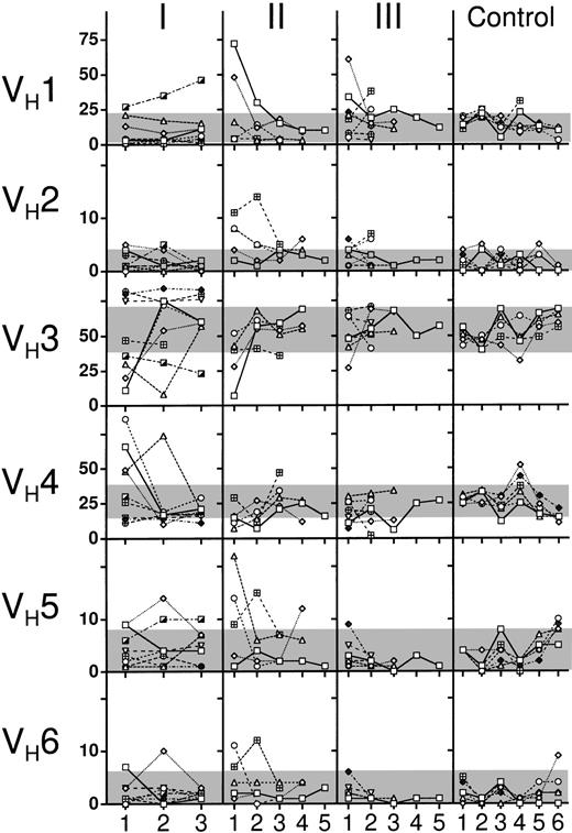
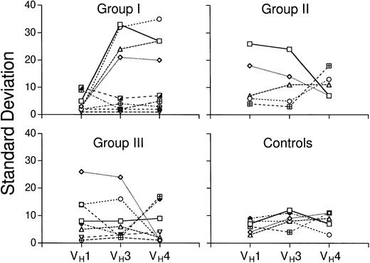
To examine the stability of the Ig VH gene subgroup distribution over time, we collected six blood samples from each of six members of the control group at 3- to 6-month intervals (Fig 1). One control subject had Ig VH gene subgroups levels that always were within the defined “normal” range. Four of the six had a single Ig VH gene subgroup level that was a few percentage points above or below the “normal” range on one or two occasions. Finally, one control subject had an elevated level of VH2 (at 5%) on two occasions, an elevated proportion of VH6 (at 9%) on another occasion, and a low level of VH3 (at 32%) associated with a high level of VH4 (at 52%) on one occasion (see lines connecting the diamonds in the graphs of the far right column, labeled “Control,” Fig 1). We calculated the mean and standard deviation of each Ig VH gene subgroup in the serial samples for each individual (Fig 2). We found that these standard deviations of the means over time for each person ranged from 3% to 9% for Ig VH1, 4% to 12% for Ig VH3, and 3% to 13% for Ig VH4, among the six members of the control group.
Expressed IgM VH gene distribution in serial blood samples of HIV infected patients. Each graph depicts the percent expression of Ig VH gene subgroups 1 through 6, over time in: (I) persons infected with HIV infection and CD4+T-cell count of more than 500 cells per mm3 blood; (II) patients with AIDS and a CD4+ T-cell count of less than 200 cells per mm3 blood; (III) patients with AIDS, as described in Group II that developed lymphoma at a later date; and Controls, healthy HIV-seronegative adults. One symbol is used for the same subject within a given group.
Expressed IgM VH gene distribution in serial blood samples of HIV infected patients. Each graph depicts the percent expression of Ig VH gene subgroups 1 through 6, over time in: (I) persons infected with HIV infection and CD4+T-cell count of more than 500 cells per mm3 blood; (II) patients with AIDS and a CD4+ T-cell count of less than 200 cells per mm3 blood; (III) patients with AIDS, as described in Group II that developed lymphoma at a later date; and Controls, healthy HIV-seronegative adults. One symbol is used for the same subject within a given group.
Variation in the proportions of the major Ig VH gene subgroups in HIV-infected persons and control subjects over time. The graphs depict the data collected on persons in Group I (top left), Group II (top right), Group III (bottom left), or HIV-seronegative controls (bottom right), as indicated at the top of each graph. The standard deviations (SD) about the mean levels of VH1, VH3, or VH4 over time for each subject are indicated by the symbols. The percent SD about the mean Ig VH gene subgroup levels are indicated on the far right axis. Lines connect the symbols that correspond to the SD of the mean levels for the same person. Each symbol corresponds to the same symbol and subject as in Fig 1.
Variation in the proportions of the major Ig VH gene subgroups in HIV-infected persons and control subjects over time. The graphs depict the data collected on persons in Group I (top left), Group II (top right), Group III (bottom left), or HIV-seronegative controls (bottom right), as indicated at the top of each graph. The standard deviations (SD) about the mean levels of VH1, VH3, or VH4 over time for each subject are indicated by the symbols. The percent SD about the mean Ig VH gene subgroup levels are indicated on the far right axis. Lines connect the symbols that correspond to the SD of the mean levels for the same person. Each symbol corresponds to the same symbol and subject as in Fig 1.
In parallel, we analyzed two to five serial blood samples that were collected at 3- to 6-month intervals from each of the HIV-infected persons in Groups I , II , and III (Fig 1). Overall, we found fluctuations of the expressed Ig VH gene distribution that were higher than the one observed in normal controls. In detail, four of the eight repetitively examined persons of Group I (HO, HQ, HR, and HP; Table 1) had extreme reciprocal variations in the levels of Ig VH3 and Ig VH4 subgroups over time (Fig 1), with standard deviations about the means over time ranging from 24% to 32% for Ig VH3, and 20% to 37% for Ig VH4 (Fig 2). Two of these four subjects also had fluctuations in the Ig VH5 and Ig VH6 subgroups that reciprocated the changes observed in Ig VH3 (open squares and diamonds in the column labeled “I”; Fig 1). Finally, one person in Group I experienced increases in the proportions of Ig VH1 and Ig VH5 that were associated with decreases in relative proportion of Ig VH3 over time (HW; Table 1 and Fig 1). On the other hand, three persons in Group I had VH gene subgroup distributions that did not vary over time, despite having abnormally high levels of Ig VH3 and low levels of Ig VH4 (HT, HU, HV; Table 1 and Fig 2). Whatever the pattern, however, the subjects of Group I generally had “abnormal” levels of Ig VH3. Only one individual (HP; Table 1) who was found initially to have abnormal expression of Ig VH3 genes had a normal Ig VH3 level on more than one occasion (Fig 1; open diamonds, Column I).
In contrast, the subjects in Groups II and III had relatively normal levels of Ig VH3 on successive testing. Those who initially had “abnormal” levels of Ig VH3 (HA, HB, and HG; Table 1) were found to have “normal” Ig VH3 levels at later time points (Fig 1). Also, the high levels of Ig VH1 that initially were observed in these subjects dropped to normal levels (Fig 1). Except for one member of Group II who had an abnormally low Ig VH3 level of 36% on one occasion (HE; Table 1), the other members of these groups (namely HC, HD, HF, HH, HI, HJ, HK, HM, and HN; Table 1) had Ig VH3 levels within the normal range at all time points tested (Fig 1). Collectively, the proportion of subjects in Groups II and III who had normal Ig VH3 levels at each time point was significantly higher than that noted for those in Group I at any time point (P < .05, Student’s ttest).
Instead, the subjects in Groups II more typically had abnormalities in the levels of some of the smaller Ig VH gene subgroups. In particular, four of the five members of this group (HB, HC, HD, and HE; Table 1) had marked fluctuations in the levels of Ig VH5, with levels of more than 10% on at least one occasion (Fig 1). Two of these (HC and HE; Table 1) at one point had levels of Ig VH6 in excess of 10% (Fig 1). Finally, three subjects in this group (HC, HD, and HE; Table 1) also had levels of Ig VH2 in excess of 8% on at least one occasion. Such abnormalities were not observed in the samples obtained from members of Group III. Nevertheless, the subjects in either Group II or III tended to have standard deviations about the means over time for the larger Ig VH gene subgroups that were higher than that of the control group (Fig 2).
DISCUSSION
We examined the relative expression of Ig VH gene subgroups in serial blood samples of persons infected with HIV and seronegative healthy adults. The blood B cells of seronegative donors had Ig VH gene subgroup repertoires that were similar to those previously reported for normal adults.14,15,18,19,23 24Furthermore, upon testing six seronegative donors at 3- to 6-month intervals, we noted that the Ig VH subgroup expression levels for each normal subject were relatively constant over time.
These observations are consistent with those made in a previous study on pairs of adult monozygotic twins, eight of which were concordant or discordant for rheumatoid arthritis (RA).19 Each pair had more similar IgM VH gene repertoires than did unrelated subjects, regardless of whether they were discordant for RA. Collectively, these studies suggest that genetic factors have a predominant influence on the distribution of Ig VH gene subgroups expressed by IgM+ blood B cells.
However, the IgM+ blood B cells of persons infected with HIV expressed VH gene subgroup repertoires that were strikingly aberrant and generally unstable. Twenty-nine of the 37 persons tested (78%) had “abnormal” repertoires, generally involving two or more Ig VH gene subgroups (Table 1). Moreover, 24 (65%) had levels of one or more Ig VH gene subgroups that differed by more than three standard deviations from the mean observed among seronegative adults (Table 1). The incidence of abnormal subgroup distributions among HIV-infected persons was significantly higher than that noted among the control group (P< .001, Χ2 analysis). In addition, persons infected with HIV generally had large fluctuations in the proportionate expression of several Ig VH gene subgroups over time (Fig2).
Prior studies noted that persons infected with HIV can have skewed Ig VH repertoires depleted of IgM+, IgM/IgD+, and IgG+ B cells that express Ig VH genes of the VH3 subgroup.10,11,25 However, these studies did not examine the proportions of the Ig VH gene subgroups relative to that of all the other VH gene subgroups, or examine the same patient on more than one occasion. In light of the current study, it seems likely that the relative deficiency noted in the expression of Ig VH3 genes by persons infected with HIV may not have been a stable phenotype. Moreover, cases with overexpression of Ig VH3 genes relative to that of the other subgroups may have been missed. Finally, the results of our study indicate that alterations in the relative expression levels of this subgroup most typically occur in persons at the early stages of HIV-infection, contrary to speculation that persons infected with HIV may develop progressive depletion of VH3-expressing B cells over time.11
Conceivably, B-cell superantigens may account for the abnormal levels of Ig VH gene subgroups observed in patients infected with HIV.26,27 B-cell superantigens are substances that bind the immunoglobulin encoded by most Ig VH genes of a given subgroup.28-30 Moreover, most Ig that react with a superantigen, such as Staphylococcal protein A, are encoded by Ig VH genes homologous to their germline counterparts,31 indicating that somatic mutation and selection are not required to develop Ig with superantigen binding activity. Such antigens can induce expansion or depletion of all B cells that express surface Ig encoded by a specific Ig VHgene subgroup. This is in contrast to conventional antigens that generally induce specific antibodies encoded by any one of several different Ig VH subgroups.
HIV gp120 binds to a conserved immunoglobulin motif unique to immunoglobulins encoded by Ig VH genes of the VH3 subgroup.32 Berberian et al25reported a threefold elevation of such B cells in some HIV-seropositive adults relative to that of seronegative control subjects. However, they noted that the number of VH3-expressing B cells were markedly reduced in patients with AIDS, as were the levels of serum anti-gp120 antibodies bearing a VH3-encoded crossreactive idiotype. As such, gp120 may function as a superantigen that can induce expansion or deletion of B cells expressing antibodies encoded by VH3 genes.
However, in the current study we found large fluctuations in Ig VH gene subgroups other than VH3 that could not be explained by alterations in the levels of Ig VH3 alone. For example, isolated expansions or contractions in the population of B cells that express Ig encoded by VH3 genes should not affect the proportion of the second largest Ig VH gene subgroup, VH4, relative to that of Ig VH1, or to that of the other Ig VH subgroups. However, we found that the proportions of these smaller Ig VH groups relative to one another also fluctuated over time. As such, patients infected with HIV do not have abnormalities restricted to B cells expressing Ig VH3 genes, particularly patients at advanced stages of infection.
This does not exclude the possibility that aberrant use of Ig VH genes in HIV-infected individuals reflects an ongoing immune response to HIV or an interaction between HIV-associated glycoproteins and B-cell surface Ig. During the early stages of HIV infection, individuals often develop an antibody response against HIV glycoproteins that diminishes as the CD4+ T-cell counts fall.33 Furthermore, Ig VH genes isolated from AIDS-associated lymphoproliferations or AIDS-associated lymphoma have evidence for somatic mutations suggestive of having been selected in an antigen-driven immune response(s).7,34,35 In addition, the instability in the expression of the various Ig VH gene subgroups may reflect changes occurring in the antigenic-epitopes of the virus in any one person over time. It is estimated that HIV undergoes over 180 generations per year,36 allowing for the emergence of a large number of different HIV mutants soon after infection.37,38 Mutations generally occur in the genes encoding the envelop glycoproteins that alter the tropism and biologic properties of the virus over time.39-42 Such alterations may account for the differences observed in the proportionate use of Ig VH genes in patients with high CD4+ T-cell counts (Group I ) versus those used by Groups II and III who had established AIDS.
The mutability and altered tropism of the HIV glycoproteins that occur during the course of infection for any one individual can provide for a complex and dynamic relationship between the virus and its host. B cells may express functional chemokine receptors that recently have been identified as being important coreceptors for HIV.43-45 This raises the possibility that HIV may interact with B cells directly via surface Ig and such receptors to induce the B-cell pathophysiology that is observed in patients infected with HIV.
In any case, the results of our study is consistent with the notion that B cells of persons infected with HIV are undergoing rapid turnover, with successive waves of stimulation, expansion, and/or depletion. Such turnover may allow for chance acquisition of somatic changes that can lead to the malignant transformation of a responding B-cell clone. Further study on the mechanism(s) underlying the dynamics within the B-cell compartment of persons infected with HIV may show the factor(s) accounting for the high rate of lymphomas that occur in patients with AIDS.
This project was funded by NIH Grants No. RO1 CA 65408-03 (T.J.K.), AI27670, and AI 36214 (Center for AIDS Research), and the Research Center for AIDS and HIV Infection of the San Diego Veterans Affairs Medical Center.
Address correspondence to Thomas J. Kipps, MD, PhD, Divisions of Hematology/Oncology and Infectious Diseases, Department of Medicine, University of California, San Diego, CA 92093-0663.
The publication costs of this article were defrayed in part by page charge payment. This article must therefore be hereby marked "advertisement" is accordance with 18 U.S.C. section 1734 solely to indicate this fact.


