To address the fundamental question of whether or not stem cell populations age, we performed quantitative measurements of the cycling status and frequency of hematopoietic stem cells in long-lived C57BL/6 (B6) and short-lived DBA/2 (DBA) mice at different developmental and aging stages. The frequency of cobblestone area-forming cells (CAFC) day-35 in DBA fetal liver was twofold to threefold higher than in B6 mice, and by late gestation, the total stem cell number was nearly as large as that of young DBA adults. Following a further ≈50% increase in stem cells between 6 weeks and 1 year of age, numbers in old DBA mice dropped precipitously between 12 and 20 months of age. In marked contrast, this stem cell population in B6 mice increased at a constant rate from late gestation to 20 months of age with no signs of abatement. Throughout development an inverse correlation was observed between stem cell numbers and the percentage of cells in S-phase. Because a strong genetic component contributed to the changes in stem cell numbers during aging, we quantified stem cells of 20-month old BXD recombinant inbred (RI) mice, derived from B6 and DBA progenitor strains, thus permitting detailed interstrain genetic analysis. For each BXD strain we calculated the stem cell increase or decrease as mice aged from 2 to 20 months. Net changes in CAFC-day 35 numbers among BXD strains ranged from an ≈10-fold decrease to an ≈10-fold increase. A genome-wide search for loci associated with this quantitative trait was performed. Several loci contribute to the trait—putative loci map to chromosomes X, 2, and 14. We conclude that stem cell numbers fluctuate widely during aging and that this has a strong genetic basis.
HEMATOPOIETIC STEM CELLS are generally believed to possess self-replicating potential that not only sustains lifelong blood cell production, but embodies a reserve potential capable of meeting needs not normally encountered. For example, serial transplantation studies have shown that not only is a small fraction of bone marrow stem cells from a single animal capable of restoring hematopoiesis in multiple lethally irradiated recipients, but that single clones are capable of doing this in secondary, tertiary, and quaternary hosts over a cumulative period that exceeds the lifespan of the donor.1-3 Thus it may seem that stem cells escape deleterious changes culminating in the senescence that befalls somatic cells of almost all other tissues during aging.4 But is this really so? The question has been addressed repeatedly over the years usually by transplantation assays, which are convenient, but bear only partial similarity to natural changes accompanying aging.2,5-8 Normal hematopoiesis and engraftment in a transplantation setting share a common dependence on the numbers of stem cells present and their proliferative potential, either of which may be independently affected by aging. As persuasive evidence of this, during ontogeny, while stem cell numbers exponentially increased, the proliferative potential of sorted human stem cells, as measured by the number of progeny generated in vitro, was shown to decrease rapidly.9 In a similar vein, mouse C57BL/6 (B6) and CBA fetal liver stem cells have a higher repopulating ability than their adult marrow counterparts.10-13Moreover, we have recently extended and confirmed data showing that during a good part of the entire mouse lifespan stem/progenitor cell cycling decreased from near a theoretical maximum to almost undetectable levels,14,15 a finding consistent with observations made in many other aging tissues.4Surprisingly, stem cell frequencies, measured either by flow cytometry in long-term bone marrow culture or by in vivo retransplantation assays, have been shown to significantly increase during aging, at least in the C57BL/6 mouse.14,16,17 This apparently counterintuitive finding is not easy to explain, but we have recently argued that the aging stem cell pool may have lower residual proliferative potential or ‘quality’, as it presumably has collectively completed more cell cycles.18 A gradual loss of proliferative capacity in dividing cells has been attributed to an erosion of telomeres, which may thus serve as a cell’s mitotic clock.19,20 Ectopic expression of telomerase in normally telomerase-negative fibroblasts has recently been shown to promote telomere lengthening and, most importantly, a not yet defined, but substantial, extension of proliferative potential.21 Other findings support the notion that a telomere-driven mitotic clock may be operative in the hematopoietic system, as it has been shown that telomeres of hematopoietic cells shorten significantly during aging and after bone marrow transplantation,20,22 despite the fact that stem cells express low levels of telomerase.23-25
It has been established, using a variety of experimental approaches, that a strong genetic component underlies the proliferation and population size of stem cells in different mouse strains, including B6 and DBA/2 (DBA).26-30 We have recently mapped to chromosome 18 a locus affecting stem cell frequency in young adult mice.30 To better understand the genetic regulation underlying stem cell dynamics during aging of B6 and DBA mice, we compared stem cell numbers obtained in young (2 months) and old (20 months) BXD recombinant inbred strains. Our data show that multiple quantitative trait loci (QTLs) contribute to variation in changes in stem cell numbers during aging.
MATERIALS AND METHODS
Mice used.
Young 6-week old female B6, DBA, and BXD recombinant inbred (RI) mice were purchased from The Jackson Laboratory (Bar Harbor, ME). Twelve- and 20-month old B6 and DBA mice were obtained from the National Institute of Aging (Bethesda, MD). For measurements using 20-month old BXD strains, 6-week old BXD mice were purchased from the Jackson Laboratory and were aged in the animal facility of the University of Kentucky. Mice were kept in microisolator cages and fed sterilized food and acidified water. Mice with obvious tumors, or otherwise not thriving, were excluded from the study. Twenty of the 26 existing BXD strains were available for this analysis. BXD mice of strains 2, 5, 8, 13, 21, and 22 all died before reaching 20 months of age.
RI strains.
RI mice are a powerful genetic tool for gene mapping. We chose to study the BXD strains because we have previously documented numerous differences in both hematopoietic stem and progenitor cell parameters and longevity parameters between the two progenitor strains from which they were derived.14,26-28 30 Twenty-six BXD strains have been generated by crossing C57BL/6 and DBA/2 inbred strains, and through continuous inbreeding beginning at the F2generation, each strain has evolved a unique combination of homozygous ‘B’ or ‘D’ alleles. The unique patterns of recombination in each RI strain are maintained through continuous inbreeding at The Jackson Laboratory and they are commercially available from this source. Over the ensuing years since their generation, numerous laboratories have used them to map either genetic markers or genes of interest. Thus, in the context of the BXD RI strains, roughly 1,700 genotypes have been mapped. As the mouse genome comprises about 1,500 to 1,600 centimorgans (cM) in total, there is an average of about one marker per cM, resulting in a high-density genetic map. It is therefore not surprising that genes contributing relatively large quantitative effects can be mapped in this set of RI strains without further genotyping of marker loci.
Genome-wide linkage statistics.
The frequencies of cobblestone area-forming cells (CAFC) day-35 (per 105 bone marrow cells) in young and old BXD mice was measured as described below. The percent change, either positive or negative, in CAFC day-35 frequency was calculated by comparing the data obtained in this study at 20 months of age with frequencies we obtained in young (2 month old) BXD mice.30 It was this parameter, percent change, that became the phenotype of interest in our study. The array of phenotypic results obtained from the set of BXD strains is often referred to as a strain distribution profile (SDP). In the case of a simple Mendelian trait, determined by a single locus, the phenotypes may be expected to fall into two groups: those characteristic of each of the two progenitor strains (C57BL/6 and DBA/2 in this case), provided they differ for the trait in question. In this case, the genotype (either ‘B’ or ‘D’) of a given BXD strain at the locus of interest can be deduced by simply comparing the phenotype with that of the two progenitor strains and determining which it matches. In the case of complex traits such as the subject of the present study, multiple loci make small individual contributions to the final phenotype and thus, as we found here, the SDP may well include values more extreme than either progenitor strain, as well as the full range of intermediate values (see Fig 4). Phenotypes more extreme than either of the progenitor strains are possible because of combinations of both positive and negative influences of individual quantitative trait loci (QTLs) on the phenotype, and the fact that neither progenitor strain may necessarily represent a pure combination of loci responsible for either all of the positive or all of the negative effects on phenotype. As discussed below, powerful computational software is now available for mapping QTLs such that the user is not required to discriminate between ‘B’ and ‘D’ phenotypes, rather the quantitative phenotypic data for each RI strain serves as the starting point for analysis.
Irrespective of the complexity, the strategy for mapping is to select the most similar, if not identical, SDP from the large list of previously mapped genotypes mapped in BXD strains. A concordance between a phenotypic SDP and an existing genotypic SDP (map location) indicates the presence of a QTL at or near that location contributing to the phenotype. However, due to the limited number of BXD strains available, the number of SDPs to be compared, and consequently the complexity of testing required to establish concordance, a close or identical match in SDPs may occur by chance.31-33 As with all statistical comparisons, it is necessary to make a calculation of the probability that the observed result was a false-positive. We therefore calculated the genome-wide probability of obtaining the observed linkages by random chance corresponding to an error threshold of P = .05; that is, one chance in 20 of such a false-positive error. We did this using the robust nonparametric permutation method developed by Churchill and Doerge,34 which is implemented in the Map Manager QT (b15) software developed by Manley.35
Interval mapping.
A subroutine of the Map Manager QT software, using computationally efficient regression equations, was used for mapping the QTLs.35 The probability of linkage between our trait (differences in CAFC frequency between young and old mice) and previously mapped genotypes was estimated at 1-cM intervals along the entire genome, except for the Y chromosome. The statistical power of linkage of the phenotype to individual genotypes (point-wise linkage statistics), such as those reported (see Table 2) for the three principal loci uncovered in our interval mapping, should attain values of between .000133 and .0000231 to reach a level of genome-wide statistical significance. Linkages approximating that level are deemed ‘suggestive’ and are worthy of reporting, although confirmation of linkage is required.32
The BXD genotype database.
The current mapping data files for BXD RI strains, compiled by R.W. Elliott and B. Taylor, were downloaded from The Roswell Park Cancer Institute (Buffalo, NY;ftp://mcbio.med.buffalo.edu/pub/MapMgr/data/).
Preparation of hematopoietic tissues and cells.
Fetal livers were harvested from day 13.5, 14.5, and 16.5 postcoitum (pc) fetuses. To obtain fetuses, three to four females were introduced into a cage with one male at 5:00 pm, and pregnant females, identified by the presence of a vaginal plug, were isolated the next morning at 9:00 am. This timepoint was considered day 0.5 pc. At the designated time, embryos were dissected and fetal livers isolated. Three to five fetal livers were pooled and disrupted by flushing through increasingly smaller gauge needles.
For experiments with bone marrow, cells were flushed from femora obtained from three to six B6 or DBA mice, or from one to three BXD mice, pooled, and used in the CAFC assay.
CAFC assay.
The CAFC assay, described by Ploemacher et al,36 was used as published earlier, with minor modifications.14,30 In brief, cells of the stromal cell line, FBMD-1, originally established by Neben et al,37 were seeded in 96-well plates (Costar, Cambridge, MA) in Dulbecco’s modified Eagle’s medium (DMEM) containing L-glutamine (GIBCO-BRL, Life Technologies, Grand Island, NY), 5% horse serum, 15% fetal bovine serum (sera from GIBCO-BRL), 10−4 mol/L β-mercaptoethanol, 10−5 mol/L hydrocortisone (Sigma, St Louis, MO), 80 U/mL penicillin, 80 μg/mL streptomycin (both from GIBCO-BRL), and 25 mmol/L NaHCO3. Plates were incubated at 33°C in 5% CO2, and used 10 to 14 days later. Bone marrow cells were seeded onto these preestablished stromal layers in six dilutions, serially in threefold increments from 333 to 81,000 cells/well (40 wells per dilution). At this time, the medium was switched from 5% horse serum and 15% fetal bovine serum to 20% horse serum. After 1 week, all wells were evaluated for the presence or absence of cobblestone areas, defined as colonies of at least five small nonrefractile cells growing beneath the stromal layer. CAFC day-7 correspond to relatively committed progenitor cells, whereas CAFC day-21 and 35 reflect increasingly more primitive cell subsets.36,38,39 CAFC frequencies were calculated using Poisson statistics.40 For calculations of total body stem cell numbers in the fetus, the frequency of CAFC day-35 was multiplied by fetal liver cellularity. For total body stem cell numbers in adult mice, the CAFC day-35 frequency was first multiplied by the femoral cellularity, and subsequently by a factor of 17, under the assumption that one femur represents ≈6% of the total marrow.41
Measurement of progenitor cell cycling.
The percentage of CAFC day-7 in S-phase of the cell cycle was determined by using a hydroxyurea suicide technique, as described previously.14 30 Briefly, 2 aliquots of marrow or fetal liver cells were diluted to a concentration of 1 × 10⁷ cells/mL. Hydroxyurea (Sigma) was added on 1 aliquot at a concentration of 200 μg/mL, and both samples were incubated at 33°C for 1 hour. After incubation, the two-cell suspensions were washed and a nucleated cell count was performed. A CAFC day-7 assay was performed with the 2 aliquots, and the fraction of cells killed by hydroxyurea was calculated by dividing the CAFC frequency in the hydroxyurea-treated cell suspension by the frequency obtained with the control cell aliquot.
RESULTS
Progenitor cell cycling in DBA and B6 mice during aging.
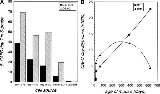
CAFC day-7 represent relatively committed progenitor cells that are actively cycling, whereas CAFC day-35 reflect primitive stem cells that are predominantly quiescent.14,36 38 To assess the steady-state proliferation status of the stem cell compartment, we measured the percentage of CAFC day-7 in S-phase (Fig 1A). At all timepoints, except at 1 year, DBA/2 cells had a much higher cycling activity than B6 cells. The percentage of cells in S-phase declined daily from gestational days 13.5 to 16.5 of fetal development.
Changes in the proliferative status and total number of hematopoietic stem cells in B6 and DBA mice during aging. (A) The percentage of CAFC day-7 in S-phase was measured at different timepoints during development in the fetal liver (FL) and adult bone marrow (BM) in B6 and DBA mice. (B) The total number of CAFC day-35 per B6 (▪) and DBA (▴) mouse was calculated in the fetal liver (first three datapoints) or adult bone marrow (other datapoints). Differences between B6 and DBA values are significant (nonoverlapping 95% confidence intervals) at all timepoints, except at 1 year.
Changes in the proliferative status and total number of hematopoietic stem cells in B6 and DBA mice during aging. (A) The percentage of CAFC day-7 in S-phase was measured at different timepoints during development in the fetal liver (FL) and adult bone marrow (BM) in B6 and DBA mice. (B) The total number of CAFC day-35 per B6 (▪) and DBA (▴) mouse was calculated in the fetal liver (first three datapoints) or adult bone marrow (other datapoints). Differences between B6 and DBA values are significant (nonoverlapping 95% confidence intervals) at all timepoints, except at 1 year.
Stem cell numbers in DBA and B6 mice during aging.
We have previously shown that CAFC day-35 numbers in the bone marrow increase twofold to threefold as both B6 and DBA mice age from 2 to 12 months.14 In this study, we made more detailed measurements of the dynamics of stem cell numbers by extending our analysis of CAFC day-35 frequencies to three times during fetal development and to 20-month old mice. Because we were interested in assessing the total hematopoietic stem cell pool size, we calculated the absolute number of CAFC day-35 per mouse (Fig 1B). For fetuses, this was done by multiplying the frequency of CAFC day-35 per 105 liver cells, as shown in Table 1, with fetal liver cellularity. Total body CAFC day-35 numbers for adult mice were calculated assuming that the femur represents 6% of total marrow.41 We did not quantify stem cell numbers in the spleen, but we have previously established that during adult steady-state hematopoiesis, the spleen contains less then 1% of the total stem cell pool.42 Similarly, the resulting number in fetuses is a modest underestimation of the total stem cell pool, as small numbers of hematopoietic stem cells may also be found in other fetal organs.43
Interestingly, the vast majority of stem cells found in young adult bone marrow is already present in the fetal liver at day 16.5, a finding supported by a recent study by Harrison and Astle.44 It is apparent from Fig 1B that the stem cell pool size is far from static during aging; rather, it is highly dynamic. Importantly, the continuously changing size of this compartment appears to be strongly influenced by genetic determinants. A seemingly ever-expanding stem cell population was observed in B6 mice during the 20 months of observation, whereas in DBA mice, a clear maximum was observed at ≈12 months, after which the stem cell compartment contracted.
Table 1 shows the relative frequency of CAFC day-35 numbers per 105 fetal liver or adult bone marrow cells that was used to calculate the total stem cell pool per mouse as presented in Fig 1B. Perhaps not surprisingly, CAFC day-35 frequency is high in the fetal liver from mid to late fetal development, and subsequently is approximately fivefold less in marrow, presumably due to the dilution accompanying the dramatic increase in overall size of the newborn, including hematopoietic tissue, and the demand for rapid blood cell production. Quantitatively similar differences between fetal liver and adult marrow stem cell frequencies have been reported using an in vivo competitive repopulating unit assay12 or by flow cytometry analysis of stem cell markers.45 Interestingly, the relative frequency of CAFC day-35 in both strains of mice drops by ≈50% from day 13.5 to day 14.5 in utero. This relative decline is probably caused by an expansion of nonhematopoietic cells in the fetal liver at this time, and it should be emphasized that the absolute number of stem cells in the fetal liver increases substantially from day 13.5 to 14.5 (Fig 1B). Once in the bone marrow, stem cell frequencies in B6 mice increased continuously during aging. Initially, a qualitatively similar observation was made in DBA mice, but at 20 months, stem cell frequencies in this strain were severely reduced to ≈25% of values observed at 12 months. Although the stem cell frequency within a strain changes significantly with age, there was no evidence for increased intrastrain variation during aging, ie, the 95% confidence limits at all timepoints remained between ≈75% and ≈140% of the estimated frequency from Poisson statistics (Table 1).
Stem cell numbers in old BXD recombinant inbred mice.
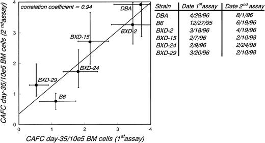
The genomes of BXD mice consist of a mosaic of homozygous B6 and DBA segments. Phenotyping BXD mice for a specific trait, followed by a linkage analysis of this trait with polymorphic markers previously mapped in these strains, may identify loci genetically linked with the observed phenotypic variation, and thus provide a map location.46 We have recently used 2-month old BXD RI mice to map loci that contribute to the observed variation in CAFC day-35 numbers between young B6 and DBA mice. The principal locus mapped to chromosome 18, approximately 19 cM from the centromere.30In the present study, we followed a similar strategy to map loci associated with changes of stem cell numbers during aging in old B6 and DBA mice. We verified the reproducibility of quantifying CAFC day-35 numbers over an extended period of time by retesting selected BXD strains and both progenitor strains. Figure2 shows that the CAFC assay as used in these experiments is reliable, as data were highly reproducible over a 2-year period. All data-points fall within the 95% confidence limits obtained by linear regression analysis.
Testing the reproducibility of the CAFC assay. CAFC day-35 frequencies were measured in two independent experiments over a 2-year interval using six different mouse strains as marrow donors. In each experiment cells from three mice were pooled. Results are given as mean values, and 95% confidence intervals are indicated.
Testing the reproducibility of the CAFC assay. CAFC day-35 frequencies were measured in two independent experiments over a 2-year interval using six different mouse strains as marrow donors. In each experiment cells from three mice were pooled. Results are given as mean values, and 95% confidence intervals are indicated.
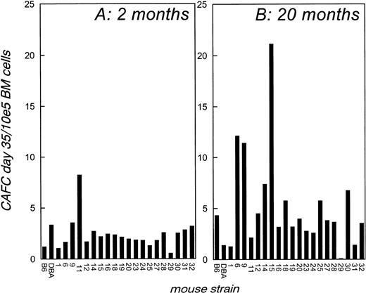
Figure 3B shows the CAFC day-35 frequency per 105 bone marrow cells in the 20-month old BXD strains that were available for analysis. Mice of the six missing strains (BXD-2, 5, 8, 13, 21, and 22) had died before 20 months. Figure 3A is shown for comparison and presents data obtained with 2-month old BXD strains, as published previously.30 It is immediately apparent that the variation in stem cell frequency among the strains is much higher in old than in young BXD mice, very much like the data obtained with the parental strains (compare with Fig 1B). The range in CAFC day-35 frequency varies approximately 16-fold in young BXD strains (BXD-29 having the lowest, and BXD-11 having highest stem cell numbers), but this increases to approximately 340-fold in old BXD mice. BXD-29 remained the lowest with .063 CAFC day-35/105 cells, and BXD-15 was the highest with 21.1 CAFC day-35/105 cells. For comparison, these values translate into a total CAFC day-35 pool size per mouse of only ≈100 for BXD-29 and of ≈82,000 for BXD-15.
CAFC day-35 frequency in bone marrow of young and old BXD mice. CAFC day-35 frequencies were determined in the bone marrow of 2-month (A) and 20-month (B) old BXD mice. The results presented in (A) are shown for comparison and have been published.30
CAFC day-35 frequency in bone marrow of young and old BXD mice. CAFC day-35 frequencies were determined in the bone marrow of 2-month (A) and 20-month (B) old BXD mice. The results presented in (A) are shown for comparison and have been published.30
Change in BXD stem cell numbers during aging.
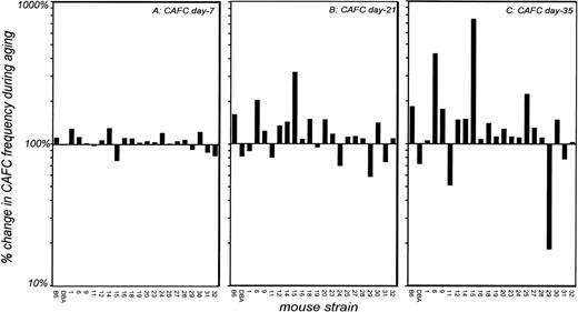
An aim of this study was to map loci that contribute to the age-dependent stem cell dynamics in B6 and DBA mice. To this end, we calculated the percentage change of CAFC day-35 frequency in old BXD mice (Fig 3B) compared with the value observed in their young counterparts (Fig 3A) to obtain the relative changes. In addition, we performed this calculation for less primitive CAFC day-7 and day-21 subsets (Fig 4). Figure 4A depicts the change in CAFC day-7 frequencies during aging. Old BXD mice have essentially similar progenitor cell frequencies as young mice, but when more primitive CAFC day-21 frequencies are compared, an increased variation is apparent (Fig 4B). Frequencies of this cell stage in old B6 mice increased to 300% compared with young, whereas this population is reduced to 50% in old DBA mice. Old BXD mice show a similar pattern; in some strains CAFC day-21 frequencies increase twofold to threefold (B6 phenotype), whereas in others, a DBA-like reduction is observed. Several strains show an intermediate phenotype, and old BXD-6 and BXD-15 mice show a greater expansion than parental B6. When primitive CAFC day-35 frequencies were measured, the variation among strains was even more extreme (Fig 4C). Old BXD-6 and BXD-15 mice exhibited an exceptional expansion of stem cell numbers, whereas the stem cell pool in BXD-29 declined more dramatically (approximately 10-fold). This variation cannot be due to experimental error in the assay, as CAFC day-7 frequencies, scored in the same plates using the same limiting-dilutions, are constant during aging and thus serve as an internal control. In addition, we find no evidence that intrastrain variation, which potentially could cause extreme outliers, increases during aging because the 95% confidence intervals of CAFC day-35 frequencies in B6 and DBA mice remained stable throughout their lifespan (Table 1).
Changes in progenitor and stem cell numbers in BXD mice during aging. CAFC day-7, 21, and 35 were measured in young (2 months) and old (20 months) BXD mice, and the percentage increase or decrease in cell numbers during aging was calculated. (A, B, and C) Show relative changes in the numbers of CAFC day-7, day-21, and day-35, respectively. Note that the Y-axis for all three panels is the same. Three young BXD mice were used for all datapoints. The number of old BXD mice per datapoint was 1 (seven strains), 2 (five strains), or 3 (eight strains). The most extreme deviations from young CAFC day-21 and 35 numbers were seen in strains from which three animals were used (BXD-6, -15, and 29).
Changes in progenitor and stem cell numbers in BXD mice during aging. CAFC day-7, 21, and 35 were measured in young (2 months) and old (20 months) BXD mice, and the percentage increase or decrease in cell numbers during aging was calculated. (A, B, and C) Show relative changes in the numbers of CAFC day-7, day-21, and day-35, respectively. Note that the Y-axis for all three panels is the same. Three young BXD mice were used for all datapoints. The number of old BXD mice per datapoint was 1 (seven strains), 2 (five strains), or 3 (eight strains). The most extreme deviations from young CAFC day-21 and 35 numbers were seen in strains from which three animals were used (BXD-6, -15, and 29).
Mapping loci that control population dynamics of stem cells during aging.
A genome-wide search for linked loci was performed using the data presented in Fig 4C as the phenotype of interest. Table 2 summarizes the results of this linkage analysis and lists the loci most strongly associated with the trait. These putative loci on chromosomes 2, 14, and X reached LOD scores of 3.1, 2.4, and 2.4, respectively, values associated with suggestive, but not significant, linkage.32 33 This is likely to result from the involvement of multiple loci at which B6 and DBA mice differ. The change in stem cell pool size during aging in these mice is probably the result of interaction of the QTLs listed in Table 2, as well as others not detected in our analysis. To resolve these loci, more meiotic events will have to be generated (and progeny tested) than can be evaluated in the limited context of the 20 BXD strains.
DISCUSSION
In this study, we have quantified stem cell populations during fetal development and adult aging in short-lived DBA and long-lived B6 mice. Because of their normally quiescent nature and their capability to extensively self-renew, large fluctuations in the size of the stem cell pool were not expected. Surprisingly however, we observed dynamic changes in the number of stem cells during the lifespan of these strains of mice, as stem cell numbers accumulated continuously in aging B6 mice, but dropped to embryonic values in old DBA mice. We have previously reported that progenitor cells from DBA mice show a marked reduction in cycling activity from 2 to 12 months of age.14Together with the present observation of declining CAFC day-35 numbers, this suggests that hematopoietic cell production in DBA mice generally enters a period of decline after 12 months, possibly leading to senescence. Although, as yet, we formally cannot prove the significance of these findings for in vivo stem cell function, the concept of sencescence is strengthened by observations made in B6↔DBA embryo-aggregated chimeras, where initially stable blood cell chimerism is established, but DBA stem cells slow and then cease blood cell production after ≈1 year.27 Thus our findings here reiterate both temporally and quantitatively what we have previously reported using chimeras and firmly establish that primitive hematopoietic stem cell characteristics are significantly affected by aging, presumably due to their replicative history. The effect of aging on stem cells is highly strain-specific however, and our present study was aimed at providing insight into the genetic predispostion of stem cell aging. Not surprisingly, the complex pattern of changes in stem cell numbers was found to be caused by multiple loci. Putative loci were shown to map to chromosomes X, 2, and 14, but the restricted genetic power of BXD analysis to map variation in traits that are influenced by many loci, precluded attaining significant linkage with any of these genomic intervals. We found no linkage to any marker on chromosome 18, to which we have previously mapped a locus affecting CAFC day-35 frequency in young mice,30 suggesting the two traits are independent. Our finding of linkage to an interval on the X-chromosome deserves further comment. Studies by Abkowitz et al47 in female heterozygous Safari cats (Geoffrey x domestic) have recently shown an X-linked locus that appears to regulate stem cell kinetics by conferring a competitive advantage to stem cells bearing a euchromatic Geoffrey X chromosome (and lyonization of the domestic-derived counterpart). This competitive growth advantage was observed indirectly as a predominance of mature blood cells with this X-inactivation pattern and became apparent only after 3 to 6 years.47 Excessive skewing of X-inactivation patterns, attributed to a declining stem cell population, has also been observed in blood cells of 40% to 60% of normal women over 60 years old who are heterozygous for X-linked G6PD alleles.48 49 While intriguing, it remains to be established whether the X-linked locus, which we report here, is involved in the dynamic regulation of stem cell numbers during aging in cats or humans, an association that requires independent confirmation of linkage.
Primitive hematopoietic stem cells, with the possible exception of those in the fetus, have a very low turnover rate and are generally believed to have a self-renewal probability (P) of .5, resulting in adequate hematopoietic blood cell replacement and a static pool size.50 It has been suggested that P, a value not possible to measure directly, can temporarily fluctuate to meet needs imposed by perturbation, such as stem cell transplantation or administration of cytotoxic drugs in cancer therapy.1,51Our data here suggest that values for P can change dramatically during normal aging, as the stem cell pool size is dynamic. In the fetal liver, it must initially be considerably higher than .5 (and may be close to 1.0), given the exponential daily expansion of stem cell numbers. Expansion of stem cells in fetal liver of DBA mice is particularly noteworthy. In adult marrow, however, a more complex pattern is observed. In B6 mice, in which the stem cell compartment expands at a constant rate (Fig 1), P appears to be slightly higher than .5 during at least 20 months of life. In DBA mice, the stem cell compartment increases less dramatically between the initial expansion in fetal liver and 1 year of age. Thus, P in DBA adult mice must be lower than P in adult B6 during the first 12 months, but nonetheless greater than .5. Second, and more importantly, it must be less than .5 thereafter, when stem cell numbers decline. It remains to be determined whether the contracting stem cell pool is responsible for the shorter lifespan of DBA mice or whether it is merely a correlative symptom of it. If a mitotic clock, potentially provided by telomere length,19,20 sets the maximal number of divisions stem cells may undergo, a prediction would be that a population of fast cycling stem cells is exhausted earlier than a population of slowly cycling stem cells. In this respect, it is interesting to note that DBA stem cells have a threefold to fourfold higher cycling activity than B6 cells, and this line of reasoning was used to explain extinction of DBA-derived hematopoiesis in B6↔DBA chimeras.14,26,30 Preliminary measurements in aged B6D2F1 mice, which are longer-lived than B6, show an even more pronounced stem cell increase during normal aging (data not shown). It should be emphasized however, that the relationship between lifespan and changes in stem cell numbers as suggested by these data is likely more complicated. For example, BXD-29 and BXD-15, the two most extreme strains analyzed here, have comparable cell cycle kinetics52 and lifespans.53 The complex relationships between these parameters suggest the existence of intricate regulatory mechanisms controlling alterations of in vivo stem cell numbers during aging and the findings reported here are but a first step in unravelling them. For example, the complexity obviously does not preclude identification of genetic mechanisms that play a role in this process, and the present study offers genetic approaches for subsequent analyses that are currently being pursued.
ACKNOWLEDGMENT
The authors thank Drs Stephen J. Szilvassy and Craig T. Jordan for critically reading the manuscript and Dr Rob Williams for invaluable assistance with MapManager software.
Supported by the Department of Internal Medicine, University of Kentucky Hospital and the Lucille P. Markey Cancer Center. G.dH. is a fellow of the Netherlands Organisation for Scientific Research (NWO).
The publication costs of this article were defrayed in part by page charge payment. This article must therefore be hereby marked “advertisement” in accordance with 18 U.S.C. section 1734 solely to indicate this fact.
REFERENCES
Author notes
Address reprint requests to Gary Van Zant, PhD, Blood and Marrow Transplant Program, Division of Hematology/Oncology, University of Kentucky, Markey Cancer Center, 800 Rose St, Lexington, KY 40536-0093; email: gvzant1@pop.uky.edu.


