Abstract
It is widely accepted that the serum of healthy individuals contains natural antibodies only against those blood group A or B antigens that are not expressed on the individual’s red blood cells. The mechanisms involved in tolerance to autologous blood group antigens remain unclear. In the present study, we show that IgM and IgG antibodies reactive with autologous blood group antigens are present in the immunoglobulin fraction of normal human serum. Natural IgG anti-A antibodies purified by affinity chromatography from IgG of individuals of blood group A exhibited an affinity for A trisaccharide antigen in the micromolar range and agglutinated A red cells at sixfold higher concentrations than those required for agglutination with affinity-purified anti-A IgG of individuals of blood group B. Whereas autoantibodies reactive with self A and B antigens are readily detected in purified IgG and IgM fractions, their expression is restricted in whole serum as a result of complementary interactions between variable regions of antibodies. These observations suggest that tolerance to autologous ABO blood group antigens is dependent on peripheral control of antibody autoreactivity.
THE PRESENCE OF antibodies directed against A and B alloantigens pre-existing in the serum of naive individuals before transfusion has been recognized for several decades.1 It has been considered that because ABO histo-blood group antigens are oligosaccharides commonly occurring in nature, individuals of blood group A become immunized to type-B carbohydrate antigen present in the bacterial environment, although they remain tolerant to A antigens.2 Thus, anti-A antibodies are found in the serum of group O and B individuals and anti-B antibodies are found in the serum of group O and A individuals. Group AB individuals are believed not to have anti-A nor anti-B antibodies because they express both antigens on their red cells.
Natural antibodies, ie, antibodies produced in the absence of overt antigenic stimulation, have been recognized in the serum of healthy individuals.3 Natural autoantibodies consist of immunoglobulinM (IgM), IgG, and IgA immunoglobulins encoded by germline genes, and react with a wide range of self-antigens including nuclear antigens, intracellular and membrane components, and circulating plasma proteins.4 Natural autoantibodies are often polyreactive, exhibit idiotypic cross-reactivity and variable (V) region connectivity.5-8 Complementary interactions between V regions regulate autoreactivity of natural autoantibodies in whole serum, thus contributing to the maintenance of immune homeostasis under physiological conditions.9 10
We now show that IgG and IgM antibodies reactive with autologous A and B antigens are present in normal serum immunoglobulin. Our results indicate that autoantibody activity to A and B antigens is controlled in whole serum by antibodies complementary to the V regions of the antibodies to autologous blood group antigens. These observations suggest that “tolerance” to autologous ABO blood group antigens as observed in serum, is dependent on peripheral control of antibody autoreactivity.
MATERIALS AND METHODS
Antibodies.
Serum was obtained from 32 healthy adult blood donors of blood groups A1 (n = 8), B (n = 8), AB (n = 8), and O (n = 8). All donors were RhD-negative. IgG was purified from serum by chromatography on protein G-Sepharose (Pharmacia Fine Chemicals, Uppsala, Sweden). When required, F(ab′)2 fragments were prepared from IgG by pepsin digestion (2% wt/wt) (Sigma Chemical Co, St Louis, MO) in acetate buffer pH 4.1 for 18 hours at 37°C and chromatography on protein A-Sepharose (Pharmacia). Concentrations of purified IgG and F(ab′)2 fragments were determined spectrophotometrically at 280 nm using an extinction coefficient of 1.4. The concentration of IgG in serum was determined by ELISA. IgM was quantitated in the flow-through of the protein G-Sepharose column by ELISA and by using the BCA protein assay (Pierce, Rockford, IL). The concentration of IgG in the IgG-depleted fraction of serum was below 20 μg/mL.
Anti-A antibodies were purified from pooled serum of six donors of blood group A1 and of six donors of blood group B by affinity chromatography on a macroporous glass matrix covalently coated with poly[N-(2-hydroxyethyl)acrylamide] (PAA)-bound synthetic A trisaccharide (Atri-PAA-MPG) (Syntesome, Moscow, Russia) and elution of the column at pH 3.0 followed immediately by neutralization of the eluted fractions using Tris 2.0 mol/L. Affinity-purified anti-A IgG antibodies were then separated from anti-A IgM by chromatography on protein G-Sepharose. Concentrations of purified anti-A IgM were determined using the BCA protein assay. The purity of the PAA-conjugates was determined by initial saccharides. Synthetic w-aminoalkyl glycosides used for coupling with the polymer were more than 95% pure as evidenced by high-performance liquid chromatography (HPLC) and 1H-NMR. M06 is a human monoclonal IgG directed against pp65 antigen of cytomegalovirus,11 a kind gift of Dr M. Ohlin (Lund, Sweden).
ABO antigens.
PAA-bound A trisaccharide glycoside (GalNAcα1-3(Fucα1-2)Galβ1) and B trisaccharide (Galα1-3(Fucα1-2)Galβ1) were obtained from Syntesome. The A and B antigen conjugates contained 0.77μmol/mg (15%) and 0.36μmol/mg (5%) of saccharide, respectively.
Enzyme immunoassays.
Anti-A and anti-B antibody activities were quantitated by ELISA. Ninety six-well polystyrene flat-bottom microtiter ELISA plates (Nunc, Roskilde, Denmark) were coated with PAA-bound A (10 μg/mL) or B (10 μg/mL) trisaccharides in carbonate buffer pH 9.0 for 2 hours at 37°C and, subsequently, for 12 hours at 4°C. The plates were then incubated with 1.0% bovine serum albumin (BSA) (Sigma) in phosphate-buffered saline (PBS) for 1 hour at room temperature. After washing with PBS, the plates were incubated with various dilutions of the antibodies to be tested in PBS containing 1.0% BSA for 1 hour at 37°C. The plates were then washed with PBS and incubated with peroxidase-labeled goat antihuman Fcγ (Jackson Laboratories, West Grove, PA) or peroxidase-labeled goat antihuman Fcμ antibodies (Southern Biotechnology, Birmingham, AL) for 1 hour at 37°C. Bound IgG or IgM were revealed using the enzyme substrate OPD. The reaction was stopped after 4 minutes in all assays. Absorbance at 495 nm was determined using an Emax ELISA reader (Molecular Devices, Menlo Park, CA). Anti-A and anti-B monoclonal antibodies (MoAbs) (clones A003 anti-A; B005 anti-B; Biotest Pharma, Dreieich, Germany) were used as positive controls for standardization of ELISA. Optical density values in ELISA < 0.3 were considered “negative.”
Agglutination assays.
Blood group A1 and B red blood cells for use in agglutination tests were prepared from freshly drawn venous blood of healthy volunteers. Indirect antiglobulin tests were performed by incubating 50 μL of a 3.0% saline suspension of indicator red cells with 100 μL of 7.0% BSA and 100 μL of the source of antibody to be tested diluted in PBS, for 45 minutes at 37°C. After incubation, the cells were washed three times with PBS, and blotted dry. One drop of antihuman globulin Coombs reagent (Organon Teknika BV, Boxtel, Holland) was then added. The tubes were mixed, centrifuged, and examined for agglutination microscopically. Agglutination was graded from − to ++++, using a conventional grading system. Direct agglutination assays were performed by incubating indicator cells with antibodies in V-bottom wells for 1 hour at 37°C. Agglutination was recorded on inclination of the plates and by microscopic examination.
Biosensor measurement of antibody activity.
Real-time analysis of antigen-antibody complex formation between A and B trisaccharides and anti-A and anti-B antibodies was performed using the BIAcore system (Pharmacia). Biotinylated PAA-antigen A (Atri-PAA-biotin) and antigen B (Btri-PAA-biotin) (Mr∼30 kD) (Syntesome) were immobilized on streptavidin-coated sensor chip SA5(Pharmacia) in HEPES complete buffer pH 7.4 (10 mmol/L HEPES, 150 mmol/L NaCl, 3.4 mmol/L EDTA, 0.05% Surfactant P20) (HBS) at a continuous flow rate of 5 μL/minute. One hundred microliters of HBS containing 1.0 mg/mL of purified IgG or 0.3 mg/mL of affinity-purified anti-A IgG to be tested were injected at a continuous flow rate of 5 μL/minute allowing a total contact time with the antigens of 6 minutes. Kinetic analysis and calculations of association and dissociation constants were performed using the BIAevaluation Software (Pharmacia).
Statistics.
When appropriate, statistical analysis was performed using analysis of variance (ANOVA), Fisher’s test, and the StatView software (SAS Institute Inc, Cary, NC).
RESULTS
IgG and IgM antibodies to autologous ABO antigens in normal human serum.
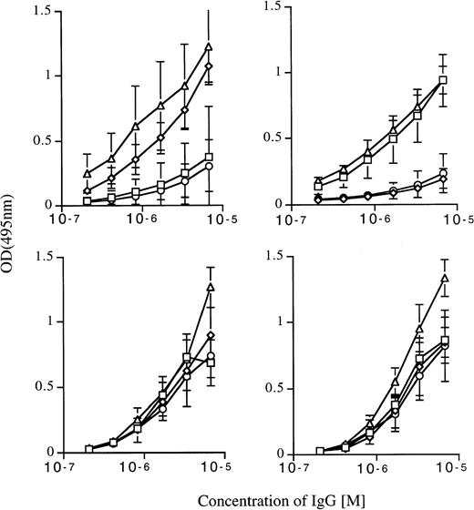
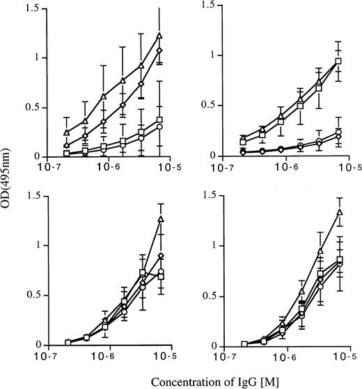
The reactivity of IgG in the serum and in the purified IgG fraction of the serum of four healthy adults of each of the blood group categories A1, B, AB, and O, with A and B antigens, was measured by means of ELISA (Fig 1). As expected, IgG in whole serum of individuals of blood groups B and O bound to A antigen in a dose-dependent fashion. The binding of serum IgG of individuals of blood groups A1 and AB was low and differed significantly from that of serum IgG of individuals B and O (P < .0001 in all instances). The mean binding of serum IgG of individuals of blood group A1 did not differ from that of individuals of blood group AB. Similarly, IgG in whole serum of individuals of blood groups A1 and O exhibited a significantly higher reactivity with B antigen than did serum IgG of individuals of blood groups B and AB (P < .0001 in all instances). The mean binding to B antigen of IgG in whole serum of individuals of blood group O was significantly higher than that of serum IgG of individuals of blood group A1 (P < .0001). In contrast to results obtained using whole serum, we observed no difference in the mean binding activity of IgG purified from the serum of donors of blood groups A1, B, AB, and O to A and B antigens. Thus, there was no difference in the mean reactivity of purified IgG of donors of blood group A1 and individuals of blood group B with antigen A (P = .8698) and antigen B (P = .3439) (Fig 1). The specificity of interaction of natural antibodies to blood group antigens was validated by using MoAbs. Thus, we showed that MoAbs directed against A antigen bound to A antigen and not to B antigen and conversely MoAbs directed against B antigen bound to B antigen and not to A antigen (data not shown).
Natural IgG antibodies to autologous A and B blood group antigens in normal human serum. Upper panels: reactivity of IgG in whole serum of individuals of blood group A (□), B (◊), O (▵), and AB (○) with immobilized A (left panel) and B (right panel) antigens, as assessed by ELISA. Lower panels: reactivity of IgG purified from the serum of individuals of blood groups A (□), B (◊), O (▵), and AB (○) with A (left panel) and B ( right panel) antigens. Each data point represents mean OD ± SD obtained on testing IgG of four individuals in each blood group category.
Natural IgG antibodies to autologous A and B blood group antigens in normal human serum. Upper panels: reactivity of IgG in whole serum of individuals of blood group A (□), B (◊), O (▵), and AB (○) with immobilized A (left panel) and B (right panel) antigens, as assessed by ELISA. Lower panels: reactivity of IgG purified from the serum of individuals of blood groups A (□), B (◊), O (▵), and AB (○) with A (left panel) and B ( right panel) antigens. Each data point represents mean OD ± SD obtained on testing IgG of four individuals in each blood group category.
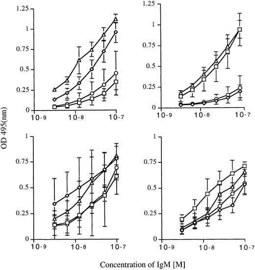
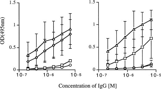
The binding of IgM in the serum of individuals of blood groups B and O to A antigen was higher than that of serum IgM of individuals of blood group A1 and AB (P < .0001 in all cases). Similarly, the binding of serum IgM of individuals of blood group A1 and O to B antigen was higher than that of serum IgM of individuals of blood group B and AB (P < .0001 in all cases). We then prepared the IgG-depleted fraction of serum of the four donors by chromatography on protein G Sepharose and used it as a source of IgM. IgM thus purified from the serum of A1 and B donors bound to autologous A and B antigens, although no or little such binding was observed on testing of IgM in whole serum (Fig 2). Thus, the difference in reactivity of purified IgM with allo- and with autologous A and B antigens was small (Fig 2).
Natural IgM antibodies to autologous A and B blood group antigens in normal human serum. Upper panels: reactivity of IgM in whole serum of individuals of blood group A (□), B (◊), O (▵), and AB (○) with immobilized A (left panel) and B (right panel) antigens, as assessed by ELISA. Lower panels: reactivity of IgM (IgG-depleted serum) of individuals of blood groups A (□), B (◊), O (▵), and AB (○) with A (left panel) and B (right panel) antigens. Each data point represents mean OD ± SD obtained on testing IgG of four individuals in each blood group category.
Natural IgM antibodies to autologous A and B blood group antigens in normal human serum. Upper panels: reactivity of IgM in whole serum of individuals of blood group A (□), B (◊), O (▵), and AB (○) with immobilized A (left panel) and B (right panel) antigens, as assessed by ELISA. Lower panels: reactivity of IgM (IgG-depleted serum) of individuals of blood groups A (□), B (◊), O (▵), and AB (○) with A (left panel) and B (right panel) antigens. Each data point represents mean OD ± SD obtained on testing IgG of four individuals in each blood group category.
Inhibition of the binding of IgG to autologous blood group antigens by autologous IgG-depleted serum.
IgG purified from the serum of individuals of blood groups A1, B, AB, and O was coincubated with IgG-depleted serum, used as a source of autologous IgM, at a ratio of IgG/IgM (wt/wt) of 1:10, for 1 hour at 37°C before assessing the reactivity of IgG with A and B antigens by ELISA. IgM (IgG-depleted serum) of individuals of blood group A1 inhibited the reactivity of autologous IgG with A antigen but not with B antigen (Fig 3). In a similar fashion, IgM (IgG-depleted serum) of individuals of blood group B inhibited the reactivity of autologous IgG with B antigen but not with A antigen. IgM of individuals O did not affect the binding of the corresponding IgG to both antigens. IgM (IgG-depleted serum) of AB blood group individuals inhibited the reactivity of autologous IgG to both antigens. These results suggest that the absence of reactivity of IgG with autologous A and B antigens in the serum of individuals of blood groups A and B, is dependent on inhibition of self-reactive IgG by factors present in IgG-depleted fraction of serum, most likely by autologous IgM.
IgM inhibits the reactivity of purified autologous IgG with autologous blood group antigens. IgG was purified from serum of individuals of blood groups A1 (□), B (◊), O (▵), and AB (○) and incubated with the respective autologous IgG-depleted fractions of serum used as sources of IgM at a ratio of IgG to IgM (wt/wt) of 1:10 for 1 hour at 37°C. The reactivity of IgG with A (left panel) and B antigens (right panel) was then assessed by ELISA. Each data point represents the mean OD ± SD of binding of IgG from four individuals of each blood group category. The binding of IgG of individuals of blood group A to A antigen in the presence of autologous IgM was significantly lower than the binding of IgG of individuals of blood group B coincubated with autologous IgM (P < .0001). Similarly, the binding of B IgG to B antigen in the presence of B IgM was significantly lower than that of A IgG in the presence of A IgM (P < .0001).
IgM inhibits the reactivity of purified autologous IgG with autologous blood group antigens. IgG was purified from serum of individuals of blood groups A1 (□), B (◊), O (▵), and AB (○) and incubated with the respective autologous IgG-depleted fractions of serum used as sources of IgM at a ratio of IgG to IgM (wt/wt) of 1:10 for 1 hour at 37°C. The reactivity of IgG with A (left panel) and B antigens (right panel) was then assessed by ELISA. Each data point represents the mean OD ± SD of binding of IgG from four individuals of each blood group category. The binding of IgG of individuals of blood group A to A antigen in the presence of autologous IgM was significantly lower than the binding of IgG of individuals of blood group B coincubated with autologous IgM (P < .0001). Similarly, the binding of B IgG to B antigen in the presence of B IgM was significantly lower than that of A IgG in the presence of A IgM (P < .0001).
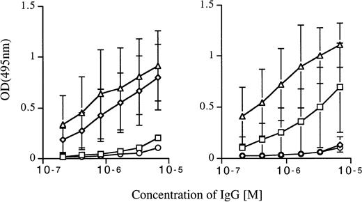
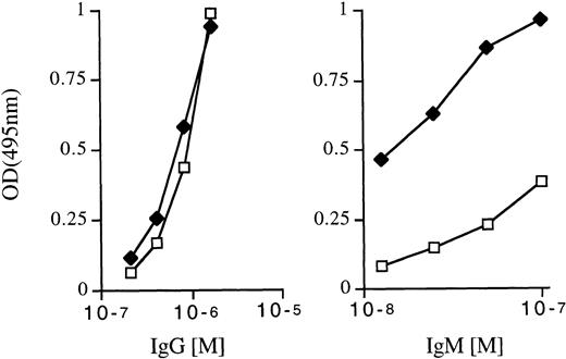
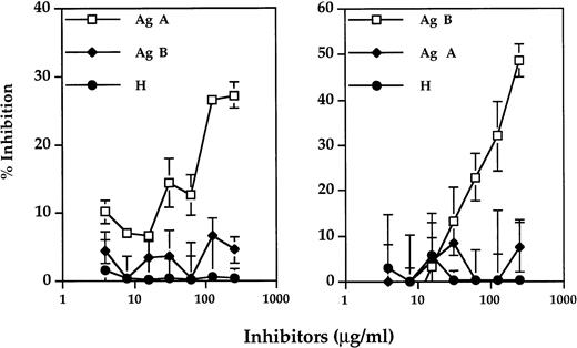
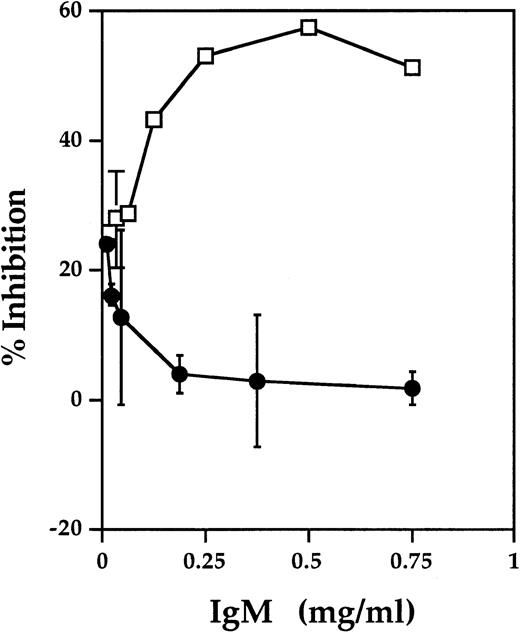
We then affinity-purified anti-A antibodies from pooled serum of six individuals belonging to blood groups A1 and B, respectively, using a macroporous glass matrix covalently coated with poly[N-(2-hydroxyethyl)acrylamide] (PAA)-bound synthetic A trisaccharide (Atri-PAA-MPG). Anti-A IgG was then isolated from the non-IgG containing fraction by chromatography on protein G Sepharose. We then assessed the reactivity of anti-A IgG of individuals of the two blood groups with A antigen by ELISA. As shown in Fig 4, we found no difference in the mean binding activity to A antigen of purified anti-A IgG of donors of blood groups A1 and B. However, the non-IgG fraction obtained from individuals of blood group B bound with higher avidity to A antigen than the non-IgG fraction of individuals of blood group A1 (Fig 4). The specificity of the binding of affinity-purified anti-A IgG to immobilized A antigen was confirmed by two additional experiments. First, we increased the concentration of BSA, used as a blocking agent from 1.0% to 4.0% and observed a dose-dependent binding of affinity-purified anti-A IgG of an individual A to the A antigen bound to an ELISA plate. Second, we showed a dose-dependent inhibition of the binding of affinity-purified anti-A IgG of individuals A and B to immobilized A antigen, by soluble A blood group antigen in an ELISA (Fig 5). We further observed that coincubation of the non-IgG fraction obtained as described above, of donors of blood group A1 with anti-A IgG, inhibited the binding of anti-A IgG of A1 donors to A antigen. In contrast, the addition of the non-IgG fraction obtained from blood group B donors did not inhibit the binding of anti-A IgG of individuals of blood group A1 to A antigen (Fig 6). Taken together, these data indicate that normal circulating IgG expresses antibody activity against autologous ABO blood group antigens and that autoreactivity of IgG to ABO antigens is masked in serum by interactions of IgG with autologous complementary molecules. A rough estimate indicates that for every antibody molecule that binds to A antigen, there are approximately 30 complementary inhibitory molecules. Conversely, it is likely that the reactivity of circulating IgM with autologous ABO antigens is masked by complementary V region-dependent interactions with autologous IgG.
Reactivity with A antigen of affinity-purified anti-A antibodies isolated from the serum of donors of group A1 and of group B. Anti-A antibodies were affinity-purified from pooled serum of six individuals belonging to each blood group category by chromatography on a macroporous glass matrix covalently coated with synthetic A trisaccharide. Anti-A IgG was then separated from anti-A IgM by chromatography on protein G Sepharose. Left panel: Binding of anti-A IgG of A1 (□) and B (⧫) individuals to A antigen, as assessed by ELISA. Right panel: Binding of anti-A IgM of A1 (□) and B (⧫) individuals to A antigen, as assessed by ELISA.
Reactivity with A antigen of affinity-purified anti-A antibodies isolated from the serum of donors of group A1 and of group B. Anti-A antibodies were affinity-purified from pooled serum of six individuals belonging to each blood group category by chromatography on a macroporous glass matrix covalently coated with synthetic A trisaccharide. Anti-A IgG was then separated from anti-A IgM by chromatography on protein G Sepharose. Left panel: Binding of anti-A IgG of A1 (□) and B (⧫) individuals to A antigen, as assessed by ELISA. Right panel: Binding of anti-A IgM of A1 (□) and B (⧫) individuals to A antigen, as assessed by ELISA.
Inhibition of the binding of anti-A IgG of an individual of blood group A and of an individual of blood group B, to A antigen by soluble A antigen. Anti-A IgG of an individual of blood group A (left panel) and anti-A IgG of an individual of blood group B (right panel) were coincubated with varying concentrations of inhibitors soluble A antigen, B antigen, and H substance. Antibody binding was determined using an ELISA.
Inhibition of the binding of anti-A IgG of an individual of blood group A and of an individual of blood group B, to A antigen by soluble A antigen. Anti-A IgG of an individual of blood group A (left panel) and anti-A IgG of an individual of blood group B (right panel) were coincubated with varying concentrations of inhibitors soluble A antigen, B antigen, and H substance. Antibody binding was determined using an ELISA.
IgM (IgG-depleted fraction of the serum) of donors of blood group A but not of donors of blood group B inhibits the reactivity of affinity-purified autologous anti-A IgG with A antigen. Affinity-purified anti-A IgG were obtained as described in the legend to Fig 3. Increasing concentrations of IgM (IgG-depleted fraction of the serum) of donors of blood group A1 (•) and of blood group B (□) were coincubated with affinity-purified, biotinylated anti-A IgG of individuals of blood group A1 before assessing the binding of IgG to A antigen by ELISA.
IgM (IgG-depleted fraction of the serum) of donors of blood group A but not of donors of blood group B inhibits the reactivity of affinity-purified autologous anti-A IgG with A antigen. Affinity-purified anti-A IgG were obtained as described in the legend to Fig 3. Increasing concentrations of IgM (IgG-depleted fraction of the serum) of donors of blood group A1 (•) and of blood group B (□) were coincubated with affinity-purified, biotinylated anti-A IgG of individuals of blood group A1 before assessing the binding of IgG to A antigen by ELISA.
Agglutination of red cells by natural anti-ABO antibodies.
We examined the capacity of purified IgG to agglutinate red cells expressing self- or allo-ABO antigens, by means of an indirect antiglobulin Coombs test. Whereas normal IgG purified from serum agglutinated cells expressing allo-ABO antigens at concentrations above 0.25 mg/mL, IgG did not agglutinate red cells expressing self-ABO antigens under the same experimental conditions (Table 1). The lack of self-agglutinating ability of purified serum IgG, even if cross-linked, suggests that natural IgG autoantibodies to autologous ABO antigens are present in small amounts and/or exhibit low affinity toward the self antigens. However, agglutination of A red cells was observed when anti-A IgG of individuals A had been affinity-purified and used instead of unfractionated IgG (Table 2). The concentration of anti-A IgG, necessary for agglutination was 3.25 mg/mL. Similar to findings with IgG, we observed that IgM in purified form (IgG-depleted fraction of serum) agglutinated allo-ABO antigen-bearing red cells but did not agglutinate autologous ABO antigen-bearing cells at similar inputs in the assay (Table 1); however, affinity-purified anti-A IgM of A individuals agglutinated A1 red cells (Table 2).
The agglutinating ability of affinity-purified anti-A IgG of A individuals was further investigated by means of a direct agglutination assay. As shown in Table 3, affinity-purified anti-A IgG of A individuals agglutinated A red cells at a concentration of IgG of 1.8 mg/mL. At an equivalent concentration, no agglutination occurred in the presence of the human monoclonal MO6 IgG directed against pp65 antigen of cytomegalovirus. Additional negative controls consisted of antibodies present in the effluent (pass-through) from the affinity column of (PAA)-bound synthetic A trisaccharide, onto which IgG of individual of blood group A had been loaded. The effluent that was checked for depletion of anti-A activity by ELISA, did not agglutinate the red blood cells of individuals of blood group A. Affinity-purified anti-A IgG of B individuals agglutinated the cells at concentrations of 400 and 200 μg/mL, in a dose-dependent fashion. Affinity-purified anti-A IgG of A individuals did not agglutinate red cells of individuals of blood groups B and O.
Analysis of real time complex formation between natural IgG antibodies and autologous ABO antigens.
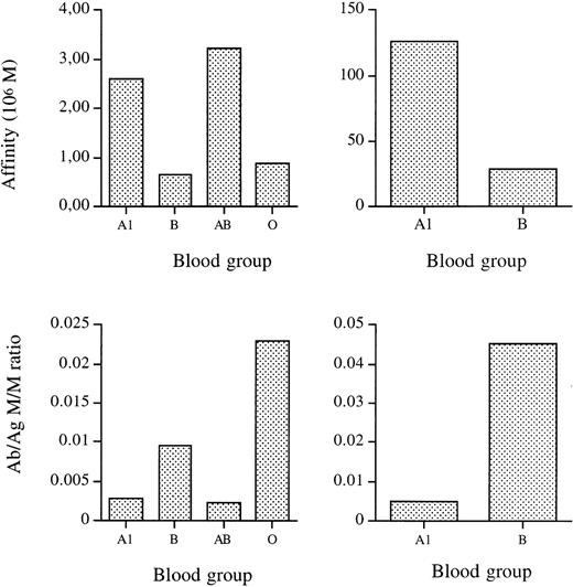
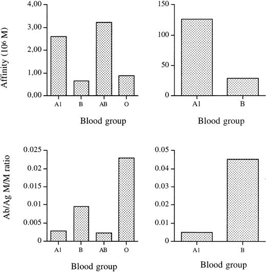
We used the BIAcore technology to further analyze the interaction of natural IgG antibodies with autologous blood group antigens. Synthetic A antigen was immobilized onto the sensor chip (1.8 ng per mm3). IgG was purified from pooled serum of six individuals of each blood group A1, B, AB, and O. The binding constants measured at equilibrium (KE) of the binding of purified IgG to A antigen were calculated from the on- and off-rate constants to be 2.60, 0.64, 3.22, and 0.88 × 10⁶ mol/L for IgG of individuals A1, B, AB, and O, respectively (Fig 7). We then affinity-purified anti-A IgG antibodies from pooled serum of six A1 and six B individuals. The binding constants for the binding of affinity-purified anti-A IgG to A antigen were 127 and 28 × 106 mol/L for IgG of individuals A1 and B, respectively (Fig 7). We then deduced mole:mole ratios of bound antibody to antigen from the amount of Resonance Units (RU) bound at equilibrium, using the BIAevaluation software that allows the calculation of theoretical amounts of bound antibody when equilibrium is reached. The results indicate that, although their association constant for A antigen is higher, much fewer molecules of specific anti-A IgG are present in IgG of individuals of blood group A than in IgG of individuals of blood group B.
Real time analysis of the binding of anti-A antibodies to immobilized A antigen. Top panels: apparent equilibrium constants (KA) for the binding of purified IgG from serum of individuals A1, B, AB, and O (left panel) and for the binding of affinity-purified anti-A IgG from individuals A1 and B (right panel) to A antigen. Lower panels: mole to mole ratios of bound antibody to antigen for the binding to A antigen of IgG of individuals A1, B, AB, and O (left panel) and for the binding to A antigen of affinity-purified anti-A IgG from individuals A1 and B (right panel).
Real time analysis of the binding of anti-A antibodies to immobilized A antigen. Top panels: apparent equilibrium constants (KA) for the binding of purified IgG from serum of individuals A1, B, AB, and O (left panel) and for the binding of affinity-purified anti-A IgG from individuals A1 and B (right panel) to A antigen. Lower panels: mole to mole ratios of bound antibody to antigen for the binding to A antigen of IgG of individuals A1, B, AB, and O (left panel) and for the binding to A antigen of affinity-purified anti-A IgG from individuals A1 and B (right panel).
DISCUSSION
It is widely accepted that the serum of healthy individuals contains natural antibodies only against those blood group A or B antigens that are not expressed on the individual’s red blood cells. The trisaccharidic terminal structures recognized by anti-ABO blood group antigen antibodies result from the sequential activity of H, A, and/or B glycosyltransferases. In individuals of blood groups A, B, O, and AB, the H transferase adds a fucose residue to a terminal galactose. In individuals of blood groups A, B, or AB, the A or B transferase then adds either an N-acetylgalactosamine (blood group A) or galactose (blood group B). The mechanisms involved in tolerance to autologous blood group antigens remain unclear. It is generally believed that healthy individuals are exposed to ABO trisaccharides expressed on bacteria of the intestinal flora, and, as a result, produce antibodies to allo-ABH antigens although they remain unresponsive to autologous ABH antigens. Natural anti-ABO blood group antigen antibodies have long been considered to be of the IgM isotype, in contrast with immune antibodies of the IgG isotype that occur after alloimmunization in pregnancy or ABO incompatible transfusions. It had been proposed by Landsteiner12 that natural antibodies had a dual origin, antigen-induced and spontaneous. Natural anti-A and anti-B antibodies believed to be heteroagglutinins are produced as a response to substances in the environment. Antierythrocyte antibodies were considered to be of spontaneous origin.13 Anti-ABO alloantibodies of the four IgG subclasses have, however, been characterized in normal serum.14 In the present study, we show that natural IgM and IgG autoantibodies reactive with self blood group antigens with affinities in the micromolar range are present in normal serum immunoglobulin. Whereas these antibodies are readily detected in purified IgG and IgM fractions, their expression is restricted in whole serum as a result of complementary interactions between V regions of antibodies.
We observed that purified IgG and IgM of healthy individuals expressed similar reactivity toward A and B trisaccharides, irrespective of the ABO blood group of the donor, when assessed by ELISA. Thus, IgG and IgM antibodies reactive with autologous A and B blood group antigens are present in serum immunoglobulin of healthy individuals. Assays using whole serum have previously detected low levels of self reactivity of natural anti-A and anti-B antibodies although the results were interpreted as being nonspecific.14 When we tested IgG and IgM in whole serum at the same concentrations as those used for testing purified IgG and IgM, little if no reactivity was observed with autologous A and B antigens. As expected, high reactivity of IgG and IgM was observed with allo-A or B antigens. The results indicate that IgG reactivity with self A and B antigens is controlled in whole serum by non-IgG serum factors. The results suggest that, under the conditions of the assay, inhibition by autologous IgM of the reactivity of anti-A IgG of individuals A1 with A antigen is dependent on complementary V region interactions between IgG and IgM rather than on a competition between immunoglobulins for the binding to the antigen. We and others have shown previously that autoreactivity of IgG in normal human and mouse serum is regulated by complementary interactions between V regions of IgG and V regions of autologous IgM antibodies.9,10 This is one of the mechanisms of peripheral control of autoreactivity that has been widely documented, both under physiological conditions9,10,15,16 and in patients in remission of autoimmune disease.17-20 Thus, the addition of autologous IgM dose-dependently inhibited the reactivity of purified human IgG with a panel of homologous antigens.10 The regulatory capacity of IgM is selective in that certain IgG self reactivities are more stringently regulated than others, as shown by using a semiquantitative immunoblotting technique using solubilized proteins from tissue extracts as sources of self antigens.21 Selectivity of control of IgG autoreactivity by IgM differs from one individual to another,22 which has led to the concept of “antibody-finger printing” referring to the individual patterns of self reactivity of IgG in serum as opposed to the highly homogeneous patterns of reactivity of purified IgG with the same antigen panels.23 Thus, the paratopic repertoire of self-reactive IgG in serum is highly dependent on the idiotypic repertoire displayed by autologous IgM. No control by IgM of IgG reactivity was found when binding of IgG to foreign antigens rather than to self antigens, was examined.10 As we added autologous IgM to purified IgG, we observed a dose-dependent inhibition of the binding of IgG to autologous A and B trisaccharides. The inhibitory effect of IgM was restricted to the autologous combination of antibodies, ie, IgM of individuals A inhibited the reactivity of IgG of individuals A with A antigen, whereas IgM of individuals B or O failed to inhibit IgG anti-A reactivity of individuals A. The inhibitory effect of IgM was not dependent on paratopic competition between IgG and IgM for the binding to the blood group antigen because the reactivity of IgM of individuals A with A antigen was much lower than that of IgM of individuals B. The results strongly suggest that, as in the case of other natural self-reactive IgG antibodies, the reactivity of natural anti-self ABO blood group antigen IgG antibodies in serum is restricted by V-region complementary interactions with autologous IgM. We speculate that, as a corollary, self reactivity of IgM with A and B blood group antigens is controlled in serum by complementary V regions expressed by autologous IgG. An additional degree of complexity determines the outcome of the binding reaction of self-reactive IgG antibodies in whole serum, in that the IgG and IgM fractions contain both autoantibodies and antiautoantibodies that are not readily detectable when the reactivity of purified IgG or IgM is tested, although these may dissociate from complexes and exert a regulatory effect when IgG, IgM, and the source of self antigen are allowed to interact in the solid phase-binding assay. Furthermore, soluble A and B (ABO) antigens may also contribute to the neutralization of autologous anti-ABO blood group antibodies in AB serum.
To further characterize natural antibodies reactive with self ABO antigens, we isolated anti-A IgG and IgM antibodies from the serum of A individuals by affinity chomatography. We observed that the binding of anti-A IgG autoantibodies to A antigen was similar to that of affinity-purified anti-A antibodies of individuals of blood group B. The calculated affinity constant for the binding of anti-A IgG autoantibodies to the A trisaccharide was in the micromolar range, similar to that of allo-anti-A IgG antibodies of B individuals. Thus, natural self-reactive anti-ABO blood group antigen antibodies are of significant affinity, as previously reported in the case of natural IgG autoantibodies to eg, cytokines and cytoskeletal proteins.4 24 The data further argues against the hypothesis that natural alloreactive antibodies exhibit the characteristics of immune antibodies that would result as a response to foreign A and B substances on microorganisms. Although autoantibodies and alloantibodies exhibited similar association constants, less autoantibody bound to the A antigen as compared with allo antibody when the mole to mole ratio was calculated from real time binding experiments using the Biacore. Furthermore, the autoantibody was found not to agglutinate A red blood cells at concentrations similar to those of allo-anti-A IgG. This may be explained by the high dissociation rate of autoantibodies as compared with that of alloantibodies and may provide a means for protecting self tissues from the potential deleterious effects of autoantibodies. However, using high concentrations of affinity-purified anti-A IgG of A individuals, we could show agglutination of A red cells, in a direct agglutination assay. The agglutinating capacity of the antibodies was specific in that equivalent concentrations of an irrelevant human MoAb had no effect.
Our results imply that the lack of self reactivity of IgG and IgM with A and B blood group antigens in serum is not dependent on central tolerance or peripheral B/T-cell anergy, in that equal amounts of autoantibodies and alloantibodies are found in the IgG and IgM fractions of normal serum. The results agree with the findings of Rieben et al,14 25 showing that B cells capable of generating antibodies to autologous blood group antigens are present in healthy subjects. However, by using a limiting dilution technique, these authors found a higher frequency of precursor B cells producing alloreactive than that of cells producing autoreactive anti-ABO antigen antibodies. Our observations further imply that normal human plasma should also contain natural IgG autoantibodies to substance H and the corresponding regulatory IgM molecules.
Taken together, our observations suggest that tolerance to autologous ABO blood group antigens is dependent on peripheral control rather than on clonal deletion or anergy at the B- or T-cell level. In this respect, autoreactivity to A and B blood group antigens appears to be controlled in a similar fashion to autoreactivity with DNA, thyroglobulin, Factor VIII, and neutrophil cytoplasmic antigens that are also subject to tight regulation by V region-connected antibodies in serum.26
Supported by Institut National de la Santé et de la Recherche Médicale (INSERM), France, and the Central Laboratory of the Swiss Red Cross, Bern, Switzerland. S.H.S. is a recipient of a grant from INSERM.
The publication costs of this article were defrayed in part by page charge payment. This article must therefore be hereby marked “advertisement” in accordance with 18 U.S.C. section 1734 solely to indicate this fact.

