Abstract
The macrophage-specific cell surface receptor sialoadhesin, which is a member of the newly recognized family of sialic acid binding lectins called siglecs, binds glycoprotein and glycolipid ligands containing a2-3–linked sialic acid on the surface of several leukocyte subsets. Recently, the sialic acid binding activity of the siglec CD22 has been demonstrated to be regulated by sialylation of the CD22 receptor molecule. In the present work, we show that desialylation of in vivo macrophage sialylconjugates enhances sialoadhesin-mediated lectin activity. Herein, we show that receptor sialylation of soluble sialoadhesin inhibits its binding to Jurkat cell ligands, and that charge-dependent repulsion alone cannot explain this inhibition. Furthermore, we show that the inhibitory effect of sialic acid is partially dependent on the presence of an intact exocyclic side chain. These results, in conjunction with previous findings, suggest that sialylation of siglecs by specific glycosyltransferases may be a common mechanism by which siglec-mediated adhesion is regulated.
NUMEROUS STUDIES have demonstrated that carbohydrates serve directly as molecular determinants responsible for mediating molecular and cellular interactions.1-4 The monosaccharide sialic acid, by virtue of both its location at the terminal position on glycans associated with cell surface glycoproteins and glycolipids, and its net negative charge at physiological pH, serves as a potentially important regulator of these interations.4 Polysialic acid (PSA)-mediated abrogation of homotypic interactions between neural cell adhesion molecules (NCAM) illustrates the potential inhibitory effect of sialic acid.5 In contrast to its role as a regulatory inhibitor, sialic acid can also promote interactions by providing a critical ligand component for various sialic acid binding lectins.4Recognition of sialic acid by lectins can be affected by a variety of modifications of sialic acid itself, and variation in the a-ketosidic linkages to the underlying sugar chain, the structure of the chain, and the structure of the underlying protein or lipid.
Recently, a subset of immunoglobulin superfamily receptors has been found to behave as sialic acid binding lectins and proposed to define a new class of adhesion receptors known as siglecs.6 Although all members of this family, which include CD22, CD33, myelin-associated glycoprotein (MAG), and sialoadhesin (Sn) share sialic acid binding properties, they differ in the manner in which they recognize sialic acid. More specifically, sialoadhesin, CD33, and MAG recognize a2-3 sialylated-, while CD22 recognizes a2-6–sialylated-glycoproteins and glycolipids.7-9 The sialylated oligosaccharides recognized by each siglec are common to many glycoproteins and glycolipids, suggesting that siglec-mediated interactions may be highly regulated. Evidence for such regulation has been provided for interactions between CD22 and its ligands. a2,6-sialylation of CD22 itself abrogates CD22-mediated adhesion,10 and 9-O-acetylation of the side chain of a2-6–linked sialic acid masks CD22 ligands in vivo.11 These observations suggest that molecular interactions between CD22 and its ligands may be intimately associated with the expression and function of at least two specific transferases, a2-6-sialyltransferase and O-acetyltransferase.
Recent evidence suggests that regulation of carbohydrate recognition by several siglec family members may be similar to that of CD22. For example, sialylation of cis ligands for CD33 and MAG modulates their adhesive interaction.12,13 In addition, modification of cell surface ligand-associated sialic acids by 9-O-acetylation has been demonstrated to abrogate sialoadhesin-mediated binding of red blood cells (RBC).14 Together, these findings suggest that siglecs may use common mechanisms to regulate their adhesive properties. In the present work, we sought to determine whether sialylation of sialoadhesin itself regulates its binding properties. We demonstrate that sialylation of sialoadhesin-associated glycans abrogates sialoadhesin-receptorglobulin (Sn-Rg) binding activity and that such negative regulation by sialoadhesin-associated sialic acid is, in part, contingent on an intact polyhydroxyl side chain. Furthermore, our data suggest that the inhibitory effect of sialoadhesin-associated sialic acid is not solely a result of charge-dependent repulsion.
MATERIALS AND METHODS
Materials.
Cell culture media (Dulbecco’s modified Eagle’s medium [DMEM]) and fetal bovine serum were purchased from Irvine Scientific (Santa Ana, CA). L-glutamine and antibiotics were from GIBCO (Grand Island, NY). Diethyl aminoethyl (DEAE)-dextran, dimethyl sulfoxide (DMSO), Nonidet P-40, aprotinin, and sodium periodate were from Sigma (St Louis, MO). Fluorescein-labeled goat antihuman affinity-purified antibodies were from Cappel (Malvern, PA). Oligonucleotide synthesis reagents were acquired from Millipore (Bedford, MA). Vibrio cholerae (VC) andarthrobacter ureafaciens (AU) sialidase were purchased from Boehringer Mannheim (Indianapolis, IN). NANase I was purchased from Glyko (Novato, CA). Sheep erythrocytes were purchased from ICN (Costa Mesa, CA). Fluorescein isothiocyanate (FITC)-conjugated polyacrylamide substituted with a2-3 sialylactose (3′-PAA-FITC) was obtained from GlycoTech (Rockville, MD).
COS-7 cell adhesion (rosetting) assay.
Rosetting assays were performed as described previously.15Briefly, COS-7 cells were transfected with full-length sialoadhesin cDNA or mock-transfected using the DEAE-dextran method as described.15 Twelve hours after transfection, cells were trypsinized and replated onto fresh 60-mm culture plates. Forty-eight hours later the transfected COS-7 cells were overlaid with human or sheep RBC, which were allowed to adhere for 20 minutes at 4°C. Nonadherent RBCs were washed away with serum-free DMEM, and rosetting was evaluated under an inverted microscope. Five separate transfections performed independently on 5 different days were evaluated. To analyze the effect of endogenous sialylation of surface expressed sialoadhesin, transfectants were treated with 50 mU/mL vibrio choleraesialidase (VC-sialidase) at 37°C in phosphate-buffered saline (PBS), pH 6.9 for 1 hour before performing the rosetting assay. The rosettes obtained under these conditions were compared with those of untreated transfectants under identical conditions.
Splenic cryostat adhesion assay.
Splenic cryostat sections (8 μm) were placed on glass slides and air dried for 60 minutes. Cryostat sections were preincubated with 50 mU/mL of VC-sialidase or heat inactivated VC-sialidase for 1 hour at 37°C, washed extensively with cold PBS, and incubated with washed sheep erythrocytes (sRBC) for 30 minutes at 4°C. The slides were washed in PBS with gentle agitation to remove unbound sRBC from the sections and immediately fixed in 4% formaldehyde. Binding was examined by light microscopy. Antibody blocking studies were performed as follows. Sialidase-treated cryostat sections were preincubated with 3 mg/mL of either the antisialoadhesin monoclonal antibody (MoAb) 3D6 (Serotec, Raleigh, NC) or the isotype-matched control anti-CD44 MoAb KM81 for 30 minutes at room temperature (RT). The slides were washed extensively with PBS and then subjected to the adhesion assay described above.
3′-PAA-FITC staining.
The lectin activity of cell surface sialoadhesin was examined using a protocol similar to that previously described.16 Briefly, Mm1 cells were washed several times in ice-cold PBS containing 0.02% sodium azide, 1% bovine serum albumin (BSA) (staining buffer), and the lectin activity of cell surface sialoadhesin examined by incubating cells (untreated and treated) for 1 hour in 100 mL staining buffer containing 1.5 mg 3′-PAA-FITC. The cells were washed in staining buffer and analyzed with a FACscan instrument (Becton Dickinson Immunocytometry Systems, Mountain View, CA). The treated cells were incubated withArthrobacter ureafaciens (AU) and VC-sialidase, 40 and 50 mU/mL, respectively, washed with staining buffer three times, and preincubated with staining buffer alone or staining buffer containing 5 mg/mL of the antisialoadhesin antibody, 3D6. Following these treatments, the cells were stained with 3′-PAA-FITC and analyzed as described above.
Development of receptorglobulin fusion proteins.
Soluble receptor-immunoglobulin fusion proteins (termed Rg for “receptorglobulin”) were prepared according to the methods of Aruffo et al.17,18 Development of murine SnRg19CD44Rg,18 and CD8Rg18 have been described previously; the SnRg contains the first four Ig-like domains of Sn. COS cells were transfected with either SnRg, CD44Rg, or CD8Rg by the DEAE-dextran method,17 and 12 hours after transfection, culture media replaced with serum-free DMEM, which was maintained for an additional 72 hours. The supernatant was harvested, and the fusion proteins purified by protein-A sepharose (PAS) chromatography, eluted with sodium citrate buffer pH 3.0, dialyzed against PBS, concentrated with ultrafree-CL filters (Millipore, Bedford, MA), and quantitated with a Bio-Rad (Richmond, CA) protein assay kit.8 In addition, SnRg and CD8Rg were also purified from two separate Chinese hamster ovary (CHO) cell lines stably transfected with the SnRg-coding and CD8Rg-coding plasmids.
Cell lines and cell culture.
The Jurkat T-cell line was obtained from American Type Culture Collection (ATCC, Rockville, MD) and grown in RPMI (Irvine Scientific) supplemented with 10% fetal bovine serum. Human erythrocytes were obtained from healthy donors, washed in PBS, and used directly in COS-7 cell rosetting assays. The Mm1 cell line was a generous gift from Paul Crocker (University of Dundee, Dundee, Scotland) and grown in RPMI (Irvine Scientific) supplemented with 10% fetal bovine serum.
Determination of the effect of sialidase treatment on sialoRg function.
COS-7 cells were transfected with cDNA coding for SnRg by the DEAE-dextran method, and supernatants were harvested 4 days posttransfection as described above. SnRg was bound batchwise to PAS beads at 4°C overnight, washed with PBS, and the PAS-bound SnRg was subjected to digestion by different types of sialidases. PAS-bound SnRg was resuspended in 0.5 mL of 0.1 mol/L sodium acetate pH 5.5, 0.1 mol/L NaCl, 1 mmol/L CaCl2 (AU sialidase buffer), and digested with 100 mU of Arthrobacter ureafaciens sialidase (AU sialidase) or VC-sialidase for 6 hours at 37°C. An equal amount of PAS-bound SnRg was resupended in 0.5 mL of 50 mmol/L sodium phosphate pH 6.0 and digested with 250 mU of NANaseI (Glyko) for 10 hours at 37°C. After the sialidase digest, the PAS beads were extensively washed with PBS, SnRg eluted, dialyzed, concentrated, and quantitated as described above. Untreated PAS-bound SnRg was resuspended in AU-sialidase buffer and incubated in the absence of sialidase for 10 hours at 37°C and purified as described above. Jurkat cells were incubated with 50 mg/mL sialidase-treated SnRg, untreated SnRg, or control (CD44Rg and CD8Rg) receptorglobulins for 1 hour on ice. Cells were washed in cold PBS, resuspended in PBS, 0.02% sodium azide and 5 mg/mL fluorescein-conjugated, affinity-purified goat antihuman antibody for 30 minutes on ice, washed, fixed with PBS containing 4% formaldehyde, and analyzed by a FACscan instrument (Becton Dickinson Immunocytometry Systems).
Coupling of glutamic acid to VC-sialidase–treated SnRg with dithiobis(sulfosuccinimidylproprionate) (DTSSP).
The amine selective reagent, DTSSP (Pierce, Rockford, IL), was used according to the manufacturer’s recommended protocol to covalently cross-link glutamic acid to VC-sialidase–treated SnRg. Briefly, VC-treated SnRg (400 mg), glutamic acid, and DTSSP were added to PBS at 1:1:50 molar ratio and incubated at 37°C for 30 minutes. The reaction was terminated with the addition of Tris-HCl to a final concentration of 50 mmol/L. The reaction products were separated and isolated by FPLC gel filtration.
Gel filtration chromatography.
Protein A-sepharose affinity-purified NAN-SnRg and glutamate cross-linked-NAN-SnRg (Glut-SnRg) were individually injected onto a Superose-6 HR 10/30 gel filtration column (Pharmacia LKB, Uppsala, Sweden) and subjected to a constant flow rate of 0.5 mL PBS/minute using an FPLC System (Pharmacia). Elution fractions were collected and protein detected by spectral absorbance at 226 nm. Elution profiles were generated by plotting protein concentration (spectral absorbance, A226nm) versus volume (mL).
Capillary electrophoresis.
Capillary electrophoresis was performed on a model 3850 capillary electropherograph (Isco, Lincoln, NE) using an uncoated silica capillary (75 μm inner diameter, 62.5 cm length, 39 cm inlet to detector window, ISCO), a 5-second vacuum injection (10 nL), 15kV (95 mA), and 210 nm ultraviolet (UV) absorbance detection in 50 mmol/L NaPO4, pH 7.4. Samples (10 mL) contained 2 mg SnRg and 480 mg/mL mesityl oxide (MO). Mobility (m) of a given molecule is defined as its anodal migration relative to MO, the neutral marker, and is determined from migration times by: m = ld(lc/V)(1/teo − 1/t); where ld is the inlet to detector window length, lcis the capillary length, V is the applied voltage, teo is the migration time for MO, and t is the migration time for SnRg.
Mild periodate oxidation of Jurkat cells and PAS bound SnRg.
A fresh stock of 100 mmol/L sodium metaperiodate in PBS, pH7.2, was prepared for each experiment. Jurkat cells were washed and resuspended in ice-cold PBS with 2 mmol/L sodium metaperiodate at 1 × 10⁶ cells/mL and incubated on ice in the dark for 15 minutes as previously described.8 11 The cells were washed with ice-cold PBS, stained with SnRg or CD44Rg, and analyzed by FACscan.
SnRg (400 mg) was batch adsorbed onto PAS beads overnight at 4°C, washed with ice-cold PBS, resuspended in 20 mL of ice-cold PBS with 2 mmol/L sodium metaperiodate and incubated on ice in the dark for 20 minutes. The beads were then extensively washed with PBS, the SnRg purified as described above, and the periodate-treated SnRg (50 mg/mL) used to stain cells.
Immunoprecipitation and silver stain.
Untreated and sialidase-treated SnRg (10 mg) were batch adsorbed onto PAS beads overnight at 4°C in PBS. The beads were washed four times in PBS/0.05% Nonidet P-40 and precipitates eluted by boiling in sample buffer in the presence of 2% 2-mercaptoethanol. Samples were subjected to sodium dodecyl sulfate (SDS)/8% polyacrylamide (PAGE), the gel fixed, and proteins detected by silver nitrate staining.20
RESULTS AND DISCUSSION
Sialoadhesin-mediated adhesion in COS-7 cells.
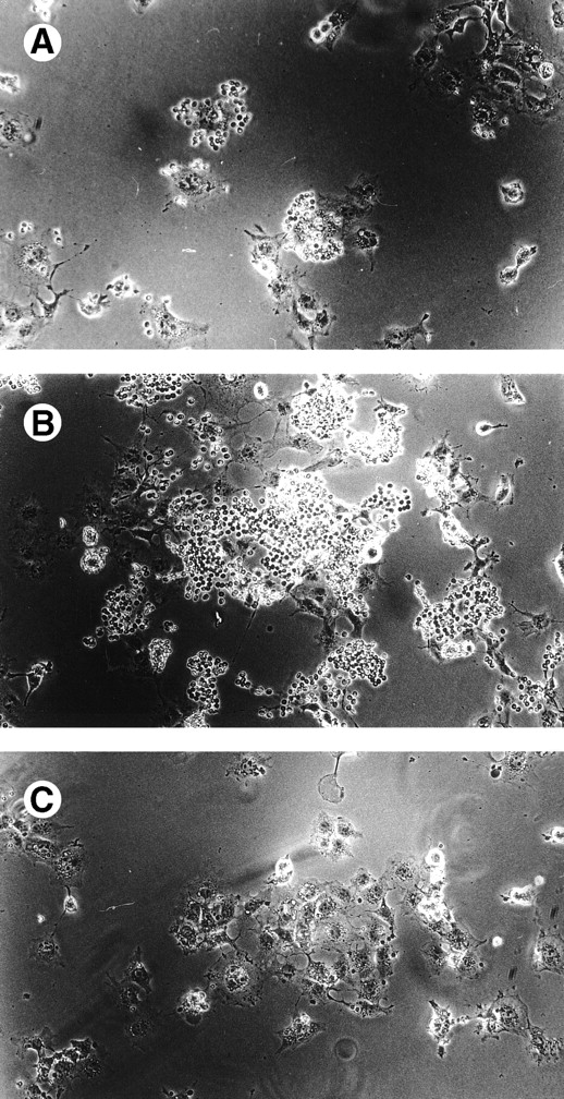
Expression of sialoadhesin (Sn) in COS-7 cells promoted minimal binding of human RBCs as demonstrated by both the small size (<20 RBCs/rosette) and number (<10% of sialoadhesin-bearing COS-7 cells) of rosettes (Fig 1A). Because sialylation of the siglec CD22 has been demonstrated to inhibit its sialic acid binding lectin activity,10 we sought to determine if sialylation of sialoadhesin by COS-7 cells modulates sialoadhesin-mediated binding of RBCs. To address this possibility, Sn-expressing COS-7 cells were pretreated with VC sialidase before the binding assay. Sialidase pretreatment resulted in high levels of RBC binding as demonstrated by numerous large rosettes (Fig 1B); greater than 85% of the sialoadhesin-expressing cells supported RBC rosettes each consisting, on average, of more than 30 cells. The specificity of the adhesion is underscored by the observation that VC sialidase-treated mock-transfected COS-7 cells did not support RBC binding (Fig 1C).
Sialidase pretreatment of sialoadhesin-transfected COS cells unmasks their ability to mediate red blood cell binding. (A) Untreated sialoadhesin-transfected COS cells. (B)Vibrio Cholerae sialidase-treated sialoadhesin-transfected COS-7 cells. (C) Vibrio Cholerae sialidase-treated mock-transfected COS cells.
Sialidase pretreatment of sialoadhesin-transfected COS cells unmasks their ability to mediate red blood cell binding. (A) Untreated sialoadhesin-transfected COS cells. (B)Vibrio Cholerae sialidase-treated sialoadhesin-transfected COS-7 cells. (C) Vibrio Cholerae sialidase-treated mock-transfected COS cells.
In vivo sialoadhesin-mediated adhesion is enhanced by sialidase.
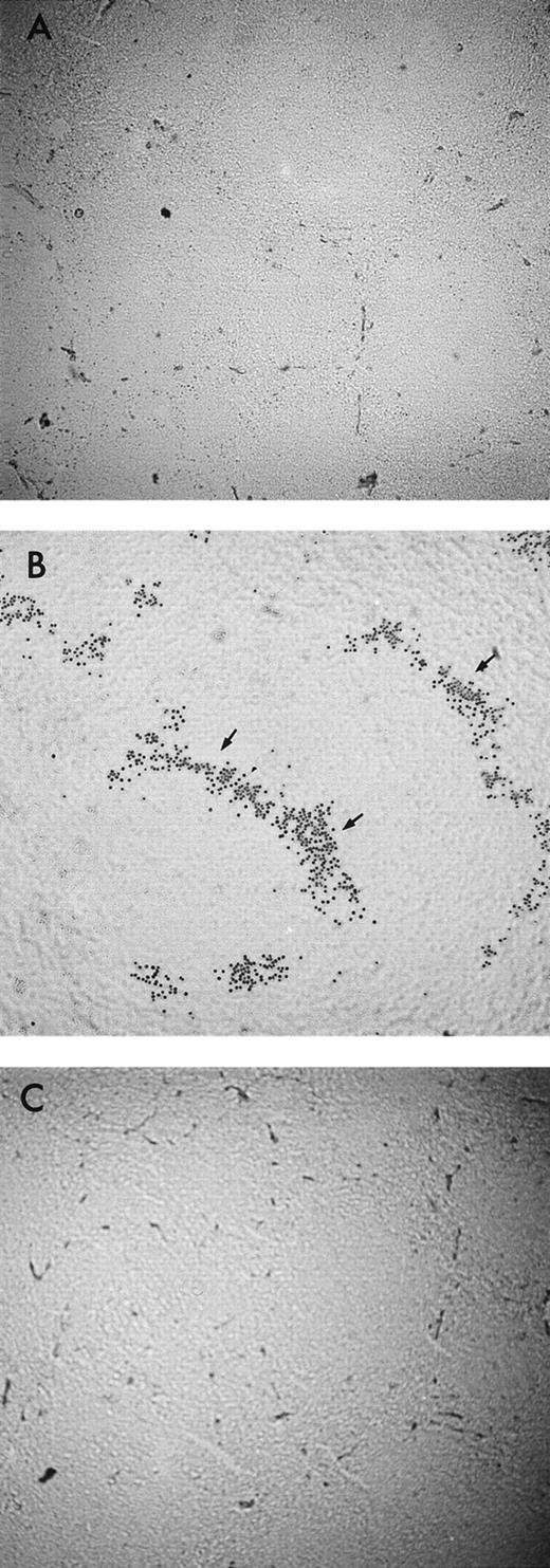
Although sialoadhesin-mediated adhesion in COS cells is augmented by sialidase pretreatment, we sought to demonstrate this phenomenon in macrophages in vivo. Therefore, we performed a sheep erythrocyte binding assay using sialidase treated- and untreated-cryostat sections of mouse spleen. We demonstrate that untreated cryostat sections of mouse spleen bind sRBC minimally under our experimental conditions (Fig 2A). Interestingly, pretreatment of cryostat splenic sections with sialidase resulted in marked erythrocyte binding to the splenic marginal zone, an area that coincides with the expression of sialoadhesin (Fig 2B).21Mouse splenic cryostat sections pretreated with heat-inactivated sialidase did not enhance sRBC binding (data not shown). Preincubation of the sialidase-treated cryostat section with the antisialoadhesin MoAb 3D6 resulted in the loss of sRBC adhesion (Fig 2C), demonstrating the sialoadhesin-dependent nature of this binding; an isotype-matched antibody did not inhibit sRBC binding under the same experimental conditions (data not shown).
Sialidase pretreatment of splenic cryostat sections enhances sialoadhesin-mediated adhesion of sheep erythrocytes. (A) Murine splenic cryostat section (untreated). (B) Murine splenic cryostat section pretreated with VC-sialidase; arrow denotes adherent sheep erythrocytes in the splenic marginal zones. (C) VC-sialidase treated splenic cryostat section pretreated with the antisialoadhesin blocking MoAb, 3D6.
Sialidase pretreatment of splenic cryostat sections enhances sialoadhesin-mediated adhesion of sheep erythrocytes. (A) Murine splenic cryostat section (untreated). (B) Murine splenic cryostat section pretreated with VC-sialidase; arrow denotes adherent sheep erythrocytes in the splenic marginal zones. (C) VC-sialidase treated splenic cryostat section pretreated with the antisialoadhesin blocking MoAb, 3D6.
Recently, a probe consisting of polyacrylamide substituted with multiple copies of Siaa2-6Galb1-4Glcb1 has been successfully used to directly measure the lectin activity of cell surface CD22.16 With this in mind, we sought to probe the lectin activity of cell surface sialoadhesin with commercially available synthetic conjugate of fluorescein-labeled polyacrylamide substituted with multiple copies of Siaa2-3Galb1-4Glc (3′-PAA-FITC); sialoadhesin has been previously demonstrated to specifically recognize the sugar moieties Siaa2-3Galb1-4GlcNAc or Siaa2-3 Galb1-3GalNAc.9 Untreated Mm1 cells, a macrophage cell line, which constitutively expresses sialoadhesin,22 did not stain with 3′-PAA-FITC, however pretreatment of such cells with sialidase unmasks the sialoadhesin-lectin activity (Fig 3). The specificity of 3′-PAA-FITC for sialoadhesin is demonstated by the observation that preincubation of Mm1 cells with the adhesion-blocking antisialoadhesin MoAb 3D623 abrogates 3′-PAA-FITC binding (Fig 3); the isotype-matched rat MoAb to CD44 fails to inhibit 3′-PAA-FITC binding. These results indicate that desialylation of in vivo sialylconjugates enhances sialoadhesin-mediated lectin activity and that 3′-PAA can be used as a specific ligand for detecting unoccupied sialoadhesin lectin molecules.
Detection of cell surface sialoadhesin lectin activity on Mm1 cells using a multivalent 3′-sialylactosylated probe (3′-PAA-FITC). Cultured Mm1 cells, a murine macrophage cell line, were washed, untreated, Arthrobacter Ureafaciens(AU)/VC-sialidase treated, or AU/VC-sialidase treated followed by preincubation with the 3D6 adhesion-blocking MoAb, and stained with FITC-conjugated 3′-PAA. Staining was detected using single color flow cytometry (fluorescence-activated cell sorting [FACS] analysis).
Detection of cell surface sialoadhesin lectin activity on Mm1 cells using a multivalent 3′-sialylactosylated probe (3′-PAA-FITC). Cultured Mm1 cells, a murine macrophage cell line, were washed, untreated, Arthrobacter Ureafaciens(AU)/VC-sialidase treated, or AU/VC-sialidase treated followed by preincubation with the 3D6 adhesion-blocking MoAb, and stained with FITC-conjugated 3′-PAA. Staining was detected using single color flow cytometry (fluorescence-activated cell sorting [FACS] analysis).
Sialylation of the sialoadhesin receptorglobulin (SnRg) modulates its binding activity.
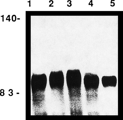
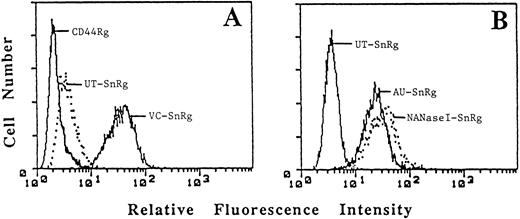
There are at least two possible interpretations of the above experimental results. First, an a2,3-sialylated COS glycoprotein or glycolipid on the same cell surface (in cis) may bind sialoadhesin, thereby preventing its interaction with ligands on adjacent cells (RBCs). Second, sialylation of sialoadhesin itself may interfere with its ability to recognize its ligands. To address the latter possibility, cDNA encoding the soluble recombinant sialoadhesin-immunoglobulin fusion protein (SnRg) was transiently and stably expressed in COS and CHO cells, respectively. The SnRg was purified from the supernatants, subjected to sialidase or mock digestion, analyzed by SDS-PAGE and assessed for binding to Jurkat cells by flow cytometry. In comparison to mock-digested SnRg, VC sialidase-treated SnRg (VC-SnRg) displayed a decrease in molecular mass, consistent with the notion that COS-7–derived SnRg is sialylated (Fig 4, lane 4) and a significant increase in Jurkat cell binding activity (Fig 5A); soluble CD44Rg (Fig 5), as well as soluble CD8Rg (Fig 6) served as negative controls. Previous work has demonstrated that the a2,6-sialic acid binding activity of CD22 is specifically inhibited by CD22-associated a2,6-linked sialic acid.8 To determine if the observed inhibition of SnRg-mediated binding may be sialic acid linkage specific, PAS-bound SnRg was treated with different sialidases, eluted, analyzed by SDS-PAGE, and tested for binding to Jurkat cell-surface ligands. SnRg treated with AU sialidase (AU-SnRg), which hydrolyzes a2-3, 2-6-and 2-8-linked sialic acid, displayed a significant decrease in molecular mass (Fig 4, lane 5) and an approximately 10-fold increase in binding activity (Fig 5B). Interestingly, SnRg treated with NANaseI (NAN-SnRg) enhanced its binding activity (Fig 5B), despite undergoing a slight decrease in molecular mass (Fig 4, lane 1); NANaseI specifically and selectively hydrolyzes a2,3-linked sialic acid. The differential sialidase hydrolysis suggests that SnRg expressed in COS-7 cells is at least a2,3- and a2-6-sialylated, but that a2,3-sialylation, rather than a2-6-sialylation, may play a dominating role in regulating ligand binding activity. The difference in binding activity of native (or untreated) SnRg in Figs 5 and 6 may be explained by the fact that the SnRg in Fig 5 was derived from transient COS cell transfectant supernatants, whereas the SnRg used for Fig 6 was derived from the supernatants of CHO cells stably tranfected with SnRg. The COS-derived SnRg was harvested 3 days after transfection, whereas CHO-derived SnRg was harvested 8 days after plating in serum-free medium. Thus, the binding differences between the COS- and CHO-derived SnRg may be explained by variable release of soluble neuraminidase from cells cultured under suboptimal nutrient conditions.24,25Neuraminidase released into the culture medium after CHO cell lysis can desialylate recombinant soluble glycoproteins.24
SnRg produced in COS-7 cells is sialylated. SnRg was precipitated with protein A-sepharose (PAS) beads and subjected to mock (lane 2), NANaseI (lane 1), Vibrio Cholerae sialidase (lane 4),Arthrobacter Ureafaciens sialidase (lane 5) digestions, or mild periodate oxidation (lane 3). Treated and nontreated SnRg (10 mg/lane) were subjected to 7% SDS-PAGE and protein detected by silver nitrate staining. Molecular mass markers (in kD) are shown.
SnRg produced in COS-7 cells is sialylated. SnRg was precipitated with protein A-sepharose (PAS) beads and subjected to mock (lane 2), NANaseI (lane 1), Vibrio Cholerae sialidase (lane 4),Arthrobacter Ureafaciens sialidase (lane 5) digestions, or mild periodate oxidation (lane 3). Treated and nontreated SnRg (10 mg/lane) were subjected to 7% SDS-PAGE and protein detected by silver nitrate staining. Molecular mass markers (in kD) are shown.
Sialidase treatment of SnRg enhances reactivity to the Jurkat T-cell line. Jurkat (1 × 10⁶) cells were incubated in (A) with 50 mg/mL of CD44Rg, untreated sialoRg (UT-SnRg) orVibrio Cholerae sialidase-treated SnRg (VC-SnRg) and in (B) with 50 mg/mL UT-sialoRg, Arthrobacter Ureafacienssialidase-treated SnRg (AU-SnRg) or NANaseI-treated SnRg.
Sialidase treatment of SnRg enhances reactivity to the Jurkat T-cell line. Jurkat (1 × 10⁶) cells were incubated in (A) with 50 mg/mL of CD44Rg, untreated sialoRg (UT-SnRg) orVibrio Cholerae sialidase-treated SnRg (VC-SnRg) and in (B) with 50 mg/mL UT-sialoRg, Arthrobacter Ureafacienssialidase-treated SnRg (AU-SnRg) or NANaseI-treated SnRg.
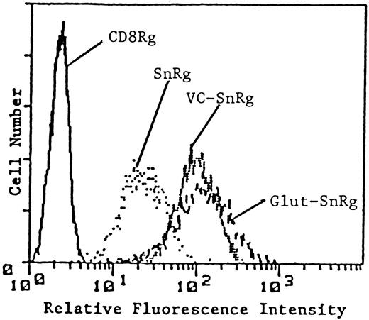
The addition of a negatively charged moiety to sialidase-treated SnRg does not inhibit its binding activity. Jurkat (1 × 10⁶) cells were incubated with 50 mg/ml of CD8Rg, SnRg, VC-SnRg, or Glut-SnRg and FACS analyzed for binding reactivity.
The addition of a negatively charged moiety to sialidase-treated SnRg does not inhibit its binding activity. Jurkat (1 × 10⁶) cells were incubated with 50 mg/ml of CD8Rg, SnRg, VC-SnRg, or Glut-SnRg and FACS analyzed for binding reactivity.
Because sialidase treatment results in the removal of negative charge, we addressed the possibility that the negative charge inherent to the carboxylate at the 1-carbon position of sialic acid (at physiological pH) may modulate binding through electrostatic repulsion, as has been demonstrated for neural cell adhesion molecule (NCAM).5Sialic acid was removed from SnRg with VC-sialidase, which hydrolyzes sialic acid irrespective of its linkage, and the net negative charge reconstituted by covalently coupling glutamic acid to the VC-sialidase treated SnRg (VC-SnRg) using the amine reactive homobifunctional N-hydoxysuccimide ester (NHS-ester), DTSSP. The cross-linking reaction was performed with an equal molar ratio of glutamic acid to VC-SnRg, and a 50 molar excess of DTSSP to optimize the coupling of glutamic acid to SnRg (Glut-sialoRg) while minimizing the production of VC-SnRg dimers. Primary amines of amino acid side chains are the principle targets for DTSSP, and while five amino acids have nitrogen in their side chains, only the e-amine of lysine reacts significantly with DTSSP.26 Theoretically, under our reaction conditions, one end of DTSSP reacts with the amine group of one molecule of glutamic acid, while the other end of the cross-linker reacts with the e-amine of a lysine residue in one molecule of VC-SnRg. Thus, as a result of this reaction, each VC-SnRg molecule will acquire a net negative charge of 3 for each molecule of glutamic acid added: one molecule of glutamic acid contains two free carboxyl groups, and the positively charged lysine of VC-SnRg is neutralized by its reaction with DTSSP. Furthermore, because DTSSP is in 50 molar excess, it will react with and neutralize the positive charge associated with additional lysine residues of VC-SnRg and result in an even greater net negative charge. The uncoupled glutamic acid, VC-SnRg dimers and Glut-SnRg products were separated and isolated using FPLC gel filtration. The Glut-SnRg eluted in the same separation fractions and displayed SDS-PAGE mobility similiar to that of VC-SnRg (data not shown). Furthermore, the isolated Glut-SnRg displayed a significant increase in net negative charge relative to native SnRg and VC-SnRg as assessed by capillary electrophoresis (Table 1), in agreement with the expected result. Interestingly, despite the greater overall negative charge, Glut-SnRg retained its ligand binding activity (Fig6), implying that charge-dependent repulsion is not likely to be solely responsible for regulating the lectin activity of SnRg. Thus, the inhibitory mechanism by which sialylation affects binding of sialoadhesin differs from that which operates on NCAM.5
Mild periodate oxidation of SnRg partially unmasks its binding activity.
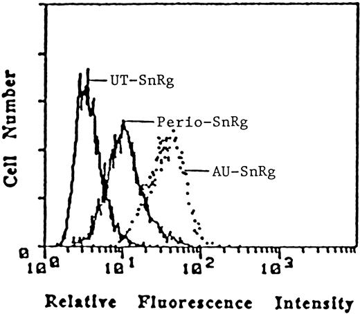
Modifications of the exocyclic side chain of sialic acid have been shown to affect a wide spectrum of biological phenomena.27We have previously demonstrated that the sialic acid side chains of B- and T-cell CD22 ligands are required for recognition by CD22,8,28 and others have demonstrated that 9-O-acetylation of CD22 ligands inhibits binding to CD22.11 Similarly, modifications of sialoadhesin ligand-associated sialic acid have been demonstrated to alter sialoadhesin-mediated recognition.22In light of these observations regarding ligand sialylation, we addressed the possibility that the sialic acid residues on the receptor, SnRg, may require an intact exocyclic side chain to provide their inhibitory activity. PAS-bound SnRg was subjected to mild sodium metaperiodate oxidation, and the resulting receptorglobulin binding activity assayed. Under these mild conditions, sodium metaperiodate selectively oxidizes the exocyclic side chain of sialic acid to produce the eight and seven carbon products, 5-acetamido-3-4dideoxy-D-galactosyloctuosonic and galactosylheptulosonic acids, respectively, while leaving the ring structure of sialic acid and the underlying oligosaccharides intact.29 The binding activity of periodate-treated SnRg was fourfold higher than that of untreated SnRg and threefold lower than AU-sialidase-treated SnRg (Fig 7), suggesting that the integrity of endogenous sialic acid side chains is required for optimal inhibition. Capillary electrophoretic mobility of the periodate-treated SnRg was similar to that of untreated SnRg (Table1) confirming that the periodate treatment did not change the charge of the molecule. This latter observation, in conjunction with the increased binding activity of periodate-treated SnRg, further supports the notion that charge-dependent repulsion alone cannot explain the inhibitory effect of sialic acid. Taken together, our results suggest that although the general mechanism of sialic acid inhibition is common among two siglec members, additional regulatory requirements based on recognition of specific sialic acid structural components (ie, side chain modifications) may exist within this family.
Mild periodate treatment of SnRg unmasks its binding reactivity to Jurkat cells. Jurkat cells were incubated with 50 mg/mL of untreated SnRg, AU-sialidase-treated SnRg, and sodium periodate-treated SnRg and tested for reactivity as assessed by indirect immunofluorescence and FACS analysis.
Mild periodate treatment of SnRg unmasks its binding reactivity to Jurkat cells. Jurkat cells were incubated with 50 mg/mL of untreated SnRg, AU-sialidase-treated SnRg, and sodium periodate-treated SnRg and tested for reactivity as assessed by indirect immunofluorescence and FACS analysis.
The in vivo regulation of siglec binding to its ligands is undoubtedly complex. In vitro studies have demonstrated that sialylation ofcis ligands regulates binding of CD22, CD33, and MAG.10,12,13 Similarly, the observation that macrophages express sialoadhesin ligands has led to speculation that the lectin binding site of sialoadhesin may be occupied in a cisconfiguration by sialo-oligosaccharides within the macrophage glycocalyx.30 It has been hypothesized that the 17 Ig-like domains of sialoadhesin project its functional domain above the cellular glycocalyx thereby reducing the inhibitory potential of thesecis ligands and allowing adhesive interactions to occur between macrophages and cells bearing appropriate counterreceptors.30 Attractive as this hypothesis may be, the relevance of cis interactions as an in vivo regulatory determinant of sialoadhesin binding remains to be resolved. Furthermore, based on the observation that sialylation of recombinant CD22 abrogates its binding activity,10 it has been hypothesized that sialylation of siglecs may be an additional mechanism by which to regulate their binding activity. Herein, we provide in vitro evidence to support this latter theory, and we now present additional evidence to suggest that sialylation of siglecs may be a common mechanism governing siglec binding.
We and others have hypothesized that the reciprocal nature of receptor-ligand sialylation appears to play an important role in defining the regulatory process among siglecs, and this reciprocity may be intimately related to expression and function of specific glycosyltransferases.7,13,31 Thus, the degree to which specific glycosyltransferase activity modulates siglec-mediated adhesion may be related to the differentiation and activation state of the cell. This concept is supported by the observation that expression of a2,6-sialyltransferase (ST) in B cells is cell cycle-dependent, and under the control of a B-cell specific a2,6ST promoter, which is induced on B-cell activation.32 Speculatively, regulation of a2,6ST expression and activity in B cells and a2,3ST expression and activity in macrophages may, in turn, regulate the ligand binding ability of CD22 and sialoadhesin as a function of the B cell and macrophage activation states, respectively. In vivo confirmation of this mechanism remains to be determined, and thus a major challenge in the future will be to understand the temporal changes in glycosyltransferase activity as they relate to siglec-ligand interactions during the growth, differentiation, and activation states of hematopoietic cells.
Supported by National Institutes of Health Clinical Scientist Development Award AI/01252 (to D.S.) and U.S. Public Health Services Grant GM/AI 48614 (to I.S.). I.S. is a scholar of the Leukemia Society of America.
The publication costs of this article were defrayed in part by page charge payment. This article must therefore be hereby marked “advertisement” in accordance with 18 U.S.C. section 1734 solely to indicate this fact.
REFERENCES
Author notes
Address reprint requests to Dennis C. Sgroi, MD, Molecular Pathology Unit, Massachusetts General Hospital, 149 13th St, 7th Floor, Charlestown Navy Yard, Boston, MA 02129.

![Fig. 3. Detection of cell surface sialoadhesin lectin activity on Mm1 cells using a multivalent 3′-sialylactosylated probe (3′-PAA-FITC). Cultured Mm1 cells, a murine macrophage cell line, were washed, untreated, Arthrobacter Ureafaciens(AU)/VC-sialidase treated, or AU/VC-sialidase treated followed by preincubation with the 3D6 adhesion-blocking MoAb, and stained with FITC-conjugated 3′-PAA. Staining was detected using single color flow cytometry (fluorescence-activated cell sorting [FACS] analysis).](https://ash.silverchair-cdn.com/ash/content_public/journal/blood/93/4/10.1182_blood.v93.4.1245/5/m_blod40409003x.jpeg?Expires=1768684994&Signature=hUWfGDVO6Y8UtjcaNIT0clwXgt5qktT8Nc7ZtaOgC76tyaGeXkg7mGivd84nZnTy-UNOS0yoNMlWnrjyES38QSicNKzS3M4XBlPO4cLhChPChloQK9glPwP~1nYKcXwd77eyotKjZap744BqvLh1TY93g~lIxAG0Af24vrE-InkRxie5IYukWBBIT-U5UTtFD0itR39X-zCP6DRmF~dwMtAoNgk11VYzEN1GaPFKJjUFSDVXhO0rq8spuCFf8Egm-aJ6Ae-tGrjx9iggUgYdAqJBkFIbbosA11MeFoJAvdeyyN4MugHiQyMYZP4LFeTAXE-o1MMg9TxMg9P~dOFisw__&Key-Pair-Id=APKAIE5G5CRDK6RD3PGA)
