Abstract
Vascular endothelial growth factor (VEGF) is a potent mitogen for vascular endothelial cells. It has been associated with angiogenesis, growth, dissemination, metastasis, and poor outcome in solid tumors. To assess cellular VEGF levels and their prognostic significance in newly diagnosed acute myeloid leukemia (AML), we used a radioimmunoassay (RIA) to quantify VEGF levels in stored samples obtained before treatment from 99 patients with newly diagnosed AML treated at the MD Anderson Cancer Center from 1996 to 1998. Outcome in the 99 patients was representative of that observed in all patients seen at this institution with this diagnosis during these years, but the 99 patients had higher white blood cell (WBC) and blast counts than the other patients. Results of the RIA were confirmed by Western blot. There was a relationship between increasing VEGF levels and shorter survival (P = .01), as well as shorter disease-free survival, both from start of treatment and from complete response (CR) date. In contrast, there was no relationship between VEGF level and WBC or blast count, or between VEGF level and such established prognostic factors as age, cytogenetics, performance status, or presence of an antecedent hematologic disorder, and multivariate analysis indicated that VEGF was still prognostic for the above outcomes after accounting for these factors, as well as treatment. Our results suggest that at least in AML patients with higher WBC and blast counts, cellular VEGF level is an independent predictor of outcome.
VASCULAR ENDOTHELIAL growth factor (VEGF) is a 34- to 42-kD dimeric multifunctional glycoprotein with a 15% to 25% homology with platelet-derived growth factor (PDGF).1,2 The gene for human VEGF is located on chromosome 6p21.3.3 It contains 8 exons and 7 introns.4Several isoforms of VEGF have been described including 121-, 165-, 189-, and 206-amino acid forms.4 The 165-amino acid isoform appears to be the most common.5 VEGF is a potent mitogen for endothelial cells isolated from arteries, veins, and lymphatics. Overexpression of VEGF is associated with increased angiogenesis,6-11 growth, invasion, and metastasis in solid tumors.12-17 Furthermore, correlations have been reported between higher VEGF levels and poor prognosis in solid tumors.18-21
VEGF was originally cloned from the leukemic cell line HL60,22 but the role of VEGF and the role of angiogenesis in leukemia have received only limited attention. Perez-Atayde et al23 observed heightened angiogenesis in the marrow in pediatric acute lymphoid leukemia and increased concentrations of basic fibroblast growth factor (bFGF), a mediator of angiogenesis, in patients with this disease. Fiedler et al24 found VEGF transcription in 23 of 33 (69%) patients with acute myeloid leukemia (AML). Leukemic cell cultures from 24 of these patients produced significantly higher VEGF levels than CD34-enriched cell cultures obtained from normal volunteer donors.24
The purpose of this study was to examine the prognostic significance of cellular VEGF levels in newly diagnosed AML.
PATIENTS AND METHODS
Patients and controls.
Pretreatment cellular VEGF concentrations were measured in peripheral blood and marrow samples obtained from 99 patients with newly diagnosed AML treated at the MD Anderson Cancer Center between 1996 and 1998. Samples were obtained at presentation and stored at −70°C until analysis. Samples were used if blasts comprised more than 70% of the mononuclear cells. VEGF was measured in blasts from 34 peripheral blood samples and 65 bone marrow samples. In 9 patients, VEGF was measured in both bone marrow and peripheral blood blasts. Table 1 compares the 99 patients with the other 167 patients treated here for newly diagnosed AML in the 1996 to 1998 period in whom samples were not stored. The study group had higher WBC and blast cell counts, leading to the decision to store the samples. Reflecting these higher counts, the study group more frequently had inv(16), but the 2 groups were similar with regard to other standard prognostic factors such as age, performance status, history of abnormal blood counts (AHD) (hemoglobin <12 g/dL, platelet count <150 × 109/L, neutrophil count <1.5 × 10⁹/L, or WBC >20 × 109/L) for ≥1 month before diagnosis of AML, and presence of chromosome 5 and/or 7 abnormalities. Most importantly, the patients who were studied and those who were not had similar outcomes.
To show the presence of VEGF, we used Western blot. Quantification of VEGF was performed with solid-phase radioimmunoassay (RIA). We describe these methods below.
Protein extraction.
Protein extraction was performed as previously described.25Cell pellets were lysed for 30 minutes on ice in TENN buffer (50 mmol/L Tris-HCL [pH 7.4], 5 mmol/L EDTA, 0.5% Nonidet P-40, and 150 mmol/L NaCl supplemented with 1 mmol/L phenylmethylsulfonyl fluoride, 2 μg/mL leupeptin, and 2 μg/mL pepstatin). Frequent vortexing was performed, and samples were then left on ice for 1 hour. Lysates were clarified by microcentrifugation for 1 hour at 14,000 rpm. Protein concentration was determined by the Bradford method, and 200 μg of cell extract was run on a 9.5% sodium dodecyl sulfate-polyacrylamide gel electrophoresis (SDS-PAGE), and stained with coomassie blue R-250 to check the protein profile and amount of protein loaded.
Western blot analysis of VEGF protein.
Two hundred micrograms of cell extract from AML patients and from normal individuals was electrophoretically separated on 12.5% SDS-PAGE gels and were transferred to nitrocellulose membrane papers. The nitrocellulose membranes were blocked for 6 to 8 hours at room temperature with 5% nonfat milk in phosphate-buffered saline (PBS) containing 0.1% Tween 20 and 0.01% sodium azide. The blots were then incubated over night at 4°C with goat anti-VEGF polyclonal antibody (R&D Systems, Minneapolis, MN), at a concentration of 1 μg/mL in PBS containing 2.5% nonfat milk, 2.5% bovine serum albumin (BSA), and 0.1% Tween 20. The membranes were washed with PBS containing 0.1% Tween 20. The blots then were incubated with 1:2,000 diluted antigoat immunoglobulin linked to horseradish peroxidase (Sigma, St Louis, MO) in PBS containing 1% nonfat milk and 0.1% Tween 20. Immunoreactive bands were developed using the ECL detection system (Amersham, Arlington Heights, IL). After ECL detection, the membranes were stripped off from primary and secondary antibodies under conditions recommended by Amersham Inc, and the stripped membranes were then blocked and probed with antiactin monoclonal antibody IgM (Amersham) to check for equal loading of protein in each lane. The Western blot bands were scanned and intensity analyzed using Scan Analysis software from Biosoft (Cambridge, UK). There was no visible band in samples with VEGF less than 2 as determined by RIA (see below). However, considering cases with visible bands, we found good correlation between Western blot and RIA with R = 0.72.
Solid-phase RIA.
VEGF protein levels were measured using solid-phase RIA as previously detailed.25 Microtiter plates were coated overnight at 4°C with 5 μg of protein extracted from AML patients and normal individuals in 50 μL of PBS. The RIA plates were then washed with PBS and blocked with 100 μL of 1% BSA (Amersham) in PBS for 1 hour at 37°C. The plates were incubated overnight at 4°C with 50 μL of rabbit anti-VEGF antibody (Santa Cruz Biotech, Santa Cruz, CA) diluted 1:1,000 in PBS containing 1% BSA. The plates were then washed with PBS and amplified with goat anti-rabbit IgG antisera (Sigma) diluted 1:1,000 in 0.1% BSA in PBS for 2 hours at 37°C. After washing, the plates were developed for 2 hours at room temperature with excess 125I-labeled protein G in 0.1% BSA in PBS per well. The specific activity in each well varied between 150,000 to 200,000 cpm dependent on the activity of the 125I. They were then washed with PBS, separated into individual wells, and the counts in each well were recorded with a gamma counter (LKB Biotechnology, Uppsala, Sweden). The assays were performed in triplicate, and the results were corrected for the nonspecific binding (1% to 2%) detected in control wells, which were not coated with a test antigen, but were blocked with BSA. A second set of plates was incubated with antiactin antibodies to confirm the use of equal amount of total cellular protein from each sample. Each sample was tested in triplicate and repeated twice in 2 different experiments. No significant difference was observed between measurements. Normal samples were repeated with each experiment and each plate contained 2 or more normal samples. The linear range of the RIA was determined by using mixing studies and purified VEGF from a commercially available kit (R&D System). The levels of VEGF detected using cell lysates (5 μg of total cell lysate) is extremely low and below the linear range of the commercially available ELISA assay. However, using diluted purified VEGF obtained from R&D, we established the linear range for our RIA as between 0.5 and 62 pg. All analyzed samples were in the linear range (1 to 10 pg). The median cpm detected in 31 normal bone marrow samples was assigned a score of 1 and the levels of the VEGF in AML samples were normalized to the median of the normal bone marrow. However, some VEGF protein expression was detected in normal individuals by solid phase RIA. The range of the VEGF in normal bone marrow samples was between 0.84 and 1.16.
Statistical methods.
Associations between patient characteristics (covariates) were assessed for pairs of numerical variables by Spearman correlation, for categorical and continuous variables by Wilcoxon-Mann-Whitney and Kruskal-Wallis test statistics, and for pairs of categorical variables by the Fisher exact test and its generalizations. Patients with acute progranulocytic leukemia (APL) were excluded from analysis of the effect of VEGF on treatment outcome. The outcomes studied were achievement of complete response (CR), survival, and disease-free survival from start of treatment and from CR date (events = relapse, death in CR). We examined the following covariates as assessed before treatment as potential predictors of outcome: age, WBC count, platelet count, performance status (Zubrod 0 to 2 v 3 to 4), treatment in the protected environment (PE), cytogenetics (normal karyotype, including patients with insufficient metaphases analysis vinv[16] or t[8;21] v −5, 5q−, −7 or 7q− [−5/−7] v other abnormalities), presence of an AHD, VEGF level, and whether the source of VEGF was marrow or peripheral blood blasts. We also examined the effect of treatment (Idarubicin + Ara-C v Idarubicin + Ara-C + Fludarabine). Logistic regression was used to assess the ability of the patients’ characteristics to predict the probability of CR, with goodness-of-fit assessed by residual and partial residual scatter plots and likelihood ratio (LR) statistics. Unadjusted time-to-event analyses were performed using Kaplan-Meier plots. The Cox proportional hazards model26 and its generalizations27 were used to assess the ability of treatment groups and patient characteristics to predict survival, with goodness-of-fit assessed by the Grambsch-Therneau test,27 Schoenfeld residual plots, martingale residual plots,28 and LR statistics. All scatterplots were smoothed using the lowess methods of Cleveland,29 with variables transformed as appropriate based on these plots. Multivariate logistic and Cox models were obtained by performing a backward elimination with P value cut-off .05, then allowing any variable previously deleted to enter the final model if its P value was <.05. All computations were performed on a DEC Alpha 2100 5/250 system computer (Digital Electronics Corp, Nashua, NH) in StatXact (Cytel Software Corp, Cambridge, MA) and S plus, using both standard S plus functions and the survival analysis package of Therneau (gift of Dr T.M. Therneau, Mayo Clinic, Rochester, MN).
RESULTS
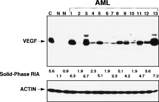
The amount of VEGF detected using Western blot was in good agreement with the amount detected using RIA (Fig 1). The RIA values of AML samples were normalized to the RIA values of normal bone marrow, which were assigned a value of 1. There was considerable variability in the patients’ RIA values (median, 2.8; mean, 2.9; average, 0.5 to 8.1). While we did not asses the effect of length of storage on VEGF levels by comparing the levels in a given sample as measured in 1996 and as measured again in 1997 or 1998, those VEGF samples obtained in 1996 had similar values as those obtained in 1997 and 1998. However, VEGF levels as measured in blood were higher than those measures in marrow (mean, 3.1 v 2.8; median, 3.6v 2.9), although the difference was only marginally significant (P = .08). There was a strong correlation between VEGF as quantified by RIA in blood and quantified in marrow in 9 patients in whom we have both samples available (P = < .01).
Western blot. In lane C is a positive control, the next 2 lanes show control from 2 normal bone marrows. Lanes 1 through 13 show a considerable variability in expression of VEGF that was confirmed with RIA.
Western blot. In lane C is a positive control, the next 2 lanes show control from 2 normal bone marrows. Lanes 1 through 13 show a considerable variability in expression of VEGF that was confirmed with RIA.
There was no relationship between VEGF concentration and such standard prognostic factors as age (P = .57), performance status (P = .12), karyotype (P = .40) (Table 2), or presence of antecedent hematologic disease probably signifying a prior myelodysplastic syndrome. There was also no correlation between VEGF concentration and pretreatment blood count. After excluding the 9 of our 99 patients who had APL, we compared CR rate and survival according to whether VEGF was above or below the median value (2.9). The CR rate was 64% in both groups and survival was also the same in both. However, it was possible that the relationship between VEGF and outcome was more complicated, and to examine the specific nature of this relationship, we used Martingale residual plots. These suggested that as VEGF continued to increase to a value of about 1.7, CR rate continued to decrease, and survival continued to shorten. At VEGF levels >1.7, there was no further effect. Hence, we compared CR rates and survival in patients who had VEGF levels above and below 1.7. Table 2 illustrates that CR rates were 59% and 84% in patients with VEGF levels above and below this value, respectively. The difference in CR rates was a result of a higher incidence of “resistance,” ie, failure to achieve CR despite living at least 35 days from the start of induction therapy.
The lack of an association between VEGF levels and conventional prognostic factors noted above suggested that VEGF was an independent predictor factor at least for CR. We tested this suggestion by fitting a logistic model for CR and Cox model for survival and disease-free survival from start of therapy and from CR date (Table 3). In these models, we examined VEGF as a continuos variable as suggested by the Martingale residual plots. Table 3 shows that VEGF was a predictor of each outcome even after accounting for the other predictors. Of note, the source of the cells used to measure VEGF (marrow v peripheral blood) was not a predictor of outcome, or was the year when the sample was obtained. This suggests that results were not biased by the length of sample storage, eg, systematically lower levels in samples obtained in 1996 than in samples obtained in 1998. Within the limits of small numbers, there was no interaction between VEGF and cytogenetic groups, ie, the effect of VEGF on outcome was not greater in any cytogenetic group. Treatment with fludarabine-containing regimens was associated with poor outcome as previously noted,30 31 but the effect of VEGF on outcome remained after accounting for treatment.
DISCUSSION
Our data suggest that VEGF levels are, on average, higher in AML than in normal bone marrow. Normal marrow contains relatively few blasts, and high expression in a small number of normal blasts could easily have been missed. However, Fiedler et al24 have reported that normal CD34+ marrow cells have low VEGF levels. The principal purpose of this report was to examine the possible prognostic significance of cellular VEGF in AML. Our results suggest that cellular VEGF levels are prognostic in newly-diagnosed AML (Tables 3) (APL patients excluded from the analysis). Although we did not study consecutive patients, the population we analyzed was representative of our newly-diagnosed AML population (Table 1) with the principal exception of higher white blood cell and blast counts and a slightly higher incidence of inv(16). The higher white blood cell counts reflect the selective use of samples containing a high number of blasts so as to facilitate VEGF measurements. Because of the selected nature of our population, our conclusions about the prognostic significance of VEGF cannot be generalized to the usual AML population, which of course has a lower white blood cell count (Table 1). Three other possibly confounding factors are the various treatments the patients received (Table 1), the possible effect of storage on VEGF levels, and the source of the cells used to measure VEGF. However, multivariate analysis found that VEGF was still prognostic after accounting for the unfavorable effect of fludarabine, and found that outcome was unaffected by the year (1996, 1997, 1998) the sample was obtained or whether the cells were obtained from blood or bone marrow.
The mechanism by which high cellular VEGF levels affect prognosis is not clear. We are currently attempting to correlate VEGF levels with marrow microvascular density and with plasma VEGF levels. Such levels were unavailable in the 99 patients studied here. Regardless of the mechanism, VEGF appears to be prognostic in at least the subset of newly diagnosed AML patients presenting with relatively high WBC and high blast count.
The publication costs of this article were defrayed in part by page charge payment. This article must therefore be hereby marked “advertisement” in accordance with 18 U.S.C. section 1734 solely to indicate this fact.
REFERENCES
Author notes
Address reprint requests to Maher Albitar, MD, Section of Hematopathology, Division of Laboratory Medicine, The University of Texas, MD Anderson Cancer Center, 1515 Holcombe Blvd, Box 72, Houston, TX 77030-4095; e-mail: malbitar@mdacc.tmc.edu.


