Abstract
Definition of the cytokine environment, which regulates the maturation of human natural killer (NK) cells, has been largely based on in vitro assays because of the lack of suitable animal models. Here we describe conditions leading to the development of human NK cells in NOD/SCID mice receiving grafts of hematopoietic CD34+ precursor cells from cord blood. After 1-week-long in vivo treatment with various combinations of interleukin (IL)–15, flt3 ligand, stem cell factor, IL-2, IL-12, and megakaryocyte growth and differentiation factor, CD56+CD3- cells were detected in bone marrow (BM), spleen, and peripheral blood (PB), comprising 5% to 15% of human CD45+ cells. Human NK cells of NOD/SCID mouse origin closely resembled NK cells from human PB with respect to phenotypic characteristics, interferon (IFN)–γ production, and cytotoxicity against HLA class 1–deficient K562 targets in vitro and antitumor activity against K562 erythroleukemia in vivo. In the absence of growth factor treatment, CD56+ cells were present only at background levels, but CD34+CD7+ and CD34-CD7+ lymphoid precursors with NK cell differentiation potential were detected in BM and spleen of chimeric NOD/SCID mice for up to 5 months after transplantation. Our results demonstrate that limitations in human NK cell development in the murine microenvironment can be overcome by treatment with NK cell growth–promoting human cytokines, resulting in the maturation of IFN-γ–producing cytotoxic NK cells. These studies establish conditions to explore human NK cell development and function in vivo in the NOD/SCID mouse model. (Blood. 2003;102:127-135)
Introduction
Human natural killer (NK) cells comprise approximately 10% of peripheral blood (PB) lymphocytes and are characterized phenotypically by the expression of CD56 and the lack of CD3 cell surface antigens.1 They are important effectors of the innate immune system and contribute to the first line of defense against infections and malignancy.2 In contrast to T lymphocytes, NK cells are able to kill cancer and virus-infected target cells without the need for prior antigen stimulation. NK cell precursors have been identified within the CD34+ hematopoietic cell population in adult bone marrow (BM) and umbilical cord blood (CB). These precursors can efficiently generate mature NK cells in vitro in the presence of interleukin-15 (IL-15) and early-acting cytokines such as flt3 ligand (FL) or stem cell factor (SCF), which increase the frequency of NK cell precursors responding to IL-15.3-5 IL-15 is a key cytokine in NK cell development.6 Targeted disruption of the IL-15 or IL-15 receptor genes in mice7,8 and spontaneously occurring mutations in the signaling components of the receptor in humans9 cause blocks in early NK cell development. FL and SCF play important roles in the early differentiation steps of NK cells and in their subsequent expansion and functional maturation as revealed in mice rendered deficient in FL or carrying mutations in the c-kit receptor.10,11 Other important regulatory cytokines are IL-12 and IL-18, which enhance cytotoxicity and trigger cytokine release by mature NK cells.12,13
Biologic responses of NK cells are controlled by a balance of signals from inhibitory and activatory receptors. The inhibitory receptors recognize epitopes shared by different alleles of HLA class 1 molecules and deliver negative signals, thereby suppressing NK cell function and blocking lysis of normal cells.14 In the absence of inhibitory signals, the ability of NK cells to lyse cells altered by virus infection and tumor transformation is mediated by the activatory receptors, including FcγRIII (CD16) and natural cytotoxicity receptors (NCRs), which were recently identified as unique NK-specific cell surface molecules.15 In human leukemia, the expression of NCRs is down-regulated and correlates with low NK cell cytotoxicity,16 whereas, conversely, antileukemic responses are enhanced by NK cell alloreactivity in mismatched stem cell transplants.17,18 Therefore, NK cells appear important for tumor surveillance and might be exploited in the immunotherapy of diseases.
However, understanding the development and function of human NK cells is largely based on in vitro analyses, and models to study human NK cells in vivo are lacking. Results of murine NK cell studies are not always applicable to the human NK cell system because of the lack of a murine homologue of CD56 and because of differences in the inhibitory and activatory receptors.14,19 Furthermore, it has not been possible to use nonobese diabetic/severe combined immunodeficiency (NOD/SCID) mice repopulated with human hematopoietic progenitors for studies on human NK cells because the lymphoid differentiation in these mice is restricted to the B-cell lineage, whereas T and NK cells are produced at a minimum level or not at all.20-22 In this study, we show that NOD/SCID mice repopulated with CB CD34+ cells contain human NK cell precursors and that the administration of recombinant human IL-15, together with other NK cell growth–promoting cytokines, leads to the in vivo development of NK cells in BM, spleen, and blood circulation. The combination of IL-15 and FL is sufficient to generate NK cells at levels comparable to the NK cell content in human PB. NK cells generated in NOD/SCID mice are CD56+CD3-, express CD16 and NKp46, and are functional with respect to interferon-γ (IFN-γ) production in response to IL-12 and IL-18. Furthermore, NK cells of NOD/SCID origin show cytotoxic activity against K562 cells in vitro and reduce the growth of K562 erythroleukemia in vivo. Establishment of human NK cell development in NOD/SCID mice provides an in vivo system to investigate NK cells in immunotherapeutic strategies against infectious diseases and cancer.
Materials and methods
Cord blood cell preparation
CB was kindly provided by the Department of Obstetrics and Gynecology, University Hospital Basel and the Department of Obstetrics and Gynecology, Kantonsspital Bruderholz, Switzerland with informed consent of the mothers. Investigations were approved by the Ethical Committee of the University Hospital Basel. CB mononuclear cells were separated by Histopaque (less than 1.077 g/cm3; Sigma, St Louis, MO) density-gradient centrifugation and subsequent red blood cell lysis. Frozen samples were pooled after thawing, and CD34+ cells were isolated with superparamagnetic MACS (magnetic cell sorting) microbeads (Miltenyi Biotec, Bergisch Gladbach, Germany) according to the manufacturer's instructions. The purity of the CD34+ cell population ranged from 80 to 95%.
Transplantation of CD34+ cells into NOD/SCID mice
NOD/LtSz-scid/scid (NOD/SCID) mice23 (The Jackson Laboratory, Bar Harbor, ME) were maintained under specific pathogen-free conditions in the animal facility of the Research Department, University Hospital Basel. CB CD34+ cells (1-2 × 10⁵) together with 1 × 10⁶ irradiated (1500 cGy) human PB mononuclear carrier cells were injected intravenously into the tail veins of 8-week-old NOD/SCID mice previously given 375 cGy from a 60Co source (2 cGy/min). Mice were kept on acidified drinking water supplemented with Bactrim (32/160 mg/L; Roche Pharma AG, Reinach, Switzerland) for the duration of the experiment.
In vivo treatment with human growth factors
Eight to 10 weeks after transplantation, NOD/SCID mice were injected intraperitoneally with phosphate-buffered saline (PBS) or the following recombinant human growth factors: IL-15, FL (both from Immunex, Seattle, WA), SCF (Amgen, Thousand Oaks, CA), PEGylated megakaryocyte growth and development factor (MGDF; Amgen and Kirin Brewery, Tokyo, Japan), IL-2 (Novartis, Basel, Switzerland), and IL-12 (Roche, Nutley, NJ). IL-15, FL, SCF, MGDF, and IL-2 were administered for 7 consecutive days, each at 10 μg daily, and IL-12 was administered at 1 μg per day for the last 2 days of the treatment. Twenty-four hours after the last injection, spleen and BM cells were harvested, and single-cell suspensions of spleen and BM were prepared. To measure circulating human NK cells, PB was drawn weekly up to 3 weeks after growth factor injections. Statistical analyses for comparison of treatment groups were performed with the Mann-Whitney U test.
Flow cytometry and cell sorting
Three-color fluorescence-activated cell sorter (FACS) analysis was used to characterize human engraftment of NOD/SCID mice that underwent transplantation. Single-cell suspensions from BM and spleen were resuspended in FACS buffer containing PBS, 2% fetal calf serum (FCS; both from Invitrogen, Carlsbad, CA), and 0.02% NaN3 (Fluka, Buchs, Switzerland) and were stained on ice for 20 minutes with fluorescein isothiocyanate (FITC)–, phycoerythrin (PE)–, or allophycocyanin (APC)–conjugated monoclonal antibodies (mAbs) against human CD2, CD3, CD7, CD16, CD19, CD33, CD34, CD38, CD45, CD56, CD62L, CD69, CD158a, CD158b, NKB1, and HLA-DR or isotype control antibodies (all from BD PharMingen, San Jose, CA). Staining with unlabeled mAb anti-CD94 (clone 39B10), anti-NKp46 (clone 9E2; each a generous gift from M. Colonna and A. Bouchon, Basel Institute for Immunology, Switzerland) and anti–IL-2Rγ chain (clone CP.B8; kindly provided by D. Baker, Biogen, Cambridge, MA) was revealed with secondary PE- or FITC-conjugated goat antimouse antibodies (Southern Biotechnology Associates, Birmingham, AL). Normal mouse serum was used to saturate free binding sites of secondary antibodies before cells were subsequently incubated with directly labeled mAbs. Propidium iodide (PI; Sigma) staining was used to exclude dead cells from the analysis. FACS analysis of circulating NK cells was performed by incubating whole blood with labeled antibodies at room temperature for 20 minutes, followed by erythrocyte lysis with FACS Lysing Solution (BD PharMingen) and subsequent washing with FACS buffer. FACS analysis was performed on a FACScalibur (Becton Dickinson), and data were analyzed using CellQuest Pro software (Becton Dickinson). For cell-sorting experiments, BM cells from NOD/SCID that underwent transplantation were resuspended in FACS buffer without NaN3 and were stained with the appropriate antibodies. Cells were washed, incubated with PI, and sorted on a FACS Vantage SE (Becton Dickinson).
NK cell differentiation in vitro
Cell suspensions from NOD/SCID mouse BM or spleen containing 20% to 90% human CD45+ cells were seeded at 1 to 2 × 10⁶/mL cells in 24-well plates in Iscove modified Dulbecco medium (IMDM) containing 5% FCS, 5% human AB+ serum (Blutspendezentrum Basel), 380 μg/mL iron-saturated human transferrin, and 1% bovine serum albumin and were supplemented with IL-15, FL, and SCF (each at 100 ng/mL). After 1 week, cells were transferred to 6-well plates, and half the medium was replaced once a week for another 2 to 5 weeks, as specified in “Results.” The development of CD56+ NK cells was determined at the indicated time points by FACS.
NK cell lines were cultured by restimulation with irradiated mono-nuclear cells from human PB and phytohemagglutinin HA 16 (2 μg/mL; Murex Biotech, Dartford, England) in the presence of 100 U IL-2 every 3 to 4 weeks.
NK cell cytotoxicity and IFN-γ production
After 4 to 5 weeks of differentiation in culture, CD56+ cells were purified by positive selection with MACS CD56-microbeads (Miltenyi Biotec). Cells were washed and resuspended in IMDM containing 2% FCS, and cytotoxicity against the NK-sensitive target K562 was determined in a 4-hour lactate dehydrogenase (LDH) release assay (CytoTox 96; Promega, Madison, WI) according to the manufacturer's instructions. The effector-target ratio ranged from 5:1 to 0.6:1.
IFN-γ production was measured by intracellular flow cytometry. MACS-purified NK cells from differentiation cultures (purity, 80%-95%) or FACS sorter-purified NK cells from BM of growth-factor–treated NOD/SCID mice were washed, and 1 × 10⁶ cells/mL were plated in 96-well plates for 36 hours in IMDM containing 5% FCS, 5% AB+ human serum, 10 U/mL IL-12, and 100 ng/mL IL-18 (PeproTech, Rocky Hill, NJ). Brefeldin A (Sigma) was added at 5 μg/mL for the final 8 hours of culture. Cells were washed with FACS buffer, stained with anti–CD56-PE mAb for 20 minutes on ice, and fixed in 2% paraformaldehyde for 15 minutes at room temperature. Cells were washed twice and permeabilized in saponin-containing FACS buffer. Anti–IFN-γ–FITC and isotype control mAb (BD PharMingen) were added for 30 minutes at room temperature. Cells were washed twice in permeabilization buffer and once in FACS buffer and were analyzed with FACScalibur.
K562 tumor formation
K562 erythroleukemia cells were resuspended in 100 μL PBS and injected subcutaneously into the dorsal lateral thorax of NOD/SCID mice. NK cells, resuspended in 200 μL PBS, were injected intravenously 1 day after tumor cell inoculation. In mice that received grafts of CB CD34+ cells, K562 cells were inoculated 1 day after 7-day-long treatment with IL-15 and FL. The effect of NK cells on tumor growth was determined in groups of 2 to 4 animals. Tumor growth was monitored weekly, and tumor surface area was calculated using the formula (a/2 × b/2) × π, where a and b are the long and short diameters (in millimeters). Statistical analyses for comparison of treatment groups were performed using the unpaired Student t test.
Results
Growth factors induce maturation of human NK cells in NOD/SCID mice
To investigate the requirements for human NK cell development in vivo, the NOD/SCID xenotransplantation system was chosen to test the effect of human growth factors known to be involved in human NK cell development in vitro. Groups of NOD/SCID mice that underwent transplantation 2 months earlier with highly enriched CB CD34+ cells were injected for 7 consecutive days with one of the following 4 combinations of cytokines: IL-15/FL, IL-15/FL/SCF, IL-15/FL/IL-12, and IL-15/FL/SCF/MGDF/IL-2. On day 8, BM and spleens were examined for the presence of human NK cells (Table 1). Among the CD45+ human lymphocytes, only background levels of 0.3% ± 0.1% human CD56+ NK cells were detected in untreated control animals (Figure 1A; Table 1). With the administration of growth factors, the frequency of CD56+ cells increased approximately 15-fold and was similar among mice tested with the 4 growth factor combinations (range, 3.9% ± 0.9% to 4.9% ± 2.0%). The content of NK cells generated in the BM of NOD/SCID mice with human growth factor treatment was comparable to that in human BM and CB (1%-10% and 4%-12% CD56+ cells, respectively, as determined using 6-8 samples from both blood sources; Figure 1C and results not shown). Human NK cells were also found in spleens of NOD/SCID mice, in which they constituted 3.2% ± 1.4% to 5.0% ± 1.4% of human CD45+ cells, depending on the growth factors used. IL-15/FL/IL-12 led to NK cell development mainly in the BM but only marginally in the spleen (Table 1). Additionally, in the blood circulation of NOD/SCID mice, 12.2% ± 0.8% (n = 8) of CD45+ human cells were CD56+ NK cells, which corresponds to the number of NK cells in human PB (7%-15%; Figure 1D-E), and they were detectable for at least 3 weeks after growth factor treatment.
Effect of growth factor administration on human hematopoietic lineages in BM and spleen of NOD/SCID mice with transplanted CD34 cord blood cells
. | Bone marrow . | . | . | Spleen . | . | . | ||||
|---|---|---|---|---|---|---|---|---|---|---|
| Treatment (no. mice) . | CD56+ . | CD19+ . | CD33+ . | CD56+ . | CD19+ . | CD33+ . | ||||
| No treatment (20) | 0.3 ± 0.1 | 87.0 ± 1.5 | 11.5 ± 1.5 | 0.3 ± 0.1 | 93.0 ± 1.2 | 3.0 ± 0.7 | ||||
| IL-15/FL (7) | 3.9 ± 0.9* | 62.0 ± 4.0* | 27.0 ± 5.0* | 3.2 ± 1.4† | 80.0 ± 5.0* | 6.6 ± 1.3† | ||||
| IL-15/FL/SCF (6) | 4.1 ± 1.1* | 64.0 ± 4.5* | 28.0 ± 3.0* | 4.4 ± 1.6* | 81.0 ± 4.0† | 9.4 ± 2.5† | ||||
| IL-15/FL/IL-12 (5) | 4.9 ± 2.0* | 86.0 ± 4.0 | 6.3 ± 2.6 | 1.1 ± 0.2† | 92.0 ± 1.0 | 2.6 ± 0.5 | ||||
| IL-15/FL/SCF/MGDF/IL-2 (3) | 4.1 ± 1.3† | 70.0 ± 3.5† | 25.0 ± 3.5† | 5.0 ± 1.4† | 80.0 ± 1.5† | 10.0 ± 2.0† | ||||
Values are mean percentages of human CD45+ cells ± SEMs.
P < .005.
P < .05.
Phenotype of human NK cells in BM of growth-factor–treated NOD/SCID mice. Two months after transplantation of CB CD34+ cells, NOD/SCID mice were treated with combinations of growth factors (A) for 7 consecutive days, and BM, spleen, and PB cells were analyzed 1 day later by flow cytometry. (A) Human NK cells in untreated control (no growth factor [GF]) and growth-factor–treated mice were detected by staining with the human leukocyte marker CD45 and the NK-cell–specific marker CD56. Analysis was performed with total mononuclear cells; numbers indicate the percentages of total BM cells. (B) Human NK cells were characterized by staining with antibodies against CD56 and the indicated NK surface marker. Numbers indicate the percentages of human CD45+ cells. For comparison, human NK cells from (C) CB and (D) PB were stained with antibodies against CD56 and CD16 (gated on lymphocytes) and NKp46 (gated on CD56+ cells). The thin line indicates staining with isotype-matched control antibody. (E) NK cells in PB of NOD/SCID mice treated with IL-15 and FL. Numbers indicate the percentages of human CD45+ cells.
Phenotype of human NK cells in BM of growth-factor–treated NOD/SCID mice. Two months after transplantation of CB CD34+ cells, NOD/SCID mice were treated with combinations of growth factors (A) for 7 consecutive days, and BM, spleen, and PB cells were analyzed 1 day later by flow cytometry. (A) Human NK cells in untreated control (no growth factor [GF]) and growth-factor–treated mice were detected by staining with the human leukocyte marker CD45 and the NK-cell–specific marker CD56. Analysis was performed with total mononuclear cells; numbers indicate the percentages of total BM cells. (B) Human NK cells were characterized by staining with antibodies against CD56 and the indicated NK surface marker. Numbers indicate the percentages of human CD45+ cells. For comparison, human NK cells from (C) CB and (D) PB were stained with antibodies against CD56 and CD16 (gated on lymphocytes) and NKp46 (gated on CD56+ cells). The thin line indicates staining with isotype-matched control antibody. (E) NK cells in PB of NOD/SCID mice treated with IL-15 and FL. Numbers indicate the percentages of human CD45+ cells.
Human NK cells that developed in NOD/SCID mice were examined for the expression of a panel of cell surface markers. Staining patterns of NK cells residing in BM and spleen were indistinguishable, and there were no marked differences in NK cell phenotype with respect to the growth factor combination used to generate them. NK cells from the BM of IL-15/FL–treated mice are shown in Figure 1B. CD56+ cells were always CD3-, indicating that these cytokine combinations promoted only the development of bona fide NK cells and not the CD56+CD3+ NK T-cell population. CD94 and the IL-2Rγ chain were expressed on all CD56+ cells from NOD/SCID mice. The early activation marker CD69 was expressed on virtually all NK cells, likely as a response to IL-15,24 whereas CD25, the low-affinity IL-2Rα chain, was not present. Of the other tested markers, CD2, CD7, and CD62L selectin were expressed by 30% to 45%, 65% to 80%, and up to 60% of NK cells, respectively (Figure 1B). Intracellular perforin was expressed by all NK cells, HLA-DR was expressed only at low levels or not at all, and c-kit was absent (not shown). Interestingly, staining of CD16 and CD56 surface markers revealed that the prevalence of the CD56dimCD16high population, which is characteristic of human CB and PB NK cells (Figure 1C-D), was not apparent in NK cells generated in NOD/SCID mice. Instead, CD16 was expressed at various cell surface densities on 50% to 70% of all CD56+ NK cells (Figure 1B, E). NKp46, the major NCR selectively expressed by NK cells, showed a heterogeneous expression pattern of 5% to 19% (Figure 1B), lower than that found in human NK cells from PB and CB (Figure 1C-D). This lower NKp46 expression was not attributed to a limiting availability of the CD3ζ chain, the coreceptor for NKp46, because FACS staining revealed CD3ζ expression in all human NK cells generated in mice that underwent transplantation (data not shown). Based on these results we conclude that human recombinant growth factors, including IL-15 and FL, specifically promoted the maturation of human NK cells in the BM and spleen microenvironments of NOD/SCID mice. The phenotype of these cells closely resembled, but was not identical to, NK cells from human PB.
Effect of growth factor treatment on human cell engraftment and lineage composition
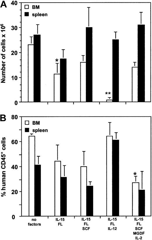
The effect of growth factor treatment on the cellularity of BM and spleen and on engraftment with human cells of NOD/SCID mice that underwent transplantation was also examined (Figure 2). The BM of 4 long bones of untreated control NOD/SCID mice contained 22 ± 4 × 10⁶ cells, of which 64% ± 5% were of human origin. Growth factor injections reduced the cellularity of BM but not of spleens. In particular, quick and dramatic 10-fold cell loss was observed with the administration of IL-15/FL/IL-12 involving 2 days of treatment with IL-12 (Figure 2A). This is in accord with the reported hypoplasia in BM but not spleen with IL-12 administration in humans and mice.25-27 The overall level of human CD45+ cells was reduced by 30% to 50% in BM and spleens in all groups of growth-factor–treated mice except the IL-15/FL/IL-12 group (Figure 2B). This reduction was associated mainly with a decrease in CD19+ B lymphocytes, which constituted most human cells in untreated and treated mice and which were reduced by 15% to 30% after growth factor injections (Table 1). Concomitantly, the proportion of myeloid cells expressing CD33 was increased up to 3-fold. The lower engraftment and changes in the proportions between B cells and myeloid lineages on growth factor treatment are reminiscent of the effects of FL alone and with IL-7 and SCF, reported to have an even greater effect when injected over several weeks.28
Cellularity and level of human engraftment in BM and spleen of NOD/SCID mice after growth factor treatment. Two months after transplantation of CB CD34+ cells, NOD/SCID mice were treated with the indicated human growth factor combinations. (A) Total cell number was determined from the BM (4 long bones, □) and from spleen (▪). (B) Human engraftment was defined by staining BM (□) and spleen (▪) cells with human anti-CD45 antibody. Mean ± SEM percentages are shown. Significant differences between untreated and treated groups are marked (*P < .05; **P < .005).
Cellularity and level of human engraftment in BM and spleen of NOD/SCID mice after growth factor treatment. Two months after transplantation of CB CD34+ cells, NOD/SCID mice were treated with the indicated human growth factor combinations. (A) Total cell number was determined from the BM (4 long bones, □) and from spleen (▪). (B) Human engraftment was defined by staining BM (□) and spleen (▪) cells with human anti-CD45 antibody. Mean ± SEM percentages are shown. Significant differences between untreated and treated groups are marked (*P < .05; **P < .005).
Lymphoid precursors with NK cell differentiation potential are present in BM and spleens of NOD/SCID mice that underwent transplantation
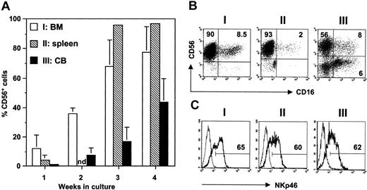
The finding that human NK cells developed within 7 days of growth factor administration prompted us to characterize the NK precursor cells in NOD/SCID mice that received grafts of human hematopoietic cells. In the first series of experiments, we investigated whether CD56+ NK cells could also be generated in vitro from BM and spleen cells obtained from NOD/SCID mice not treated with growth factors (Figure 3). In cultures containing IL-15, FL, and SCF, human CD56+ cells developed within the first week and reached 68% to 96% by 3 weeks (Figure 3A). This was faster and more efficient than the development of NK cells from CB CD34+ progenitors cultured under the same conditions (17% ± 10% CD56+ cells at week 3), particularly considering the fact that the purity of CB CD34+ cells at the start of culture was at least 80%, whereas CD34+ human cells constituted only 19.0% ± 1.1% and 6.4% ± 0.9% of all cells in NOD/SCID BM and spleen, respectively (Table 2). The phenotypes of NK cells of NOD/SCID mice origin and those generated from CB CD34+ cells were similar (Figure 3B). CD16 was expressed only at low levels in 2% to 15% of all NK cells. Remarkably, NKp46 was expressed by up to 60% of NK cells generated in vitro (Figure 3C), indicating that NKp46 is present on CD16- and CD16+ NK cells. The surface markers CD7, CD62L, CD69, and CD161 were expressed at various levels on 5% to 30% of NK cells generated in vitro from NOD/SCID BM. The killer immunoglobulin-like receptors CD158a, CD158b, and NKB1 were not detected (data not shown), in agreement with reports that NK cells differentiated in vitro from CD34+ cells are mainly negative for these receptors.3 CD56+CD3+ NK T cells or CD3+ T cells were never found in these cultures (data not shown).
In vitro differentiation of human NK cells from NOD/SCID BM and spleen and from human CB. (A) Single-cell suspensions of BM (I: n = 13, □) and spleen (II: n = 5, ▧) of NOD/SCID that underwent transplantation and purified CB CD34+ cells (III: n = 7, ▪) were cultured for 4 weeks in NK differentiation medium containing recombinant human IL-15, FL, and SCF. The appearance of CD56+ NK cells was determined by FACS analysis. Shown are the mean ± SEM percentages of total cells in the cultures. nd indicates not determined. (B) Expression of CD16 and CD56 and of (C) NKp46 on CD56+ NK cells generated in cultures I, II, and III at 4 weeks. Thin line indicates staining with isotype-matched control antibody. Numbers indicate the percentages of NKp46+ NK cells.
In vitro differentiation of human NK cells from NOD/SCID BM and spleen and from human CB. (A) Single-cell suspensions of BM (I: n = 13, □) and spleen (II: n = 5, ▧) of NOD/SCID that underwent transplantation and purified CB CD34+ cells (III: n = 7, ▪) were cultured for 4 weeks in NK differentiation medium containing recombinant human IL-15, FL, and SCF. The appearance of CD56+ NK cells was determined by FACS analysis. Shown are the mean ± SEM percentages of total cells in the cultures. nd indicates not determined. (B) Expression of CD16 and CD56 and of (C) NKp46 on CD56+ NK cells generated in cultures I, II, and III at 4 weeks. Thin line indicates staining with isotype-matched control antibody. Numbers indicate the percentages of NKp46+ NK cells.
Effect of growth factor administration on hematopoietic progenitor and lymphoid precursor cells in BM and spleen of NOD/SCID mice with transplanted CD34+ cord blood cells
. | Bone marrow* . | . | . | . | . | Spleen* . | . | . | . | |||||||
|---|---|---|---|---|---|---|---|---|---|---|---|---|---|---|---|---|
| . | . | CD34+ . | CD34+ . | CD34+ . | CD34− . | . | CD34+ . | CD34+ . | CD34− . | |||||||
| Treatment (no. mice) . | CD34+ . | CD19+ . | CD19− . | CD7+ . | CD7+ . | CD34+ . | CD19− . | CD7+ . | CD7+ . | |||||||
| No treatment (20) | 19.0 ± 1.1 | 13.2 ± 1.2 | 4.9 ± 0.4 | 0.9 ± 0.1 | 0.8 ± 0.1 | 6.4 ± 0.9 | 2.7 ± 0.6 | < 0.1 | 1.1 ± 0.2 | |||||||
| IL-15/FL (7) | 7.7 ± 1.8* | 2.1 ± 0.4* | 5.5 ± 1.8 | 1.3 ± 0.5 | 4.0 ± 1.1* | 7.3 ± 0.7 | 2.4 ± 0.4 | < 0.1 | 5.3 ± 2.0* | |||||||
| IL-15/FL/SCF (6) | 9.7 ± 1.8† | 2.7 ± 0.7* | 7.1 ± 1.4 | 1.1 ± 0.4 | 4.5 ± 1.3* | 5.5 ± 0.3 | 3.0 ± 0.5 | < 0.1 | 4.5 ± 0.9* | |||||||
| IL-15/FL/IL-12 (5) | 8.3 ± 0.6† | 4.2 ± 1.1* | 4.2 ± 0.5 | 0.7 ± 0.15 | 3.9 ± 1.3† | 7.3 ± 2.4 | 1.5 ± 0.3 | 0.15 ± 0.02 | 2.4 ± 1.1 | |||||||
Values are mean percentages of human CD45+ cells ± SEMs.
P < .005.
P < .05.
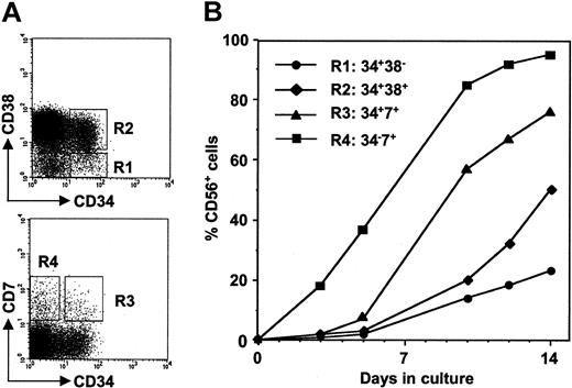
To define the precursor cell populations able to give rise to NK cells in vivo and in vitro, we performed FACS analysis of human cells from BM of growth-factor–untreated NOD/SCID mice (Figure 4A). CD34+CD38- and CD34+CD38+ hematopoietic progenitor cells and CD34+CD7+ and CD34-CD7+ cells, all reported to contain NK precursor cells,29-31 were cultured in the presence of IL-15, FL, and SCF, and NK cell development was examined. The fastest differentiation toward CD56+ cells, apparent after 3 days, was obtained with CD34-CD7+ cells, suggesting the highest frequency of NK precursors in this cell fraction (Figure 4B). CD34+CD7+ cells also gave rapid rise to 75% of CD56+ cells after 14 days. In contrast, in cultures initiated with primitive hematopoietic CD34+CD38- progenitors, only 20% of all cells expressed CD56 by day 14. These results indicate that even in the absence of growth factor treatment, human NK cell precursors had progressed in the NOD/SCID microenvironment to an advanced differentiation stage, providing an explanation for the faster response of unseparated NOD/SCID-derived cells compared with CB CD34+ cells cultured under the same conditions (Figure 3A). Despite different kinetics, NK cells generated in vitro from sorted cell populations (R1-R4) of NOD/SCID mouse BM acquired the same phenotypic characteristics as those from bulk cultures described in Figure 3 (results not shown).
In vitro differentiation of human NK cells derived from FACS sorter-purified progenitor cells of NOD/SCID mouse BM. (A) Hematopoietic progenitor cells and NK precursor cells were identified in the BM of untreated NOD/SCID mice by staining with CD34/CD38 (top panel) and CD34/CD7 (bottom panel). Cells falling into the gates R1 (CD34+CD38-: 1.5% of total BM cells), R2 (CD34+CD38+: 12%), R3 (CD34+CD7+: 0.5%), and R4 (CD34-CD7+: 1.5%) were sorted and cultured for 14 days in NK differentiation medium (see “Materials and methods”). (B) The development of CD56+ NK cells in vitro was followed by FACS analysis at the indicated time points.
In vitro differentiation of human NK cells derived from FACS sorter-purified progenitor cells of NOD/SCID mouse BM. (A) Hematopoietic progenitor cells and NK precursor cells were identified in the BM of untreated NOD/SCID mice by staining with CD34/CD38 (top panel) and CD34/CD7 (bottom panel). Cells falling into the gates R1 (CD34+CD38-: 1.5% of total BM cells), R2 (CD34+CD38+: 12%), R3 (CD34+CD7+: 0.5%), and R4 (CD34-CD7+: 1.5%) were sorted and cultured for 14 days in NK differentiation medium (see “Materials and methods”). (B) The development of CD56+ NK cells in vitro was followed by FACS analysis at the indicated time points.
Treatment of NOD/SCID that underwent transplantation with IL-15/FL, IL-15/FL/SCF, and IL-15/FL/IL-12 combinations in vivo specifically increased the population of human CD34-CD7+ NK cell precursors up to 5-fold in BM and spleens (Table 2). This increase is in accord with the increase in CD56+ cells observed after growth factor injections (Table 1), some of which were CD56+CD7+ (data not shown). The proportion of total CD34+ cells was significantly decreased in BM, primarily because of a reduction in CD34+CD19+ pre–B-cell levels. Neither the CD34+CD19- nor the CD34+CD7+ cell population was affected by the cytokine treatment in either organ. We conclude that the tested growth factors acted on a CD7+ NK precursor at the transition from the CD34+ to the CD56+ stage.
IFN-γ production by human NK cells generated in NOD/SCID mice
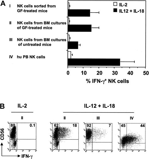
A hallmark of NK cell function is the ability to produce IFN-γ. To assess this feature, CD56+ NK cells from the BM of growth-factor–treated NOD/SCID mice were isolated by cell sorting and were stimulated in vitro for 36 hours with IL-12 and IL-18. Additionally, NK cells were isolated after 4 weeks of culture of BM from NOD/SCID mice, both growth factor treated and untreated. The proportion of IFN-γ–producing NK cells was measured by intracellular FACS analysis. NK cells differentiated in vivo and in vitro responded equally well to the cytokine stimulus (Figure 5). Of the 3 types of tested NK cells, production of IFN-γ was found in 14% ± 6% and 15% ± 5% of cells purified directly or after culture of growth-factor–treated mouse BM and in 6.4% ± 1.7% of NK cells generated in vitro from the BM of untreated NOD/SCID mice. For comparison, purified NK cells from human PB were included in the assay and responded more readily to IL-12 and IL-18 (33% ± 10%). The differences in IFN-γ production may be related to the maturation stages of the NK cell populations generated under these different experimental conditions.
IFN-γ production by NK cells from NOD/SCID mice. (A) FACS sorter-purified CD56+ NK cells from BM of growth factor (GF)–treated NOD/SCID mice (I: n = 3), NK cells generated in vitro within 4 weeks from BM of GF-treated (II: n = 3) and untreated control (III: n = 5) NOD/SCID mice, and purified NK cells isolated from human PB mononuclear cells (IV: n = 2) were stimulated with medium containing IL-2 (□) or IL-12 and IL-18 (▪) for 36 hours. Synthesis of IFN-γ was measured by intracellular FACS analysis, and the mean ± SEM percentages of IFN-γ+ NK cells are shown. (B) Representative FACS pictures of IFN-γ–producing NK cells generated in cultures II, III, and IV are shown. Numbers represent the percentages of total cells in culture.
IFN-γ production by NK cells from NOD/SCID mice. (A) FACS sorter-purified CD56+ NK cells from BM of growth factor (GF)–treated NOD/SCID mice (I: n = 3), NK cells generated in vitro within 4 weeks from BM of GF-treated (II: n = 3) and untreated control (III: n = 5) NOD/SCID mice, and purified NK cells isolated from human PB mononuclear cells (IV: n = 2) were stimulated with medium containing IL-2 (□) or IL-12 and IL-18 (▪) for 36 hours. Synthesis of IFN-γ was measured by intracellular FACS analysis, and the mean ± SEM percentages of IFN-γ+ NK cells are shown. (B) Representative FACS pictures of IFN-γ–producing NK cells generated in cultures II, III, and IV are shown. Numbers represent the percentages of total cells in culture.
Cytotoxic activity of NOD/SCID-mouse–derived human NK cells
Another characteristic of NK cells is the capacity to kill target cells without prior sensitization. To study the cytotoxic activity of NOD/SCID-mouse–derived human NK cells, we used HLA class 1–deficient human K562 erythroleukemia cells, which represent a sensitive NK cell target in vitro and, on subcutaneous inoculation in vivo, form a solid tumor whose growth can be easily followed over time.32
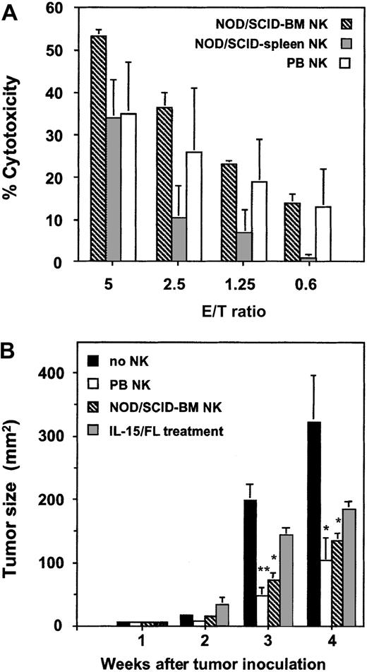
For the analysis of K562 lysis in vitro, NK cells generated in growth-factor–treated mice were not directly examined because of the limited number of cells that could be isolated by FACS sorting. Instead, we tested the cytotoxicity of human NK cells obtained in higher numbers in differentiation cultures of BM and spleen cells of growth-factor–untreated NOD/SCID mice (Figure 6A). These NK cells were highly cytotoxic; in particular, the killing potential of BM-derived cells was even higher than that of human PB NK cells. The well-pronounced cytotoxic properties of human NK cells of NOD/SCID origin generated in vitro are in accord with the high levels of activatory receptor NKp46 expressed by these cells (Figure 3C).33 To study the cytotoxic effect of human NK cells in vivo, NOD/SCID mice were inoculated subcutaneously with K562 cells and challenged intravenously 1 day later with NOD/SCID-BM–derived NK cells that showed strong cytotoxicity in vitro. Tumor growth, apparent 3 to 4 weeks after K562 injection, was significantly (60%-70%) suppressed by adoptively transferred NK cells (Figure 6B). This retardation of tumor growth was comparable to the effect of IL-2–stimulated NK cells obtained from human PB. We also inoculated K562 cells into NOD/SCID mice that had received transplanted CB CD34+ cells and had been injected with IL-15 and FL for 7 days. Under these conditions, which led to the generation of endogenous human NK cells, a marked reduction (up to 50%) of tumor growth was observed (Figure 6B). A possibly lower yield of NK cells generated in vivo may account for a lesser antitumor effect than a controlled number of cotransplanted NK cells. Based on these results, we conclude that human NK cells derived from NOD/SCID mice are functional with respect to cytotoxicity against HLA class 1–deficient targets in vitro and in vivo.
Cytotoxicity of NK cells from NOD/SCID mice. (A) NK cells were harvested after 28-day culture of NOD/SCID BM (n = 2, ▧) or spleen (n = 3, ▦) cells in the presence of IL-15, FL, and SCF. For comparison, NK cells were purified from human PB mononuclear cells (n = 2, □). Their cytotoxic activity was tested against the NK-sensitive K562 target cell line for 4 hours with an enzymatic LDH release assay. (B) NOD/SCID mice were injected subcutaneously with K562 cells (1 × 10⁷) alone (▪) or 1 day later by NK cells (2-5 × 10⁶) from human PB (□) or generated in vitro from NOD/SCID BM (▧). NOD/SCID mice that had undergone transplantation with CB CD34+ cells were injected with human IL-15 and FL for 7 days and were inoculated with K562 cells 1 day later (▦). Tumor size is expressed as mean ± SEM areas of 2 to 4 animals per treatment group. Significant differences compared with the group without NK cells are marked (*P < .05; **P < .005).
Cytotoxicity of NK cells from NOD/SCID mice. (A) NK cells were harvested after 28-day culture of NOD/SCID BM (n = 2, ▧) or spleen (n = 3, ▦) cells in the presence of IL-15, FL, and SCF. For comparison, NK cells were purified from human PB mononuclear cells (n = 2, □). Their cytotoxic activity was tested against the NK-sensitive K562 target cell line for 4 hours with an enzymatic LDH release assay. (B) NOD/SCID mice were injected subcutaneously with K562 cells (1 × 10⁷) alone (▪) or 1 day later by NK cells (2-5 × 10⁶) from human PB (□) or generated in vitro from NOD/SCID BM (▧). NOD/SCID mice that had undergone transplantation with CB CD34+ cells were injected with human IL-15 and FL for 7 days and were inoculated with K562 cells 1 day later (▦). Tumor size is expressed as mean ± SEM areas of 2 to 4 animals per treatment group. Significant differences compared with the group without NK cells are marked (*P < .05; **P < .005).
Discussion
The NOD/SCID mouse transplantation model has been widely used to study the biology of human hematopoietic stem cells of diverse ontologic origin.34-36 These studies indicate that the BM microenvironment of NOD/SCID mice supports the survival and multilineage development of human hematopoietic progenitors but that it can be selective with regard to the maturation of individual blood cell lineages. Within the lymphoid compartment, the B-cell lineage develops most efficiently.20,37 On the other hand, human T and NK cells have never been reproducibly detected, and only the transplantation of human thymic tissue or the use of IL-2R–blocking antibodies allowed T lymphopoiesis in vivo.38,39 In this study, we identified CD34+CD7+ and CD34-CD7+ NK progenitor cells in BM and spleen of NOD/SCID mice transplanted with CB CD34+ cells, and we achieved NK cell differentiation after in vivo administration of human cytokines. The establishment of human NK cell development in NOD/SCID mice provides for the first time an in vivo system to study the mechanisms governing human NK cell differentiation and function.
Several studies have demonstrated that lineage development of the human graft in NOD/SCID mice can be modulated by the administration of human cytokines. The effects seen in vivo often did not reproduce those predicted from in vitro cultures, such as a decrease in B-cell lineage development in mice receiving FL and IL-728 or an inhibition of platelet production by a growth factor combination known to support megakaryopoiesis from cultured progenitors.40 In contrast, all the cytokine combinations used in our study—namely IL-15/FL, IL-15/FL/SCF, IL-15/FL/IL-12, and IL-15/FL/SCF/MGDF/IL-2—enabled in vivo maturation of NK cells, thus recapitulating the previously reported effects of these cytokines on the generation of NK cells from cultured CD34+ progenitors. Previous studies identified IL-15 as the crucial factor for NK cell development, along with FL and SCF as cytokines that increase the frequency of NK cell precursors through the up-regulation of expression of the IL-15R complex.3,5 With human NK cells differentiated in vitro from CD34+ progenitors, a functional redundancy of FL and SCF on IL-15 responsiveness has been reported.3 Accordingly, the combination of IL-15 and FL was found to be sufficient to induce NK cells in NOD/SCID mice. Similarly, the number and phenotype of the NK cells did not differ depending on the cytokines used to generate them, though subtle differences of additional parameters cannot be excluded. Cytokine administration was associated with a moderate reduction in cellularity and with the level of human cell engraftment in BM and spleens. This is in agreement with previous studies,28,40,41 though the opposite effect was also seen in mice receiving only limited doses of stem cells.42 A rapid and nearly total disappearance of murine and human cells in NOD/SCID BM was observed when IL-12 was combined with IL-15 and FL. This is perhaps associated with the activation of newly formed NK cells given that an IL-12–dependent release of IFN-γ by NK cells, resulting in the suppression of hematopoiesis in the BM, has been postulated in humans and mice.25,26
In growth-factor–untreated mice, we detected human CD34+CD38-, CD34+CD38+, CD34+CD7+, and CD34-CD7+ cell populations, all of which responded to IL-15, FL, and SCF in vitro and gave rise to CD56+ NK cells. Judging from the kinetics of CD56+ cell generation in vitro, these populations may represent early, intermediate, and late stages in NK cell development. These NK cell precursors were found as late as 5 months after transplantation, indicating that they can persist for a long time in the NOD/SCID microenvironment. Similarly, undifferentiated, common lymphomyeloid CD34+CD38-CD19- progenitors capable of generating human B, T, NK, and granulomonocytic cells under appropriate culture conditions in vitro were maintained over the long-term in NOD/SCID mice that underwent transplantation.43 Altogether, these results indicate that the microenvironment of chimeric NOD/SCID mice harbors human NK progenitors, but an insufficient concentration or lack of cross-reactivity of endogenous murine NK cell–promoting cytokines, in particular IL-15, prevents progression to mature CD56+ NK cells above the background levels observed in untreated control NOD/SCID mice.
Human PB NK cells can be divided into 2 subsets, according to the expression of cell surface markers and functional characteristics.44-46 Approximately 10% of all NK cells are CD56highCD16-/low, and they produce abundant cytokines after activation. The prominent CD56dimCD16high subset, representing at least 90% of NK cells, secretes less IFN-γ but is more naturally cytotoxic. This phenotypic distinction was not apparent in the NK cell population that developed in NOD/SCID mice after growth factor injections. CD16 was expressed at various surface densities with a large proportion of NK cells being CD16-/low. The fact that CD56dimCD16high cells were not present as a predominant NK cell population in growth-factor–treated NOD/SCID mice suggests that the development of this subset requires the presence of other, possibly unknown, cytokines or that cellular selection is important and cannot be mimicked in the NOD/SCID mouse microenvironment. Despite this difference in phenotypic characteristic, human NK cells of NOD/SCID origin produce IFN-γ, are strongly cytotoxic against HLA class 1-deficient K562 targets in vitro, and are capable of reducing K562 erythroleukemia tumor formation in vivo, thus functionally resembling human PB NK cells.
Although inhibitory and activatory receptors have defined functions in mature PB NK cells, their roles in developing immature NK cells is unclear. In growth-factor–treated NOD/SCID mice, we consistently found fewer human NK cells expressing the major triggering receptor NKp46 than CD16+ NK cells. This is different from NK cells generated in vitro from CD34+ cells, in which NKp46 is expressed by most CD56+ cells and CD16 by only a small proportion. Likewise, 2 other groups also observed NKp46 preceding CD16 expression during NK cell differentiation in vitro.47,48 The NOD/SCID-derived NK cells acquired the NKp46bright phenotype only after isolation of the progenitors and after further culture. The mechanism that controls the level of NKp46 expression during NK cell development is unknown. Noteworthy, however, is that the surface density of NKp46 on mature NK cells from human PB is heterogeneous, with different donors harboring different proportions of NKp46bright and NKp46dim cells that display high and low cytotoxic activity, respectively.33 Contrary to the NKp46 activatory receptor, the inhibitory receptors CD158a, CD158b, and NKB1 were not detected on NOD/SCID-derived human NK cells after culture. This may result in the absence of inhibitory signals and may enhance the killing capacity of NK cells against neoplastic targets, as shown by the important graft-versus-leukemia effect in haploidentical transplantation.17
NK cells, through the production of immunoregulatory cytokines and cytotoxic effects, are candidates for immunotherapy of transformed or infected tissues. It remains to be seen whether adoptively transferred NK cells of NOD/SCID origin that recognized and killed HLA-deficient targets can also suppress the growth of primary human tumors. Notably, cytokines that boost the maturation of NK cells in chimeric NOD/SCID mice have been shown to generate potent antitumor responses. Treatment of tumor-bearing mice with FL leads to tumor regression through the activation of dendritic and NK cells.49 IL-15 activates T and NK cells, and preclinical studies show enhanced protection against viral challenge and a potential for IL-15 as a tumor vaccine adjuvant.50,51 Interestingly, we recently found that IL-15 up-regulates FL expression and that after stem cell transplantation, FL levels are strongly increased,52,53 suggesting that these 2 growth factors contribute to fast recovery of NK cells in patients who have undergone transplantation.17 Ongoing discoveries of NK-specific receptors and increasing knowledge about their respective ligands54 provide the means to modulate the responses of mature NK cells, using either specific antibodies55 or genetic modification. NOD/SCID mice with transplanted hematopoietic progenitors represent an important model for evaluating the immunotherapeutic efficacy of human NK cells and for investigation of the factors and signals orchestrating formation of the NK cell compartment in humans.
Prepublished online as Blood First Edition Paper, March 13, 2003; DOI 10.1182/blood-2002-07-2024.
Supported by grants from the Swiss National Science Foundation (31-67072.01 and 4046-058689), Stiftung zur Krebsbekämpfung and the Foundation Cord Stem Cell Therapy, Switzerland.
The publication costs of this article were defrayed in part by page charge payment. Therefore, and solely to indicate this fact, this article is hereby marked “advertisement” in accordance with 18 U.S.C. section 1734.
We thank Elena Chklovskaia for help with the NK cytotoxicity assay and Verena Jäggin for cell sorting; S. D. Lyman for recombinant human cytokines IL-15 and FL; U. Gubler for recombinant human IL-12; W. Holzgreve, D. Surbek, and S. Heinzl for providing cord blood samples; M. Colonna, A. Bouchon, and D. Baker for antibodies; and G. De Libero, A. Gratwohl, A. Rolink, and S. Bridenbaugh for critically reading the manuscript.

![Figure 1. Phenotype of human NK cells in BM of growth-factor–treated NOD/SCID mice. Two months after transplantation of CB CD34+ cells, NOD/SCID mice were treated with combinations of growth factors (A) for 7 consecutive days, and BM, spleen, and PB cells were analyzed 1 day later by flow cytometry. (A) Human NK cells in untreated control (no growth factor [GF]) and growth-factor–treated mice were detected by staining with the human leukocyte marker CD45 and the NK-cell–specific marker CD56. Analysis was performed with total mononuclear cells; numbers indicate the percentages of total BM cells. (B) Human NK cells were characterized by staining with antibodies against CD56 and the indicated NK surface marker. Numbers indicate the percentages of human CD45+ cells. For comparison, human NK cells from (C) CB and (D) PB were stained with antibodies against CD56 and CD16 (gated on lymphocytes) and NKp46 (gated on CD56+ cells). The thin line indicates staining with isotype-matched control antibody. (E) NK cells in PB of NOD/SCID mice treated with IL-15 and FL. Numbers indicate the percentages of human CD45+ cells.](https://ash.silverchair-cdn.com/ash/content_public/journal/blood/102/1/10.1182_blood-2002-07-2024/5/m_h81334557001.jpeg?Expires=1765939177&Signature=KirPSvt1f6Z0s4Mgqs5fIjDrgYwtor3zHhRpvnx046QdFqmWtJylfgUVlCB9M-zIZf1th6HX7n8HkoualCSwQ27YLy0aTh3LNe3lgIJULLZGo6SmNyWdnq6GoxMeQCR3RNd~bTMK0FnENIbNzjsmBkfzaZ-4uBduc~aM34HScTVj78ogafdMKrrkBsHeZKfCZotpY58cBfjV-ivzS7oNNECt4Ek0wvoo0IaqLwo1BdmrLN3ZolzT9FFfqtsnbgP19OALE39QKANqF4bshFqgub2YdF4Z5~AhzX4~RUuuM~7aPGrHahaoEsrzNWTOSpJfc6GE2cWJjp0-FcB6TnhRLQ__&Key-Pair-Id=APKAIE5G5CRDK6RD3PGA)
This feature is available to Subscribers Only
Sign In or Create an Account Close Modal