Abstract
Paroxysmal nocturnal hemoglobinuria (PNH) is an acquired hemolytic anemia in which venous thrombosis is the most common cause of death. Here we address the risk factors for thrombosis and the role of warfarin prophylaxis in PNH. The median follow-up of 163 PNH patients was 6 years (range, 0.2-38 years). Of the patients, 29 suffered thromboses, with a 10-year incidence of 23%. There were 9 patients who presented with thrombosis, and in the remainder the median time to thrombosis was 4.75 years (range, 3 months-15 years). The 10-year risk of thrombosis in patients with large PNH clones (PNH granulocytes > 50%) was 44% compared with 5.8% with small clones (P < .01). Patients with large PNH clones and no contraindication to anticoagulation were offered warfarin. There were no thromboses in the 39 patients who received primary prophylaxis. In comparison, 56 patients with large clones and not taking warfarin had a 10-year thrombosis rate of 36.5% (P = .01). There were 2 serious hemorrhages in more than 100 patient-years of warfarin therapy. Large PNH granulocyte clones are predictive of venous thrombosis, although the exact cut-off for clone size is still to be determined. Primary prophylaxis with warfarin in PNH prevents thrombosis with acceptable risks. (Blood. 2003;102:3587-3591)
Introduction
Paroxysmal nocturnal hemoglobinuria (PNH) is an acquired hemolytic anemia characterized by intravascular hemolysis, cytopenias, and a high incidence of life-threatening venous thrombosis (up to 40% in previous series1,2 ).
Somatic mutation of the phosphatidylinositol glycan complementation class A (PIG-A) gene in a multipotent hematopoietic stem cell is present in all cases so far described.3-5 This results in a defect of biosynthesis of the glycosylphosphatidylinositol (GPI) anchor,6,7 which links numerous antigens to the cell surface. PNH cells are therefore deficient in all GPI-linked proteins, such as CD55 (decay accelerating factor, DAF) and CD59 (membrane inhibitor of reactive lysis, MIRL).8,9 Flow cytometry to ascertain the absence of these surface antigens is the most sensitive method to diagnose the disorder. CD59 is critical because its role is to protect cells from complement-mediated attack. It is the deficiency of CD59 and CD55 from PNH red cells that renders them susceptible to complement-mediated lysis and subsequent intravascular hemolysis. PNH platelets, also deficient in GPI-linked proteins, are more easily activated by complement, and this is thought to be a factor in the predisposition to thrombosis. ADP released by intravascular lysis of red cells may play a part in platelet activation and the reduced expression of urokinase plasminogen activator receptor (u-PAR) on PNH granulocytes is possibly relevant.10-12 Venous thrombosis is the most common cause of death in PNH resulting in the death of up to a third of patients1 and, as there is a particular propensity for involvement of hepatic and cerebral veins, the associated morbidity is also high. The aim of this study was to identify factors predictive of thrombosis and to investigate the role of primary prophylaxis with warfarin in those patients found to be at particular risk in order to prevent thrombosis.
Materials and methods
Clinical data were obtained from 163 of 179 consecutive patients with PNH clones investigated in our laboratory. In view of previous reports of the extremely high incidence of thrombosis in PNH,1,2 it has been our practice over the past 6 years to recommend primary warfarin prophylaxis for patients considered at high risk of developing thrombosis. Warfarin prophylaxis was recommended to patients with granulocyte PNH clone sizes larger than 50% (a cut-off that was somewhat arbitrary but seems to identify those patients at risk), without significant thrombocytopenia (platelet count > 100 × 109/L) and with no other contraindications to anticoagulation. Uptake was dependent on patient and physician preference and was influenced by whether our advice was sought on the management of individual patients. Thrombophilia testing was not routinely requested.
Where feasible, information was updated from clinical notes. Alternatively, individual physicians were contacted by telephone. Data were gathered on clinical and drug histories, episodes of thrombosis, anticoagulation, causes of death, and full blood count parameters.
Red cells
Fluorescein isothiocyanate (Fitc)-conjugated anti-CD59 and phycoerythrin(PE)-conjugated anti-CD55 (Cymbus Biosciences, Chandler's Ford, Hampshire, United Kingdom) were used. Identification of type I, II, and III cells was derived from CD59 expression only as this provides superior resolution to CD55.
Granulocytes
Leukocytes were stained by a whole blood lysis method using a 3-color combination of monoclonal antibodies directly conjugated to the GPI-linked antigens; Fitc-conjugated CD66 (Dako, Ely, United Kingdom), PE-conjugated CD55 (Cymbus Biosciences), and CD16-PE/CyChrome 5 (PE/Cy5; HMDS). The PNH clone was identified by deficiency of all 3 GPI-linked antigens.
Patient groups were compared using the log-rank test and survival was estimated according to the Kaplan-Meier method. Analysis was performed with the use of SPSS software, version 10.1 for Windows (Claritas, Derry, North Ireland).
Results
Follow-up studies and causes of death
Clinical and flow cytometry data were available in 163 patients. The median follow-up period, from time of first symptoms of hemolysis or first presentation with cytopenias, was 6 years (range, 0.2-38 years). The median age at diagnosis was 33 years (range, 6 months-85 years).
Granulocyte flow cytometry data were available in 158 patients, with 97 patients having 50% or more PNH granulocytes and 61 with less than 50% PNH granulocytes. Of those with large clones, 32 received primary prophylaxis with warfarin. A further 7 patients had primary prophylaxis, in which the granulocyte PNH clone size was either unknown or smaller than 50%.
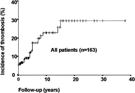
There have been 20 deaths (12.5%), of which 14 were attributable to PNH or aplasia. Of the deaths, 4 were secondary to thrombosis, all involving the liver (Table 1). There were 29 patients who had one or more episodes of venous thrombosis (Table 2), giving an overall 10-year cumulative incidence rate of thrombosis of 23% (Figure 1). The median age at time of thrombosis was 32 years (range, 1-76 years). The median platelet count was 125 × 109/L (range, 12-418 × 109/L). At the time of presentation with PNH, 9 patients had thrombosis, 2 of these dying as a result (both hepatic vein thromboses). During the course of the disease, 20 individuals developed thrombosis, with a median time from presentation of 4.75 years (range, 3 months-15 years). Only 2 patients suffered their first thrombosis more than 10 years from presentation of PNH.
Causes of death in 20 of 163 patients with PNH
Causes of death . | Number . |
|---|---|
| Attributable to PNH | 8 |
| Thrombosis (hepatic/portal) | 3 |
| Hepatic failure (probably due to thrombosis) | 1 |
| Intravascular hemolysis causing renal failure | 1 |
| Iron overload | 1 |
| Cerebral hemorrhage secondary to warfarin | 1 |
| Stem cell transplantation-related | 1 |
| Related to aplasia | 6 |
| Acute myeloid leukemia | 2 |
| Hypoplasia | 1 |
| Fungal infection | 1 |
| Stem cell transplantation-related | 1 |
| Cerebral hemorrhage secondary to thrombocytopenia | 1 |
| Probably unrelated to PNH | 5 |
| Suicide | 1 |
| Myocardial infarction | 1 |
| Malignant melanoma | 1 |
| Renal failure | 1 |
| Cerebrovascular accident | 1 |
| Unknown | 1 |
| Total | 20 |
Sites of thrombosis in 29 out of 163 cases of PNH
UPN . | Site of thrombosis . | Years after diagnosis . | Granulocyte PNH clone size, % . | Red cell PNH clone size, %* . |
|---|---|---|---|---|
| Liver-related | ||||
| 5 | Portal vein (probable cause of death) | 5 | Not known | 64 |
| 6 | Hepatic vein | 4 | 99.4 | 33.4 |
| 8 | Hepatic vein | 0 | 97.96 | 30.88 |
| 10 | Portal vein + sagittal sinus (pregnant) | 15 | Not known | 52 |
| 17 | Hepatic + portal veins | 0 | Not known | 100 |
| 27 | Portal vein | 8 | 99.6 | 89.59 |
| 33 | Hepatic vein, shunt thrombosis | 14 | 97.8 | 64 |
| 40 | Portal vein | 5 | 97 | 57 |
| 53 | `Liver' (cause of death) | 4 | 78 | 23 |
| 80 | Portal vein (cause of death) | 0 | 98 | 15.1 |
| 95 | Hepatic vein—2 events | 0 | 80 | 22.99 |
| 98 | Hepatic vein | Not known | 68.1 | 67.66 |
| 104 | Hepatic vein | Not known† | Not known† | Not known† |
| 107 | Hepatic vein (cause of death) | 0 | 99.5 | 49.81 |
| 109 | Portal vein | 0 | 99.9 | 5.73 |
| 149 | Hepatic vein | 0 | 98.3 | 83.4 |
| 153 | Portal vein, pulmonary embolus | 0.25 | 96.3 | 67.8 |
| 165 | Hepatic vein—2 events | 2.25 | 98.9 | 21.1 |
| Other | ||||
| 3 | Deep venous thrombosis—2 events | 7 | 9.7 | 4.4 |
| 22 | Pulmonary embolus (Factor V Leiden heterozygote) | 2 | 90.82 | 20.9 |
| 26 | Superficial thrombophlebitis | 1 | 55 | 24 |
| 34 | Deep venous thrombosis | 5 | 73 | 45 |
| 48 | Cerebral vein thrombosis | 8.75 | 92.9 | 77.4 |
| 61 | Line thrombosis | 1 | 67.62 | 21 |
| 70 | Retinal vein | 4 | 76.04 | 11.66 |
| 119 | Thrombosis right cerebellum (Factor V Leiden heterozygote) | 0 | 15.3 | 7.21 |
| 144 | Deep venous thrombosis | 0 | 96.6 | 43.2 |
| 146 | Pulmonary embolus | 0.8 | 84 | 57.4 |
| 177 | Pulmonary embolus | 7 | 97.1 | 81.5 |
Includes both type II (partial deficiency) and type III (complete deficiency) cells.
Diagnosed in the 1960s.
Cumulative incidence of venous thrombosis in 163 patients with PNH clones. The 10-year cumulative incidence of thrombosis in the whole group of 163 patients was 23%. This includes patients on primary thromboprophylaxis with warfarin. The curve starts at 5.4%, reflecting the incidence of patients presenting with thrombotic events. One episode of hepatic vein thrombosis was excluded from the analysis because when the diagnosis was established by abdominal ultrasound scan it was clearly extremely longstanding (probably years previously) and an accurate estimate of the timing of thrombosis could not be made.
Cumulative incidence of venous thrombosis in 163 patients with PNH clones. The 10-year cumulative incidence of thrombosis in the whole group of 163 patients was 23%. This includes patients on primary thromboprophylaxis with warfarin. The curve starts at 5.4%, reflecting the incidence of patients presenting with thrombotic events. One episode of hepatic vein thrombosis was excluded from the analysis because when the diagnosis was established by abdominal ultrasound scan it was clearly extremely longstanding (probably years previously) and an accurate estimate of the timing of thrombosis could not be made.
In PNH there is a continuum of clinical manifestations, from overtly hemolytic (associated with a large PNH clone size) with episodes of macroscopic hemoglobinuria and a thrombotic tendency, to hypoplastic (often related to aplastic anemia) with a small PNH clone and no obvious hemoglobinuria. We therefore studied thrombosis rate according to granulocyte PNH clone size, taking 50% as an arbitrary division between large and small.15
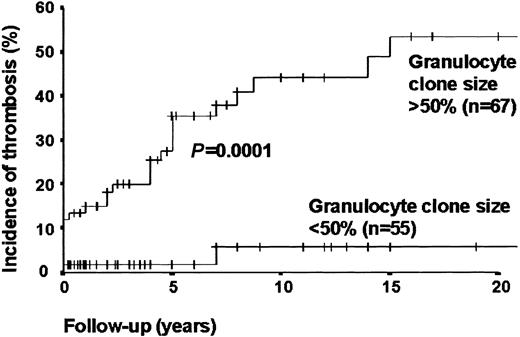
Patients with PNH granulocytes greater than 50% (including those on primary warfarin prophylaxis) were found to have a 10-year cumulative incidence rate of thrombosis of 34.5% compared with those with clone sizes smaller than 50% who had a thrombosis rate of 5.3% (P < .01). When patients on primary thromboprophylaxis were excluded, the 10-year cumulative incidence rate of thrombosis in those with large clones was 44%, compared with a thrombosis rate of 5.8% in those with clone sizes smaller than 50% (P < .01) (Figure 2).
Effect of GPI-deficient granulocyte clone size on incidence of venous thrombosis (primary prophylaxis patients excluded). When primary prophylaxis patients are excluded, the 10-year cumulative incidence rate of thrombosis in patients with PNH granulocyte clone size larger than 50% is 44%, compared with a thrombosis rate of 5.8% in those with clone size smaller than 50% (P < .01). P value calculated with use of the log-rank test.
Effect of GPI-deficient granulocyte clone size on incidence of venous thrombosis (primary prophylaxis patients excluded). When primary prophylaxis patients are excluded, the 10-year cumulative incidence rate of thrombosis in patients with PNH granulocyte clone size larger than 50% is 44%, compared with a thrombosis rate of 5.8% in those with clone size smaller than 50% (P < .01). P value calculated with use of the log-rank test.
Of the 26 patients with large clone sizes who suffered thromboses, 17 had hepatic thromboses, with 3 fatalities.
There were 2 patients with less than 50% PNH granulocytes who had thromboses. Neither was clinically hemolytic. The first presented with a cerebellar cerebrovascular accident and was found to have a small PNH clone (19.45% granulocytes) associated with heterozygosity for the Factor V Leiden mutation and a normal platelet count. The second patient had a deep venous thrombosis 7 years after presenting with pancytopenia. The platelet count at the time of thrombosis was 120 × 109/L and the granulocyte PNH clone size 9.7%.
Warfarin prophylaxis in patients at high risk of thrombosis
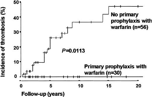
There were 32 patients with large PNH clones and no contraindication to anticoagulation who had primary thromboprophylaxis with warfarin. Target international normalized ratio (INR) was 2-3. Also taking primary prophylaxis were 7 other patients with smaller or unknown clone size (in most cases reflecting spontaneous reductions in granulocyte clone size over time), giving a total number of 39. The median age was 44 years (range, 3-76 years). The median time on warfarin was 23 months (range, < 1 month-148 months). The median platelet count was 182 × 109/L (range, 80-633 × 109/L). In this group there were no thrombotic events (Figure 3). In comparison, 56 patients did not present with thrombosis, had clone size larger than 50%, and had no warfarin primary prophylaxis. The median age at diagnosis was 32 years (range, 5-85 years). The median platelet count was 122 × 109/L (range, 19-338 × 109/L). This group had a 10-year cumulative incidence of venous thrombosis of 36.5% (P = .01).
Effect of warfarin prophylaxis on venous thrombosis in patients with PNH granulocyte clone sizes larger than 50% (patients presenting with thrombosis excluded). The 10-year cumulative incidence rate of venous thrombosis in patients with PNH granulocyte clones larger than 50%, not presenting with thrombosis, and not taking warfarin is 36.5%. In comparison, the current thrombosis rate is 0% in patients taking primary prophylaxis (P = .01). Of the 39 patients on primary prophylaxis, 32 had granulocyte clone sizes larger than 50% and could therefore be included in this analysis. A further 2 of these patients were excluded because, having stopped warfarin (1 through personal choice and 1 because of warfarin-associated hemorrhage), they went on to suffer venous thrombosis. Time 0 was the time of presentation with PNH. P value calculated with use of the log-rank test.
Effect of warfarin prophylaxis on venous thrombosis in patients with PNH granulocyte clone sizes larger than 50% (patients presenting with thrombosis excluded). The 10-year cumulative incidence rate of venous thrombosis in patients with PNH granulocyte clones larger than 50%, not presenting with thrombosis, and not taking warfarin is 36.5%. In comparison, the current thrombosis rate is 0% in patients taking primary prophylaxis (P = .01). Of the 39 patients on primary prophylaxis, 32 had granulocyte clone sizes larger than 50% and could therefore be included in this analysis. A further 2 of these patients were excluded because, having stopped warfarin (1 through personal choice and 1 because of warfarin-associated hemorrhage), they went on to suffer venous thrombosis. Time 0 was the time of presentation with PNH. P value calculated with use of the log-rank test.
The median time from presentation with PNH to starting warfarin prophylaxis was 1 year (range, 0-19 years). In order to compensate for this bias, we have also analyzed the risk of thrombosis for all patients with large PNH granulocyte clones when not on warfarin (ie, prior to commencing primary prophylaxis, prior to thrombosis and subsequent anticoagulation, and patients who never received warfarin) and compared this with patient-years taking primary prophylaxis. There were 19 thromboses in 511.5 patient-years off warfarin (3.7 thromboses per 100 patient-years not on warfarin) compared with no thromboses in 117.8 patient-years on primary warfarin prophylaxis (P < .05).
Complications of warfarin
There were 2 episodes of hemorrhage in the 39 patients taking primary thromboprophylaxis. In the first, a 51-year-old woman suffered a spontaneous, and subsequently fatal, cerebral hemorrhage after 9 months on warfarin. Her INR had been erratic (INR, 1.2-8.1), and her platelet count varied between 50 and 144 × 109/L. In the second case, a 44-year-old woman had a spontaneous chronic subdural hematoma after 8 months on warfarin. The INR was controlled between 1.9 and 3.1 and the platelet count remained normal. After surgery, she made a good recovery but 2 months later developed multiple pulmonary emboli and had to be reanticoagulated. She is currently well.
Discussion
In this unselected group of 163 patients followed over a median of 6 years, the rate of venous thrombosis was comparable with previous studies.1,2 The overall cumulative incidence (at 20 years) of thrombosis in patients with granulocyte clone sizes larger than 50%, excluding those taking warfarin as primary thromboprophylaxis, was 53.5%.
Granulocyte clone sizes larger than 50% were found to be highly predictive of thrombotic risk, with an incidence of thrombosis of only 5.8% in patients with small PNH granulocyte clones. A cut-off was taken at 50% as this provided the best delineation between aplastic and hemolytic PNH in this group of patients. Lead-time bias may affect interpretation of data comparing these 2 groups as the capacity to detect small PNH clones is a relatively new development. Although it is the PNH platelet clone that is likely to be involved in the pathogenesis of thrombosis, techniques to evaluate CD55 and CD59 expression on platelets are insensitive. However, previous studies have shown the percentage of PNH platelets to be highly correlated with the percentage of PNH granulocytes.16 Other thrombotic risk factors might be important too, such as periods of immobility and inherited thrombophilias, but this was not found to be the case in a previous report.17 Platelet counts in thrombotic patients varied widely, influenced by multiple factors including hypoplasia and liver failure.
PNH is an uncommon disorder that often results in life-threatening complications. In view of the infrequency of the disease and the high risk of thrombosis, conducting a randomized trial of warfarin prophylaxis is impractical and raises ethical concerns. It is therefore of considerable importance that in this series the 10-year incidence of venous thrombosis fell from 36.5% (patients with thrombosis at presentation excluded) to 0% when warfarin was given as primary prophylaxis. However, there are significant limitations to such a retrospective analysis. In particular, the unequal delineation between anticoagulated and nonanticoagulated groups might influence results. The reasons for rejecting anticoagulation usually reflected patient and primary physician preference, but there might be other underlying contraindications that intrinsically carry a worse prognosis and could be an unwitting source of bias. However, the finding that the patients who chose to be anticoagulated were older (median age 44 years compared with 32 years for those electing not to be anticoagulated) and had a higher platelet count (median 182 × 109/L compared with 122 × 109/L) suggests that the thrombotic risk in the anticoagulated patients might have been greater than those who were not anticoagulated. In addition, the follow-up for anticoagulated patients is relatively short in comparison with the nonanticoagulated group.
It is a sobering fact that in the group of 39 patients on primary thromboprophylaxis there were 2 warfarin-associated hemorrhages, 1 of which led to the patient's death. Bleeding rates for non-PNH patients on warfarin are reported as follows: fatal, 0.1% to 1% per year; major, 0.5% to 6.5% per year; and minor, 6.2% to 28% per year.18-21 In PNH, thrombocytopenia is particularly common, and it is of note that the fatal hemorrhage occurred in the only patient on warfarin in whom the platelet count had been erratic. In addition, both of the patients who experienced a hemorrhage were in their first year of warfarin, which is known to be the period when hemorrhagic complications are most common. Palareti et al20 found that in a prospective cohort of 2745 consecutive patients on oral anticoagulants the risk of hemorrhagic events was higher during the first 90 days of treatment (relative risk, 1.75; 95% CI, 1.27-2.44; P < .001). Thus the incidence of warfarin-induced hemorrhage appears to be no higher than other patients on warfarin.
Based on these findings, we present strong evidence that primary prophylaxis with warfarin should be considered in PNH patients if the granulocyte clone size is larger than 50%, the platelet count is stable at higher than 100 × 109/L, and there is no known contraindication to anticoagulation. The optimal therapeutic range for the INR is still to be assessed but the target in this series of patients was to have an INR between 2.0 and 3.0, which appears to prevent the occurrence of thrombosis. The observation that only 2 patients had their first thrombosis in excess of 10 years after diagnosis should be taken into consideration when deciding on anticoagulation in this group of patients (Table 3). In each case, the risk-benefit ratio must be examined carefully, and any decision to anticoagulate must be made with the patient's informed consent.
Summary of characteristics of PNH patients who should be considered for primary warfarin prophylaxis
1 | Granulocyte clone size larger than 50% |
| 2 | Platelet count stable at higher than 100 × 109/L |
| 3 | No known contraindication to anticoagulation |
| 4 | The possibility that thrombotic risk may change with time should be taken into consideration |
1 | Granulocyte clone size larger than 50% |
| 2 | Platelet count stable at higher than 100 × 109/L |
| 3 | No known contraindication to anticoagulation |
| 4 | The possibility that thrombotic risk may change with time should be taken into consideration |
Our findings clearly demonstrate that the selective use of primary warfarin prophylaxis at diagnosis can reduce morbidity and mortality in PNH.
Prepublished online as Blood First Edition Paper, July 31, 2003; DOI 10.1182/blood-2003-01-0009.
The publication costs of this article were defrayed in part by page charge payment. Therefore, and solely to indicate this fact, this article is hereby marked “advertisement” in accordance with 18 U.S.C. section 1734.
We would like to thank all hematologists who registered patients and provided samples, HMDS staff, and Professor Lucio Luzzatto for his support and encouragement.


This feature is available to Subscribers Only
Sign In or Create an Account Close Modal