Abstract
The activator protein 1 (AP-1) member JunB has recently been implicated in leukemogenesis. Here we surveyed human lymphoma samples for expression of JunB and other AP-1 members (c-Jun, c-Fos, Fra1, JunD). JunB was strongly expressed in T-cell lymphomas, but non-Hodgkin B-cell lymphomas do not or only weakly express JunB. We therefore asked whether JunB acted as a negative regulator of B-cell development, proliferation, and transformation. We used transgenic mice that expressed JunB under the control of the ubiquitin C promoter; these displayed increased JunB levels in both B- and T-lymphoid cells. JunB transgenic cells of B-lymphoid, but not of T-lymphoid, origin responded poorly to mitogenic stimuli. Furthermore, JunB transgenic cells were found to be less susceptible to the transforming potential of the Abelson oncogene in vitro. In addition, overexpression of JunB partially protected transgenic mice against the oncogenic challenge in vivo. However, transformed B cells eventually escaped from the inhibitory effect of JunB: the proliferative response was similar in explanted tumor-derived cells from transgenic animals and those from wild-type controls. Our results identify JunB as a novel regulator of B-cell proliferation and transformation. (Blood. 2003;102:4159-4165)
Introduction
Non-Hodgkin lymphomas (NHLs) comprise a heterogeneous group of malignancies that are classified based on immunophenotyping, morphologic, and clinical characteristics.1 During the last decades NHLs have shown a steadily increasing incidence. Currently, approximately 4% of cancers in industrial states are NHLs, with 55 000 new patients every year solely in the United States.2 During the last several years, genetic and epigenetic alterations were defined that underlie lymphoma development. These allowed for a better understanding of lymphoid tumor formation. The members of the activator protein 1 (AP-1) transcription factor family figure prominently among the molecules that have been implicated in lymphoid transformation.3-6
The transcription factor AP-1 converts extracellular signals into transcriptional changes of specific target genes and functions in cell proliferation, differentiation, apoptosis, and cellular transformation. AP-1 consists of dimers between members of the Fos (c-Fos, FosB, Fra-1, Fra-2), Jun (c-Jun, JunB, JunD), and activating transcription factor (ATF) families of proteins. Extensive analyses of mice and of cell lines have indicated that each family member has distinct biologic functions. These are elicited via the cell-type-specific regulation of particular subsets of target genes (reviewed in Jochum et al7 and Shaulian and Karin8,9 ). Components of the AP-1 transcription factor are involved in tumorigenesis by regulating oncogenic transformation, apoptosis, and angiogenesis. In particular, c-Jun has been shown to cooperate with oncogenic ras in cellular transformation10,11 and to block the proapoptotic activity of p53 during the development of liver tumors in mice.12 Moreover, alterations of AP-1 family members have been linked to malignant transformation in several types of human cancer.13-15
The c-Jun and JunB proteins are similar in their primary structure and their DNA binding specificity but differ in their transcriptional capacity. In contrast to c-Jun, a transcriptional activator, JunB acts both as a transcriptional activator and repressor, depending on the promoter context and on the heterodimerization partner.16,17 In certain tissues, JunB antagonizes the function of c-Jun18,19 and has been shown to negatively regulate tumor formation.20 The molecular function of JunB has been studied both in vivo and in vitro. JunB-deficient embryos die during embryonic development from vascular defects.21 In contrast, constitutive overexpression of JunB from the human ubiquitin C (Ubi-junB) promoter has no major consequences in vivo in transgenic mice.22 In transgenic fibroblasts, JunB directly activates the cyclin-dependent kinase inhibitor p16 and inhibits cyclin D1 activity, resulting in reduced retinoblastoma protein (pRB) phosphorylation and delayed progression from G1 to S phase.18,20 The negative effect of JunB on cell proliferation has also been observed in malignant mouse keratinocytes stably overexpressing JunB.23
Most recently, JunB was identified as an important factor in hematopoietic transformation.24,25 In fact, mice lacking JunB in the myeloid lineage develop a myeloproliferative disease resembling human chronic myeloid leukemia (CML).24 The increased levels of granulocyte-macrophage colony-stimulating factor α (GM-CSFα) receptor, Bcl2, and Bclxl and a decreased expression of p16 within JunB-deficient myeloid progenitors most likely account for the expansion of the myeloid compartment.24 These findings suggest a role for JunB as a tumor suppressor. In line with this concept, human CML cells display reduced JunB levels due to increased DNA methylation of its promoter, with a further decrease of JunB levels occurring in blast crisis.25 However, JunB can promote tumor growth by cooperating with c-Jun in the development of murine fibrosarcoma.26 Finally, JunB is overexpressed in human Hodgkin lymphoma but it is apparently transcriptionally inactive.27 Thus, given the dual function of JunB and its cell-type-specific action, it is difficult to predict the role of JunB in transformation of lymphoid cells. In this study we have investigated the expression pattern of JunB in non-Hodgkin lymphomas and addressed the functional role of JunB within the B-lymphoid compartment.
Materials and methods
Patient samples and protein analysis
Samples of lymph nodes of patients with lymphoid malignancies were obtained from the Institute of Pathology, University Hospital Graz (Graz, Austria). The lymphomas were classified according to the Revised European-American Classification of Lymphoid Neoplasms (REAL) classification.1
Cells were lysed in a buffer containing protease and phosphatase inhibitors (50 mM HEPES [N-2-hydroxyethylpiperazine-N′-2-ethanesulfonic acid], pH 7.5; 0.1% Tween-20; 150 mM NaCl; 1 mM EDTA [ethylenediaminetetraacetic acid], 20 mM β-glycero-phosphate; 0.1 mM sodium vanadate; 1 mM sodium fluoride; 10 μg/mL each aprotinin and leupeptin; and 1 mM PMSF [phenylmethylsulfonyl fluoride]). Protein concentrations were determined using a bicinchoninic acid (BCA) kit as recommended by the manufacturer (Pierce, Rockford, IL).28
One hundred micrograms total protein/sample was electrophoretically resolved on polyacrylamide gels containing sodium dodecyl sulfate (SDS) and transferred onto Immobilon (Millipore, MA) membranes. Membranes were probed with the antibodies indicated in the figure legend (Figures 1, 2, 4, 6, 7). The antisera directed against abl, p16, p21, and p27 were obtained from Santa Cruz Biotechnology (Santa Cruz, CA), the antibody against JunB was a generous gift from M. Yaniv (Pasteur Institute, Paris, France), and the c-Jun and Bclx antibodies were obtained from Transduction Laboratories (Lexington, KY). Sites of antibody binding were detected using protein A-conjugated horseradish peroxidase (EY Laboratories, San Mateo, CA) with chemiluminescent detection (enhanced chemiluminescence [ECL] detection kit; Amersham, Arlington Heights, IL).
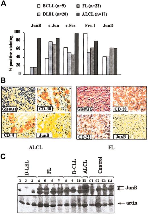
Expression of the AP-1 family members JunB, c-Jun, c-Fos, Fra-1, and JunD in human lymphomas. (A) Tissue array analysis from 77 human lymphomas samples including B-cell chronic lymphoid leukemia (B-CLL; n = 9), diffuse large-cell lymphomas (D-LBLs; n = 28), follicular lymphomas (FLs; n = 23), and anaplastic large-cell lymphomas (ALCLs, n = 17). Histogram bars indicate the percentage of each lymphoma entity with high expression of JunB, c-Jun, c-Fos, Fra-1, and JunD. (B) Immunohistologic staining of an ALCL (left) and of an FL (right). Transformed T cells in ALCL show a strong staining for CD4, CD30, and JunB. The tumor cells in FL stain positive for CD20 and CD23 but not for JunB. Original magnification, × 600. Stains were performed using DAB (3,3 Diaminobenzidin) for JunB and APAAP for all other antibodies. (C) Western blot analysis of lymph nodes from patients suffering from different B-cell malignancies for JunB expression. Lanes 1-3 indicate diffuse large-cell lymphomas (D-LBLs); lanes 4-7, follicular lymphomas (FLs); lanes 8-10, B-cell chronic lymphoid leukemia (B-CLL); lane 11, ALCL; and lanes C1-C4, control lymph nodes.
Expression of the AP-1 family members JunB, c-Jun, c-Fos, Fra-1, and JunD in human lymphomas. (A) Tissue array analysis from 77 human lymphomas samples including B-cell chronic lymphoid leukemia (B-CLL; n = 9), diffuse large-cell lymphomas (D-LBLs; n = 28), follicular lymphomas (FLs; n = 23), and anaplastic large-cell lymphomas (ALCLs, n = 17). Histogram bars indicate the percentage of each lymphoma entity with high expression of JunB, c-Jun, c-Fos, Fra-1, and JunD. (B) Immunohistologic staining of an ALCL (left) and of an FL (right). Transformed T cells in ALCL show a strong staining for CD4, CD30, and JunB. The tumor cells in FL stain positive for CD20 and CD23 but not for JunB. Original magnification, × 600. Stains were performed using DAB (3,3 Diaminobenzidin) for JunB and APAAP for all other antibodies. (C) Western blot analysis of lymph nodes from patients suffering from different B-cell malignancies for JunB expression. Lanes 1-3 indicate diffuse large-cell lymphomas (D-LBLs); lanes 4-7, follicular lymphomas (FLs); lanes 8-10, B-cell chronic lymphoid leukemia (B-CLL); lane 11, ALCL; and lanes C1-C4, control lymph nodes.
JunB overexpression inhibits proliferation of mature peripheral B lymphocytes. (A) Spleens from Ubi-junB transgenic animals (JunB tg) and their littermate wild-type controls (wt) were used for MACS purification of T and B cells. Whole-spleen extracts (S), CD3-purified T lymphocytes (T), and CD19-purified B lymphocytes were subsequently subjected to Western blotting using an antibody directed against JunB. Extracts of wild-type and Ubi-junB transgenic fibroblasts were used as controls. (B) CD3+ T lymphocytes were MACS purified from spleens of Ubi-junB transgenic animals (n = 3; ▪) and their littermate wild-type controls (n = 3; □). Cells (2 × 10⁵/well) were subsequently subjected to a [3H]thymidine proliferation assay using increasing concentrations of concanavalin A. (C-D) CD19+ B-lymphoid cells were MACS sorted from spleen of wild-type (n = 3; □) and Ubi-junB transgenic (n = 3; ▪) mice. Cells (2 × 10⁵/well) were stimulated with the factors indicated for 48 hours and subjected to a [3H] thymidine incorporation assay. Co indicates control, cells that were plated in medium without the addition of growth factors. Data represent means ± SDs from 6 individual wells. One representative experiment is shown (n = 3).
JunB overexpression inhibits proliferation of mature peripheral B lymphocytes. (A) Spleens from Ubi-junB transgenic animals (JunB tg) and their littermate wild-type controls (wt) were used for MACS purification of T and B cells. Whole-spleen extracts (S), CD3-purified T lymphocytes (T), and CD19-purified B lymphocytes were subsequently subjected to Western blotting using an antibody directed against JunB. Extracts of wild-type and Ubi-junB transgenic fibroblasts were used as controls. (B) CD3+ T lymphocytes were MACS purified from spleens of Ubi-junB transgenic animals (n = 3; ▪) and their littermate wild-type controls (n = 3; □). Cells (2 × 10⁵/well) were subsequently subjected to a [3H]thymidine proliferation assay using increasing concentrations of concanavalin A. (C-D) CD19+ B-lymphoid cells were MACS sorted from spleen of wild-type (n = 3; □) and Ubi-junB transgenic (n = 3; ▪) mice. Cells (2 × 10⁵/well) were stimulated with the factors indicated for 48 hours and subjected to a [3H] thymidine incorporation assay. Co indicates control, cells that were plated in medium without the addition of growth factors. Data represent means ± SDs from 6 individual wells. One representative experiment is shown (n = 3).
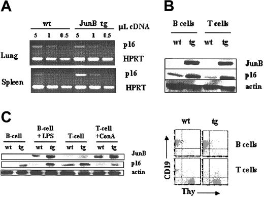
JunB overexpression in B lymphocytes is accompanied by an increased expression of the cyclin-dependent kinase inhibitor protein p16. (A) Semiquantitative RT-PCR analysis of p16 mRNA levels in lung and spleen from Ubi-junB transgenic animals and their wild-type littermate controls. (B) B- and T-lymphoid cells were MACS sorted from spleen of wild-type and Ubi-junB transgenic mice and subjected to Western blot analysis (upper panel) for JunB, p16, and β-actin (loading control). One representative FACS analysis of the purified B- and T-lymphoid cells after MACS purification is depicted (lower panel). (C) B- and T-lymphoid cells were MACS sorted from spleen of wild-type and Ubi-junB transgenic mice, stimulated with growth stimuli as indicated, and subjected to Western blot analysis for JunB, p16, and β-actin (loading control).
JunB overexpression in B lymphocytes is accompanied by an increased expression of the cyclin-dependent kinase inhibitor protein p16. (A) Semiquantitative RT-PCR analysis of p16 mRNA levels in lung and spleen from Ubi-junB transgenic animals and their wild-type littermate controls. (B) B- and T-lymphoid cells were MACS sorted from spleen of wild-type and Ubi-junB transgenic mice and subjected to Western blot analysis (upper panel) for JunB, p16, and β-actin (loading control). One representative FACS analysis of the purified B- and T-lymphoid cells after MACS purification is depicted (lower panel). (C) B- and T-lymphoid cells were MACS sorted from spleen of wild-type and Ubi-junB transgenic mice, stimulated with growth stimuli as indicated, and subjected to Western blot analysis for JunB, p16, and β-actin (loading control).
Analysis of explanted JunB transgenic and wild-type tumor cells.(A) Tumor-derived cells were subjected to a [3H]thymidine incorporation assay to investigate their proliferative potential. No difference between tumor cells from Ubi-junB transgenic and wild-type animals was observed. Data represent means ± SDs from 6 individual wells. One representative experiment is shown (n = 2). (B) Western blot analysis of JunB, p16, and actin in tumor-derived cells.
Analysis of explanted JunB transgenic and wild-type tumor cells.(A) Tumor-derived cells were subjected to a [3H]thymidine incorporation assay to investigate their proliferative potential. No difference between tumor cells from Ubi-junB transgenic and wild-type animals was observed. Data represent means ± SDs from 6 individual wells. One representative experiment is shown (n = 2). (B) Western blot analysis of JunB, p16, and actin in tumor-derived cells.
Transformed B-lymphoid cells have escaped growth suppression by JunB. (A-B) Three clonal populations of v-abl-transformed pro-B cell lines (termed 26, 27, and 47) were cocultivated with retroviral JunB-producer cell lines for 48 hours and subsequently subjected to a [3H]thymidine proliferation assay (A) or cloned in cytokine-free methylcellulose (B). Data represent means ± SE from 6 individual wells or 4 individual plates. (C) Western blot analysis of the 3 cell lines infected with pB-junB. Mock-infected cells were used as controls. (D) Induction of JunB by UV irradiation in 4 different v-abl-transformed cell lines expressing various expression levels of p16. Western blot analysis of JunB, p16, and β-actin. (E) Injection of v-abl-transformed cells in nude mice caused tumor formation in 72% of the JunB transgenic cells and in 67% of the wild-type cells. Shown are tumor weights 2 weeks after injection. Data represent means ± SE from 6 individual wells (A) or 4 individual plates (B).
Transformed B-lymphoid cells have escaped growth suppression by JunB. (A-B) Three clonal populations of v-abl-transformed pro-B cell lines (termed 26, 27, and 47) were cocultivated with retroviral JunB-producer cell lines for 48 hours and subsequently subjected to a [3H]thymidine proliferation assay (A) or cloned in cytokine-free methylcellulose (B). Data represent means ± SE from 6 individual wells or 4 individual plates. (C) Western blot analysis of the 3 cell lines infected with pB-junB. Mock-infected cells were used as controls. (D) Induction of JunB by UV irradiation in 4 different v-abl-transformed cell lines expressing various expression levels of p16. Western blot analysis of JunB, p16, and β-actin. (E) Injection of v-abl-transformed cells in nude mice caused tumor formation in 72% of the JunB transgenic cells and in 67% of the wild-type cells. Shown are tumor weights 2 weeks after injection. Data represent means ± SE from 6 individual wells (A) or 4 individual plates (B).
Histology and immunohistochemistry
Paraffin- and acrylate-embedded specimens were obtained from the Institute of Pathology, University Hospital Graz. Tissue array technology was employed to compare samples using antibodies against Ki-67, CD20, CD23, CD30, CD4 (Dako, Glostrup, Denmark), JunB, c-Jun, c-Fos, Fra-1, JunD (Santa Cruz Biotechnology), and the alkaline phosphatase antialkaline phosphatase technique.29 Samples were rated positive for the individual AP-1 members, when the staining intensity of the tumor cells was consistently higher than the surrounding untransformed cells. In addition, normal lymph nodes were used as controls and stained negative for the AP-1 members investigated under our technical conditions. A further negative control consisted of the staining of tissue sections with the secondary antibody alone.
The livers and spleens of the diseased mice were transferred into 4% phosphate-buffered saline (PBS)-buffered formalin for histologic examinations. Serial sections were performed and stained with hematoxylin and eosin (H&E). Three individual spleens and livers from each genotype were analyzed by light microscopy for the infiltration of leukemic cells. The majority of portal fields were investigated.
Mice and genotypes
Ubi-junB transgenic mice were generated as described22 and were maintained in a mixed 129/svxC57Bl/6xMF1xHimOF1 genetic background.
Tissue-culture conditions and virus preparation
Transformed fetal liver cells and cell lines derived from tumor tissue were maintained in RPMI medium containing 5% heat-inactivated fetal calf serum (FCS), 100 U/mL penicillin-streptomycin, and 5 μM β-mercaptoethanol. NIH3T3 cells, T220-29 cells (NIH3T3 cell engineered to produce interleukin-7), and A010 cells were maintained in Dulbecco modified Eagle medium (DMEM) containing 10% heat-inactivated FCS and 100 U/mL penicillin-streptomycin. A010 cells produce an ecotropic replication-deficient form of the Abelson virus. For collection of the viral supernatant, A010 cells were plated in 100-mm dishes precoated with gelatin (1%) and grown to confluence. Supernatant was harvested every 8 hours for 40 hours, pooled, and filtered through a 0.45-μm filter.30 For UV irradiation the cells were exposed to 0.006 J/cm2 in RPMI medium. Ninety minutes thereafter the cells were harvested and lysed for Western blot analysis.
B- and T-cell purification, [3H]thymidine incorporation
Splenic B and T cells were sorted for the expression of CD19 and CD3, respectively, by magnetic-activated cell separation (MACS) according to the manufacturer's instruction (Miltenyi Biotech, Bergisch Gladbach, Germany). The purity of the cells after MACS was controlled by fluorescence-activated cell sorter (FACS) and was between 85% and 95% in the individual experiments. For thymidine incorporation assays, the cells were then plated at a density of 2 × 10⁵ cells in 96 round-bottom wells and stimulated with the cytokines as indicated. The concentrations used were 100 ng/mL interleukin 4 (IL-4), 20 μg/mL α-immunoglobulin M (α-IgM), and 1 μg/mL α-CD40. [3H]thymidine was added 48 hours after stimulation for another 12 hours. To test the proliferation of cytokine-independent tumor cells, 2 × 10⁵ cells were plated in round-bottom 96-well plates; 18 hours thereafter [3H]thymidine was added and incubated for another 12 hours.30,31
Analysis of pro-B cells
JunB transgenic and wild-type bone marrow was prepared (n = 3 of each genotype). One aliquot was subjected to FACS analysis immediately after preparation. The remaining cells were transferred to RPMI medium as described above and cocultured with IL-7-producing NIH3T3 cells as feeder layers. Seven days thereafter the cell culture consisted of 80% to 90% CD19+ cells as analyzed by FACS and was subjected to a [3H]thymidine incorporation assay.
FACS
Single-cell suspensions of cells were preincubated with αCD16/CD32 antibodies (Pharmingen, Hamburg, Germany) to prevent nonspecific Fc receptor-mediated binding. Thereafter, aliquots of 5 × 10⁵ cells were stained with monoclonal antibodies conjugated with fluorescent markers and analyzed by FACS (Becton Dickinson, San Jose, CA). The antibodies used for lineage determination included the B-cell lineage markers B220, CD19, and CD43; the T-cell markers CD4, CD8, and Thy1.2; the myeloid markers Gr1 and Mac1; and the erythroid-lineage marker Ter119 (all from Pharmingen).
Infection of fetal livers, in vitro transformation assays, and establishment of cell lines
For the preparation of fetal liver cells, heterozygous animals were set up for breeding and vaginal plugs were checked daily. Fifteen days after conception the pregnant animals were killed and the fetal livers prepared. The embryo tails were used for genotyping by polymerase chain reaction (PCR). Single-cell suspensions from fetal livers were infected for 30 minutes with viral supernatant derived from A010 cells enriched with 5 ng/mL IL-7, 7 μg/mL polybrene, and β-mercaptoethanol. The virus-infected as well as the mock-infected cells were then maintained for 15 to 20 hours on IL-7-producing feeder layers (T220-29 cells). Thereafter, cells were washed several times and plated in cytokine-free methylcellulose at a density of 3 × 10⁵ cells/mL in 35-mm dishes. An aliquot of the cells was used to determine numbers of B-cell precursors in the individual fetal livers by FACS analysis. After 7 to 10 days the cloning efficiency was evaluated by counting colonies by light microscopy. The assays were performed in triplicate. Mock-infected cells did not result in growth factor-independent colonies. The ability to form cell lines was tested by transferring an aliquot of the transduced cells (1 × 10⁶) to growth factor-free medium. The medium was changed twice a week and the culture observed for the outgrowth of stable clones.32
Injection of tumor cells into nude mice
Ten days after infection, 1 × 10⁶ cells were resuspended into 300 μL of PBS and injected subcutaneously into nude (nu/nu) mice. At the time point of injection the cells had been in growth factor-free medium for at least a week and consisted of CD19+/CD43+ pro-B cells. Mice were checked daily for the development of tumors. Tumors bigger than 2 cm in diameter were excised for further analysis.32
Infection of neonatal mice with the Abelson oncogene (Ab-MuLV)
Newborn mice were injected retroperitoneally with 50 μL of replication-incompetent ecotropic retrovirus encoding for v-abl. The mice were then checked daily for onset of diseases. Sick mice were killed and analyzed carefully for signs of disease.30
Results
Low expression of JunB in human B-lymphoid malignancies
We investigated the expression of JunB, c-Jun, c-Fos, Fra-1, and JunD in human non-Hodgkin lymphomas by tissue-array technology using a collection sample covering the majority of non-Hodgkin B-lymphoid malignancies. The tissue samples comprised lymph node biopsies of patients suffering from follicular lymphomas (FLs), diffuse large-cell lymphomas (DLCLs), B-cell chronic lymphoid leukemia (B-CLL), and the highly malignant large-cell anaplastic T-cell lymphomas (ALCLs). Normal resting lymphoid cells that surrounded the tumor cells were taken as internal controls and stained negative for all 4 of the AP-1 factors analyzed. We detected positive staining for c-Jun, c-Fos, Fra-1, and JunD in the majority of the cases. ALCL was strongly positive in more than 80%. Interestingly, JunB showed a characteristic expression pattern: JunB staining was absent or low in B-cell malignancies (0% in B-CLL, between 10% and 15% for DLBL and FL), whereas the T-cell lymphomas, ALCLs, expressed JunB strongly in 15 of 17 cases (Figure 1A-B). To confirm this observation using a different technique, we complemented our study by immunoblots directed against JunB. Samples of tumor cells derived from patients suffering from FL, DLCL, B-CLL, and ALCL were analyzed by Western blot for JunB expression (biopsies of normal lymph nodes were used as controls). Again, a strong expression of JunB was detected in ALCL, whereas a normal or reduced expression of JunB was found by Western blot analysis in B-cell lymphomas (Figure 1C).
Overexpression of JunB blocks proliferation in murine B- but not T-lymphoid cells
In order to investigate the mechanism accounting for the differences in the expression of JunB in B- and T-cell malignancies, we used a transgenic mouse model that allows us to study the impact of JunB in both lymphoid compartments in vivo. As can be seen in Figure 2A, Ubi-junB transgenic animals showed an increased expression of JunB in B- and T-lymphoid cells. The numbers of B-lymphoid cells in the peripheral blood of junB transgenic animals were found to be reduced to 60% when compared with age-matched wild-type littermate controls, whereas the numbers of peripheral T-lymphoid cells were in the same range (data not shown). Stimulation of T cells with increasing concentrations of the lectin concanavalin A resulted in comparable mitogenic responses for wild-type and transgenic cells (Figure 2B). In contrast, the proliferative response in peripheral B cells (ie, CD19+ cells) was severely impaired. This effect was observed regardless of the proliferative stimulus for it was observed in the presence of increasing concentrations of lipopolysaccharide (LPS) and of the B-cell-specific stimuli IgM, anti-CD40 and IL-4 (Figure 2C-D).
We next addressed the proliferative capacity of pro-B cells isolated from bone marrow of JunB transgenic animals. It is noteworthy that the numbers of B-cell progenitors were slightly elevated in bone marrow preparations from JunB transgenic mice. This could reflect a positive growth-promoting role of JunB in early lymphoid progenitors (Figure 3A). However, stimulation with the pro-B-cell-specific interleukin-7 (IL-7) revealed a reduced proliferative capacity compared with wild-type controls (Figure 3B). Based on these 2 sets of observations we conclude that JunB exerts a growth-suppressing activity on cells starting from the pro-B-cell stage.
The growth inhibitory effect of JunB on B lymphocytes extends to CD19+/CD43+ B-cell precursors. (A-B) Bone marrow cells of wild-type (n = 3) and Ubi-junB transgenic (n = 3) mice were analyzed by FACS to evaluate the numbers of B-cell precursors. Numbers in the right corners show the amount of B-cell precursors in the bone marrow. One representative experiment is shown (n = 6) (A). The remaining cells were cocultivated on IL-7-producer cells for 7 days to enrich for B-cell precursors. After 7 days, the bulk cultures, which now consisted of 80% CD19+/CD43+ cells, were subjected to [3H]thymidine incorporation assay after stimulation with 10 ng/mL IL-7 (B). Data represent means ± SDs from 6 individual wells. One representative experiment is shown (n = 2).
The growth inhibitory effect of JunB on B lymphocytes extends to CD19+/CD43+ B-cell precursors. (A-B) Bone marrow cells of wild-type (n = 3) and Ubi-junB transgenic (n = 3) mice were analyzed by FACS to evaluate the numbers of B-cell precursors. Numbers in the right corners show the amount of B-cell precursors in the bone marrow. One representative experiment is shown (n = 6) (A). The remaining cells were cocultivated on IL-7-producer cells for 7 days to enrich for B-cell precursors. After 7 days, the bulk cultures, which now consisted of 80% CD19+/CD43+ cells, were subjected to [3H]thymidine incorporation assay after stimulation with 10 ng/mL IL-7 (B). Data represent means ± SDs from 6 individual wells. One representative experiment is shown (n = 2).
JunB overexpression is accompanied by an increased expression of the cyclin-dependent kinase inhibitor protein p16 in B cells
In fibroblasts maintained in culture, overexpression of JunB resulted in up-regulation of the cell cycle kinase inhibitor p16, which was proposed to mediate the growth-inhibitory effect of JunB.20 However, it is not known whether this effect is also seen in vivo and if it occurs in all cell types. Hence, we selected 4 tissues that reportedly expressed p16 in wild-type adult mice33 to screen for p16 mRNA levels by reverse transcriptase-PCR (RT-PCR; Figure 4A). Interestingly, transcripts encoding p16 were elevated in whole-spleen preparations from JunB transgenic animals but not in other organs tested (lung, kidney, testis). We further confirmed by Western blot that expression of p16 protein was augmented in resting splenic B and T cells from JunB transgenic mice compared with control B cells (Figure 4B). Mitogenic stimulation of B- and T-lymphoid cells resulted in an increased expression of JunB in both cell lineages (Figure 4B). However, only B cells reacted with elevated expression levels of p16 to the mitogenic stimulation, whereas p16 was expressed at low levels in T-lymphoid cells, irrespective of the levels of JunB protein (Figure 4C). These results indicate that JunB may repress B-cell proliferation by up-regulating the cell cycle kinase inhibitor p16.
JunB overexpression inhibits transformation by the B-cell-specific v-abl oncogene
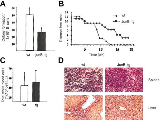
To test whether JunB might also be able to counteract transformation in the B-cell lineage, we used the Abelson oncogene (Ab-MulV).34,35 Primary fetal liver cells were infected with a replication-incompetent ecotropic Abelson retrovirus. After 3 days, which allows for expression of the Abelson oncogene, the cells were suspended in growth factor-free methyl cellulose and the numbers of pro-B-cell progenitors were determined in parallel by FACS analysis. As can be seen in Figure 5A, the v-abl-transduced JunB transgenic fetal liver preparations gave rise to lower numbers of growth factor-independent colonies. Some of the fetal liver preparations were maintained in growth factor-free medium and cell lines expressing the B-lymphoid markers CD19 and CD43 grew out from every JunB transgenic and wild-type fetal liver investigated (9/9 JunB transgenic and 7/7 wild type).
JunB overexpression reduces Ab-MuLV-induced colony formation in vitro and tumor formation in vivo. (A) Clonal outgrowth of Abelson-infected fetal liver cell suspensions. Fetal livers from wild-type (n = 6) and JunB transgenic (n = 9) mice were infected with Abelson retrovirus and subsequently cloned in cytokine-free methylcellulose. The initial number of B cells was determined by FACS. Results show the average numbers ± SD of colonies obtained per 1 × 10⁵ B cells plated (n = 13 for JunB tg and n = 8 for wild-type fetal liver cells). Mock-infected cells were used as controls and did not give rise to colonies. (B) Newborn mice were injected intraperitoneally with replication-deficient v-abl retrovirus and observed over a period of 8 months. During this time, 3 of 11 JunB transgenic mice remained free of any leukemia/lymphoma. (C) White blood cell count in wild-type (n = 6) and Ubi-junB transgenic animals (n = 7) at the time point of fully developed disease. Data represent means ± SDs. (D) Histologic sections of spleen and liver from diseased wild-type and Ubi-junB transgenic animals. Leukemic cells heavily infiltrate wild-type liver and spleen but to a lesser degree JunB transgenic organs. One representative example of each genotype is depicted (n = 3). Panels stained with H&E; original magnification, × 100.
JunB overexpression reduces Ab-MuLV-induced colony formation in vitro and tumor formation in vivo. (A) Clonal outgrowth of Abelson-infected fetal liver cell suspensions. Fetal livers from wild-type (n = 6) and JunB transgenic (n = 9) mice were infected with Abelson retrovirus and subsequently cloned in cytokine-free methylcellulose. The initial number of B cells was determined by FACS. Results show the average numbers ± SD of colonies obtained per 1 × 10⁵ B cells plated (n = 13 for JunB tg and n = 8 for wild-type fetal liver cells). Mock-infected cells were used as controls and did not give rise to colonies. (B) Newborn mice were injected intraperitoneally with replication-deficient v-abl retrovirus and observed over a period of 8 months. During this time, 3 of 11 JunB transgenic mice remained free of any leukemia/lymphoma. (C) White blood cell count in wild-type (n = 6) and Ubi-junB transgenic animals (n = 7) at the time point of fully developed disease. Data represent means ± SDs. (D) Histologic sections of spleen and liver from diseased wild-type and Ubi-junB transgenic animals. Leukemic cells heavily infiltrate wild-type liver and spleen but to a lesser degree JunB transgenic organs. One representative example of each genotype is depicted (n = 3). Panels stained with H&E; original magnification, × 100.
JunB transgenic mice are partially protected from v-abl-inflicted leukemia/lymphomas
An additional possibility to test for tumor formation by the Abelson oncogene is the direct injection of a replication-incompetent retrovirus into newborn mice. In vivo, this single exposure to the v-abl oncogene provokes the development of a B-cell lymphoma/leukemia in early adulthood. We injected 11 JunB transgenic and 12 wild-type mice with v-abl oncogene to investigate whether the overexpression of JunB protects the mice from the oncogenic challenge. Most interestingly, while all of the wild-type mice died within 3 months, 3 of the transgenic animals survived the oncogenic challenge for more than 9 months (Figure 5B). In addition, the diseased transgenic animals had an increased mean survival time compared with their wild-type littermates (12.1 ± 0.8 versus 8.4 ± 1.7 weeks; P = .04).
Nevertheless, the basic phenotypic hallmarks of the disease did not differ between JunB transgenic and wild-type animals: all tumors were of B-lymphoid origin (CD19+/CD43+), the white blood cell counts were elevated in the same range (Figure 5C), and liver and spleen were the organs most affected by infiltration with tumor cells. Histologic sections of spleen and liver revealed that the parenchyma of the spleen and the portal fields in wild-type animals were heavily infiltrated by leukemic cells. In the wild-type liver, leukemic cells were spilling out past the limiting plates of the portal tracts. In contrast, in JunB transgenic animals, spleen and liver were affected to a lesser degree such that the regular organ architecture was maintained (Figure 5D). We therefore conclude that overexpression of JunB mitigated the severity of v-abl-induced disease. This resulted in a prolonged median survival time of JunB transgenic animals.
Abelson-transformed pro-B cells have escaped the growth inhibition by JunB
In order to understand why protection by JunB was incomplete, we explanted tumors and propagated the cells in vitro. It was evident that the tumor cells obtained from JunB transgenic animals had overcome the inhibitory effect of the transgene for we failed to detect any differences between the wild-type and JunB transgenic tumor cells in response to mitogenic stimulus (Figure 6A). Western blot showed a considerable variation between cell lines derived from JunB transgenic tumors. Regardless of this variability, the levels were consistently higher than in tumor cells from wild-type animals (Figure 6B, top). The discrepancy between high levels of JunB and normal proliferative capacity suggested that during transformation and expansion in vivo the tumor cells had overcome the inhibitory effect of the junB transgene. In order to verify this conjecture, we determined the levels of p16, an established downstream target of JunB.20 In 2 of 4 transgenic tumor cells the levels of p16 were dramatically reduced. This was also the case for 6 of 10 wild-type tumors (Figure 6B). Western blot analysis of the cell cycle regulators D2, E, and A and the cell cycle inhibitor protein p21 did not show any major change (data not shown).
v-abl-transformed B cells escape growth inhibition by JunB
We therefore speculated that the transformation process might render the cells insensitive to the growth-suppressing effect of JunB by inactivating the JunB-dependent pathway that is involved in cell cycle control. To prove this hypothesis, 3 clonal Abelson (v-abl)-transformed pro-B cell lines were infected with JunB-expressing retrovirus resulting in a 5- to 8-fold overexpression of JunB (Figure 7C). As predicted, JunB overexpression affected neither proliferation nor cloning efficiency in growth factor-free methylcellulose of the transformed pro-B cell lines (Figure 7A-B).
A strong induction of JunB can be achieved by UV irradiation. Eight wild-type cell lines expressing various levels of p16 were chosen and UV irradiated (Figure 7D). Irrespective of the expression levels of p16, no further increase was observed by the UV-induced JunB induction. In addition, we injected v-abl-transformed JunB transgenic and wild-type cell lines into nude mice and analyzed tumor formation after 2 weeks. Tumor onset was observed in 72% of the mice that had received JunB transgenic tumor cell lines and in 67% of the nude mice that had obtained v-abl-transformed wild-type cells. Also, the tumor weight was comparable between both groups (Figure 7D). These experiments prove that v-abl-transformed cells had escaped growth inhibition by JunB and have lost the ability to react with elevated p16 levels to JunB expression.
Discussion
Hodgkin lymphoma cells express JunB (and c-Jun) to high levels.27 In contrast, loss of JunB leads to murine chronic myeloid leukemia.24 Thus, JunB may have Janus-like properties in hematologic cells. Here we have addressed the role of JunB in the development of T- and B-cell lymphoma. Our observations show that (1) non-Hodgkin lymphomas of B-lymphoid origin do not or only weakly express JunB, whereas a strong expression is found in lymphomas that originate from the T-lymphoid lineage; (2) JunB selectively blocks B-lymphoid, but not T-lymphoid, cell proliferation ex vivo; and (3) overexpression of JunB blunts the transforming ability of the B-lymphoid-specific form of the Abelson oncogene in vivo and in vitro.
It has long been known that AP-1 members, and JunB in particular, exert their actions in a cell-type-specific manner because they impinge on a large set of target genes that are coregulated by other transcription factors (eg, by the glucocorticoid receptor).36 An additional level of complexity is brought about by the cell-type-specific expression of AP-1 members that results in a diverse set of dimeric combination partners.37 Our findings are consistent with this concept of cell-type-specific action, for JunB only exerted antiproliferative actions in B cells. In T cells, JunB is necessary for the induction of Th2-dependent cytokines,38 but the mitogenic response of T cells, in contrast, proved resistant to elevated JunB levels. Whereas a tight correlation of JunB and p16 expression levels is observed in B-lymphoid cells, T cells react with an up-regulation of JunB but a decrease of p16 protein to mitogenic stimulation.
Immature common lymphoid precursors may also be resistant to growth inhibition by JunB. In fact we suspect that high levels of JunB may have a growth-promoting effect in early lymphoid progenitors because the numbers of (CD19+/CD43+) B-cell precursors are elevated in the bone marrow of junB transgenic mice. In contrast, we found that the levels of (CD19+/CD43-) B cells were reduced by 40% in peripheral blood. Taken together, these observations are indicative of a partial block starting at the level of the pro-B-cell stage. Finally, it is worth mentioning that the variation in AP-1 members was also readily evident in human lymphoid malignancies. Tissue array staining showed that cells from B-CLL samples were negative for JunB but highly positive for Fra1 (28 of 28 samples). By comparison, anaplastic large-cell lymphoma (ALCL), a T-cell-derived malignancy, was characterized by the highest levels of JunB. Thus, 15 of 17 samples were strongly positive by tissue-array staining. These were also highly positive for c-Jun. Our data are consistent with a recent report that shows high AP-1 activity in 3 cell lines derived from ALCL.27
Two additional findings support the argument that JunB is an inhibitor of B-lymphoid transformation. (1) Ex vivo transformation by a retrovirus-encoding v-abl is blunted if B cells overexpress JunB. We stress that this experimental strategy relies on the use of primary hematopoietic precursors. These fetal cells are unlikely to have already acquired any mutations that predispose them to transformation. (2) Accordingly, the difference between junB transgenic and wild-type B cells can be recapitulated in vivo. Overexpression of JunB mitigates the course of the disease. Three of 11 transgenic animals survived the oncogenic challenge for more than a year. In the remaining 8 Ubi-junB transgenic animals JunB overexpression delayed the development of leukemia/lymphoma resulting in an increased survival time. In contrast, all 12 wild-type littermate controls died within 12 weeks. Overexpression of JunB did not, however, completely prevent the development of leukemia/lymphoma in Ubi-junB transgenic animals. This is due to the fact that transformed cells can eventually overcome growth inhibition by JunB. This conclusion is supported by the following experimental findings. (1) When established JunB transgenic and wild-type tumor cell lines were injected into nude mice, we did not observe any difference in tumor onset, latency, or tumor weight. (2) Moreover, no differences in proliferation were observed in primary tumor cells explanted from the diseased Ubi-junB and diseased wild-type mice. This strongly indicates that the transgenic tumor cells had acquired the ability to circumvent growth inhibition by JunB. (3) Most conclusively, forced expression of JunB in established tumor cell clones did not affect cloning efficiency or cell proliferation. This finding unequivocally demonstrates that the transformed cells had overcome the JunB-dependent growth control. In murine B-lymphoid cells, the escape from growth inhibition by JunB is achieved by uncoupling of JunB elevation from p16 up-regulation. Even strong induction of JunB by UV light did not alter p16 levels, irrespective of its expression levels.
Further experiments need to determine whether this effect is specific for v-abl or whether elevated JunB levels are capable of interfering with transformation induced by other oncogenes. According to the high expression levels of JunB in ALCL and the lack of p16 induction by mitogenic stimulation in T cells, we expect the tumor-suppressing action of JunB to be limited to the B-lymphoid lineage.
To the best of our knowledge, there is no comparable systematic survey of AP-1 members in a large set of non-Hodgkin lymphoma by tissue array. However, JunB expression was assessed in a limited number of human cutaneous lymphomas by DNA microarray. JunB expression was lost in 3 of 4 primary cutaneous B-cell lymphomas,39 whereas JunB was amplified in primary cutaneous T-cell lymphomas.40 Similarly, the level of JunB has been determined in tissue samples from patients with Hodgkin disease.27 The vast majority of Hodgkin lymphomas are thought to originate from B cells, although the pathogenesis has remained elusive.41,42 It has been noted that Hodgkin lymphomas express c-Jun and JunB at high levels.27 Regardless of the different status of Hodgkin and non-Hodgkin lymphoma, it is worth pointing out that these cells also escape from growth inhibition by JunB. In spite of the very high levels of JunB that presumably arise from constitutive activation of nuclear factor-κ B (NF-κB), JunB is transcriptionally inactive in Hodgkin lymphoma.27 These observations again support our conclusion, namely that high levels of transcriptionally active JunB are incompatible with B-lymphoid transformation.
We have not systematically investigated all possible mechanisms that may underlie the escape. However, in several cell lines derived from explanted murine tumors, we observed a loss of p16 expression, which is a target gene for JunB.20 This loss is per se sufficient to account for the escape of the murine v-abl-transformed B cells from JunB-mediated growth inhibition. A loss of p16 leads to deregulation of cyclin D-dependent kinase activity and thus promotes progression through G1.43,44 However, there are various possibilities to overcome growth inhibition by p16 and alterations of cyclin D-dependent complexes are reported in more than 90% of all human lymphomas.45,46 One of the underlying molecular mechanisms is indeed loss of p16 protein expression due to hypermethylation of cytosine-phosphate-guanosine (CpG) islands.46 Additional escape mechanisms are likely to exist. Promoter methylation of JunB was shown to be a mechanism to down-regulate JunB in human CML cells.25 Taken together, both the earlier observations and the current findings highlight the tumor-suppressing role of JunB in B-lymphoid cells. In all murine v-abl-transformed cell lines we investigated, the ability of JunB to induce elevated levels of p16 was lost, indicating the loss of a normal cell cycle control in these cells.
Given the various escape mechanisms that are likely to be operative at the time point of diagnosis, it is at present, however, difficult to predict the prognostic role of JunB.
Prepublished online as Blood First Edition Paper, August 7, 2003; DOI 10.1182/blood-2003-03-0915.
Supported in part by grants P15033 and P15865 from the Austrian Science Foundation (FWF, V.S.) and by grant 1/2001 from the Styrian Cancer Foundation (L.K.). The IMP is supported by Boehringer Ingelheim.
A.P.S., L.K., and E.W. contributed equally to this study.
The publication costs of this article were defrayed in part by page charge payment. Therefore, and solely to indicate this fact, this article is hereby marked “advertisement” in accordance with 18 U.S.C. section 1734.
We are indebted to Latifa Bakiri and Peter Valent for helpful comments and critical reading of the manuscript. We are grateful to H. Drobny and R. Liebminger for technical help and to N. Rosenberg and M. Yaniv for sharing of reagents. We also want to thank U. Losert and the staff of the Institute for Biomedical Research at the Vienna University Clinics (AKH) for the good collaboration in maintaining our mouse colony.


![Figure 2. JunB overexpression inhibits proliferation of mature peripheral B lymphocytes. (A) Spleens from Ubi-junB transgenic animals (JunB tg) and their littermate wild-type controls (wt) were used for MACS purification of T and B cells. Whole-spleen extracts (S), CD3-purified T lymphocytes (T), and CD19-purified B lymphocytes were subsequently subjected to Western blotting using an antibody directed against JunB. Extracts of wild-type and Ubi-junB transgenic fibroblasts were used as controls. (B) CD3+ T lymphocytes were MACS purified from spleens of Ubi-junB transgenic animals (n = 3; ▪) and their littermate wild-type controls (n = 3; □). Cells (2 × 10⁵/well) were subsequently subjected to a [3H]thymidine proliferation assay using increasing concentrations of concanavalin A. (C-D) CD19+ B-lymphoid cells were MACS sorted from spleen of wild-type (n = 3; □) and Ubi-junB transgenic (n = 3; ▪) mice. Cells (2 × 10⁵/well) were stimulated with the factors indicated for 48 hours and subjected to a [3H] thymidine incorporation assay. Co indicates control, cells that were plated in medium without the addition of growth factors. Data represent means ± SDs from 6 individual wells. One representative experiment is shown (n = 3).](https://ash.silverchair-cdn.com/ash/content_public/journal/blood/102/12/10.1182_blood-2003-03-0915/6/m_h82335303002.jpeg?Expires=1765909146&Signature=fSEdnXciWDIJ7bp3ozKHy60GjALsWDBbeQ1xa0sWvR23Y-cJQHpgyO-jiwpFqQdDWfjxr7OJ6uE-HiJx8RDAvSL71HLeLSQIU3O5f8jK3024cF7H9uXEsxgnEgBcM4T3OGjJ6SHwfJ9KDRwrgUkRokRyt7gersKj0JjitUsxz9K4u7fKgIelLgmp0-zxM7obIG7oZEg29wsx6P08sgJ6tAdqtegWvJ2xYLk6CFZ8MQ9z8ouZddd6Y3AP4JwKG8xFsMAqWbYopymrzWte-E-8QRXW3o2nlJ0h~0jY-PczHZY6QOfM1of1RRbKzX-Sdv0i8TVzjtFbdKLzzOX3q6N5wg__&Key-Pair-Id=APKAIE5G5CRDK6RD3PGA)

![Figure 6. Analysis of explanted JunB transgenic and wild-type tumor cells.(A) Tumor-derived cells were subjected to a [3H]thymidine incorporation assay to investigate their proliferative potential. No difference between tumor cells from Ubi-junB transgenic and wild-type animals was observed. Data represent means ± SDs from 6 individual wells. One representative experiment is shown (n = 2). (B) Western blot analysis of JunB, p16, and actin in tumor-derived cells.](https://ash.silverchair-cdn.com/ash/content_public/journal/blood/102/12/10.1182_blood-2003-03-0915/6/m_h82335303006.jpeg?Expires=1765909146&Signature=U77XIHfcdl~PcE~Y~Eb-sU0VoB~s30lPVSu3L0xd25U7DLGZs9KRSpss7m3mW11fp-uqC7fkHuNkX1GgmpnYGDboBwv3AEkP86SxcUFtP4veonydTqzY4RdCLnm2Ljsu-Qkg8BkE3gjYyfDRfpiV7msYABUm5LxmZNASZr435g9YvCvcDsFYSo2w~0oZympBekUfLSah8l4OHKAlZT~BdXpVGNa4c9f9ejw28Mu7YmXyIT6lArUdg1RSGbJERzPAAdRU7DMPIPf1QzJIXD6RX13bTFytVrJvLLmBSSQw1W1A~dwMC9GYwvhbnO5jA5ZV42DZeNpF4R6hT3quKQolbA__&Key-Pair-Id=APKAIE5G5CRDK6RD3PGA)
![Figure 7. Transformed B-lymphoid cells have escaped growth suppression by JunB. (A-B) Three clonal populations of v-abl-transformed pro-B cell lines (termed 26, 27, and 47) were cocultivated with retroviral JunB-producer cell lines for 48 hours and subsequently subjected to a [3H]thymidine proliferation assay (A) or cloned in cytokine-free methylcellulose (B). Data represent means ± SE from 6 individual wells or 4 individual plates. (C) Western blot analysis of the 3 cell lines infected with pB-junB. Mock-infected cells were used as controls. (D) Induction of JunB by UV irradiation in 4 different v-abl-transformed cell lines expressing various expression levels of p16. Western blot analysis of JunB, p16, and β-actin. (E) Injection of v-abl-transformed cells in nude mice caused tumor formation in 72% of the JunB transgenic cells and in 67% of the wild-type cells. Shown are tumor weights 2 weeks after injection. Data represent means ± SE from 6 individual wells (A) or 4 individual plates (B).](https://ash.silverchair-cdn.com/ash/content_public/journal/blood/102/12/10.1182_blood-2003-03-0915/6/m_h82335303007.jpeg?Expires=1765909146&Signature=YrlIQKG2wzjycRVDMzxf7o9mQPCtlX01WnGDHsP14Z3RUt0Z3yThqiFtuyuRjvsRwBVTLV7RSmV3fy0tYgEJqYT5~rOwwtX1d96oQR3-U4PovXxu4ih-u10eWYO73uG4yIxufIHoYE9Yjhaqs2qa0tCy1Lxm61kuG1LvtDy4TzwVV83GqSuO0fh3kvDla7HoYTf-vpaCptJOfL3FvzdaJ~a8LUCckhXDArfL~Uj-dan6oToCVVa3qM~5qYOUKGjKjdNEIaCCgFH3FIRpBzAL13Bj9YEm8LWIVRm9yBYi8ICSciWYxf-4RWflqilzG~RuhsHV6ZVBEj35az1ABCXqYQ__&Key-Pair-Id=APKAIE5G5CRDK6RD3PGA)
![Figure 3. The growth inhibitory effect of JunB on B lymphocytes extends to CD19+/CD43+ B-cell precursors. (A-B) Bone marrow cells of wild-type (n = 3) and Ubi-junB transgenic (n = 3) mice were analyzed by FACS to evaluate the numbers of B-cell precursors. Numbers in the right corners show the amount of B-cell precursors in the bone marrow. One representative experiment is shown (n = 6) (A). The remaining cells were cocultivated on IL-7-producer cells for 7 days to enrich for B-cell precursors. After 7 days, the bulk cultures, which now consisted of 80% CD19+/CD43+ cells, were subjected to [3H]thymidine incorporation assay after stimulation with 10 ng/mL IL-7 (B). Data represent means ± SDs from 6 individual wells. One representative experiment is shown (n = 2).](https://ash.silverchair-cdn.com/ash/content_public/journal/blood/102/12/10.1182_blood-2003-03-0915/6/m_h82335303003.jpeg?Expires=1765909146&Signature=c01~qNBsNOQ0XN7R4omkyDZ9o0Ds7ijRmzOEM8jT1itn4AHDKhWTb9UdunwDjQ53rOmPliGS6n6LwBpneimHjk8FsA8IMM1mqRg6VpfEukKFJznHii5W9Rd7vmj8g6rAfrRxcnMHXj-pII2rFzKQl6ZhJzngMMDZPFc-ENa0DCinQexaOEpXTOVqod8yZoh1-ykaawKTWKtvZK6wxWn3hD1uxzGAhiIyVsonlXe7bw6UH~JgSonstasHimLOC0rbNtQALM8rOVtKOgO54zseRs1YxYDiJMuQ8soVtHYijixrJifZGw5tXku5xqhBE7Q3lfBmj1tpUgloGJQkNvid0Q__&Key-Pair-Id=APKAIE5G5CRDK6RD3PGA)

This feature is available to Subscribers Only
Sign In or Create an Account Close Modal