Abstract
The nature and exact function of selectin ligands involved in hematopoietic progenitor cell (HPC) homing to the bone marrow (BM) are unclear. Using murine progenitor homing assays in lethally irradiated recipients, we found that the P-selectin glycoprotein ligand-1 (PSGL-1) plays a partial role in HPC homing to the BM (a reduction of about 35% when the P-selectin binding region is blocked). Blockade of both PSGL-1 and α4 integrin did not further enhance the effect of anti-α4 integrin (a reduction of about 55%). We suspected that E-selectin ligands might contribute to the remaining homing activity. To test this hypothesis, HPC homing assays were carried out in E-selectin–deficient recipients and revealed a profound alteration in HPC homing when E-selectin and α4 integrin were inactivated (> 90% reduction). Competitive assays to test homing of long-term repopulating stem cells revealed a drastic reduction (> 99%) of the homed stem cell activity when both α4 integrin and E-selectin functions were absent. Further homing studies with PSGL-1–deficient HPCs pretreated with anti-α4 integrin antibody revealed that PSGL-1 contributes to approximately 60% of E-selectin ligand–mediated homing activity. Our results thus underscore a major difference between mature myeloid cells and immature stem/progenitor cells in that E-selectin ligands cooperate with α4 integrin rather than P-selectin ligands.
Introduction
The nature of adhesive events leading to the extravasation of leukocytes has been well characterized in a past decade.1-5 In postcapillary and collecting venules of the systemic circulation, rolling of mature leukocytes is largely mediated by P-selectin and E-selectin expressed on activated endothelium, and their glycoconjugated ligands expressed on leukocytes.6 The 2 endothelial selectins exert cooperative functions in leukocyte recruitment since the defects in doubly deficient animals are much more severe than in single-deficient mice.7-10
Recent advances have partially elucidated the adhesion pathways that mediate hematopoietic progenitor cell (HPC) homing to the bone marrow (BM). Early studies suggested that HPC homing was a specific phenomenon mediated by unique receptors and that glycoconjugates containing mannosyl and galactosyl residues might participate in the homing process.11 Subsequently, α4 integrin (VLA4 or α4β1) and its counterreceptor vascular cell adhesion molecule 1 (VCAM-1) were shown to participate in HPC homing in the mouse since antibody-blocking studies inhibited progenitor homing by approximately 50%,12 although mice chimeric for the presence or absence of α4 integrin expression from the blastocyst stage revealed that these integrins are not required to establish bone marrow hematopoiesis.13 We have shown that the defect in HPC homing to bone marrow was greater when VCAM-1 function was blocked in mice lacking both endothelial selectins.14 Although the individual role of each selectin was not assessed in HPC homing, intravital microscopy studies revealed that both P- and E-selectins contribute to progenitor rolling in the bone marrow microvasculature and that the vast majority of rolling interactions was inhibited when all 3 pathways were blocked.15 These results suggested that homing of HPCs to BM was mediated by multiple HPC-endothelial adhesion receptor pairs. It is interesting that the endothelial selectins and VCAM-1 are constitutively expressed in the BM microvasculature whereas they are only expressed upon inflammatory cytokine stimulation in other organs.15,16 The regional expression of these endothelial adhesion molecules may provide some specificity to the trafficking of progenitor cells toward the BM compartment.
Among the vast number of adhesion receptors expressed on HPCs,17 α4 integrin, α5β1, and αLβ2 have previously been suggested to participate in HPC homing.12,18,19 The main glycoprotein-bearing selectin ligand function, P-selectin glycoprotein ligand-1 (PSGL-1), can mediate rolling of mature leukocytes in murine cremasteric venules20,21 and human CD34+ cell rolling in the bone marrow microvasculature.22 Although PSGL-1 was shown to be the sole P-selectin ligand on human CD34+ cells,22,23 the identity of E-selectin ligands on HPCs is currently unclear. CD44 was suggested to represent a candidate E-selectin ligand since human CD34+ cells express a CD44 glycoform that can bind E-selectin in vitro.24 The glycoprotein CD44 may also participate in murine HPC homing in the mouse via its interaction with hyaluronic acid,18 though CD44-deficient HPCs did not display any homing defects.25 PSGL-1 represents a prime E-selectin ligand candidate on HPCs since leukocyte PSGL-1 can bind E-selectin in vitro26,27 and in vivo,28,29 and recent data suggest that mast cell progenitors can bind E-selectin through PSGL-1 in vitro.30
Here, we show that E-selectin ligands and α4 integrin expressed on murine hematopoietic progenitor/stem cells represent major adhesion receptors mediating homing to bone marrow. In addition, we have identified PSGL-1 on HPCs as a functional E-selectin ligand in vivo and show that this pathway contributes to the majority of the E-selectin–mediated homing activity when α4 integrin function is blocked. Our results thus suggest that HPCs express more than one E-selectin ligand and underscore a major difference between mature leukocytes and immature hematopoietic cells in that E-selectin ligands on HPCs cooperate with α4 integrin rather than P-selectin ligand for their recruitment into the bone marrow parenchyma.
Materials and methods
Antibodies
Rat antimouse α4 integrin monoclonal antibody (mAb) (PS/2; American Type Culture Collection, Manassas, VA) and rat antimouse P-selectin (clone RB40.34) were purified from supernatants of hybridoma cell lines. Rat antimouse PSGL-1 mAb (4RA10) immunoglobulin G 1 (IgG1), which blocks binding of PSGL-1 to P-selectin, was purified from supernatants of a 4RA10-producing hybridoma cell line which was raised using recombinant PSGL-1 and recognizes the functional 19-amino acid NH2-terminal peptide of PSGL-1.31,32 Antibodies were purified using a protein G column (Amersham Pharmacia Biotech, Uppsala, Sweden). Potential endotoxin contamination was removed using polymixin B endotoxin–removing gel (Pierce, Rockford, IL). Rat IgG control antibody was purchased from Sigma (St Louis, MO). Rat antimouse E-selectin (clone 9A9) was a generous gift of Dr Barry Wolitzky (Mitokor, San Diego, CA). For flow cytometric analysis, rat antimouse CD16/CD32 (clone 2.4G2), fluorescein isothiocyanate (FITC)–conjugated CD45.2 (clone 104), and biotinylated CD45.1 (clone A20) were purchased from BD Pharmingen (San Diego, CA). Phycoerythrin (PE)–conjugated streptavidin was purchased from Jackson Immunoresearch (West Grove, PA).
Animals
E-selectin–deficient (E–/–) mice were generated by gene targeting10 and were backcrossed 7 generations into the C57BL/6 background. PSGL-1–deficient (PSGL-1–/–) mice were also generated by gene targeting21 and were used after backcrossing 3 or 5 generations into C57BL/6 background. For PSGL-1–/– (N3 backcross) mice, wild-type (WT) N3 counterparts were used as controls and WT C57BL/6 controls were used for PSGL-1–/– N5 backcrossed mice. Wild-type C57BL/6 and C57BL/6-CD45.1 congenic mice were purchased from Charles River Laboratories (Frederick Cancer Research Center, Frederick, MD). All animals used in this study were matched for sex and age (7-14 weeks). Mice were housed at Mount Sinai School of Medicine in the East Building barrier facility. Experimental procedures performed on the animals were approved by the Animal Care and Use Committee of Mount Sinai.
Assays for hematopoietic progenitor homing
Donor BM cells were harvested from WT or PSGL-1–/– mice and were incubated with 2 μg/106 BM nucleated cells (BMNCs) rat IgG as control, PS/2 (anti–α4 integrin), 4RA10 (anti–PSGL-1), and both PS/2 and 4RA10 in RPMI on ice for 30 minutes. Five million nucleated cells in 300 μL volume were injected into lethally irradiated (12 Gy, single dose) WT or E–/– recipient mice. An IgG control group was included in each experiment testing adhesion-blocking antibodies. One or 2 irradiated mice (age-, genotype-, and gender-matched) in each experiment did not receive a transplant, to assess the numbers of residual host-derived progenitors. At 3 hours after injection, blood, BM, and spleen were harvested and transferred to colony-forming units in culture (CFU-C) assay. The number of homed CFU-Cs per femur was corrected to represent the whole BM (multiplied by 16.9 because one femur represents approximately 5.9% of the total murine BM33 ). Donor cells treated with Abs were also plated for CFU-C assay to assess the numbers of CFU-Cs injected (average input CFU-Cs: 12 467 ± 953 for Figure 1; 12 370 ± 1462 for Figure 4 WT donor; 12 161 ± 987 for Figure 4 PSGL-1–/– donor). The numbers of CFU-Cs from mice that did not receive transplants (background) were subtracted from those that received BMNCs to calculate the percentage of homed HPCs to BM. Very few background CFU-Cs were recovered after irradiation: out of 11 control mice used in the experiments shown in Figures 1 and 4, one background colony was observed in BM samples from 2 mice (one WT and one E–/– mouse; plated cellular content of 0.25 femur). For all other animals, the background colony in BM was 0. No colonies were observed in the spleen and blood from mice that did not receive a transplant.
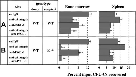
Role of α4 integrin, PSGL-1, and E-selectin ligands in HPC homing to bone marrow. Lethally irradiated wild-type (WT) or E-selectin–deficient (E–/–) mice were injected with antibody-treated wild-type donor BM cells. CFU-Cs were determined from the recipient BM and spleen 3 hours after injection. (A) Transplantation of donor cells treated with rat IgG, anti–α4 integrin (PS/2), anti–PSGL-1 (4RA10), or both PS/2 and 4RA10 into WT recipient mice. n = 6 mice per group. (B) Transplantation of antibody-treated donor cells into E–/– recipient mice. n = 5 per group. *P < .05, **P < .01 compared with rat IgG control group.
Role of α4 integrin, PSGL-1, and E-selectin ligands in HPC homing to bone marrow. Lethally irradiated wild-type (WT) or E-selectin–deficient (E–/–) mice were injected with antibody-treated wild-type donor BM cells. CFU-Cs were determined from the recipient BM and spleen 3 hours after injection. (A) Transplantation of donor cells treated with rat IgG, anti–α4 integrin (PS/2), anti–PSGL-1 (4RA10), or both PS/2 and 4RA10 into WT recipient mice. n = 6 mice per group. (B) Transplantation of antibody-treated donor cells into E–/– recipient mice. n = 5 per group. *P < .05, **P < .01 compared with rat IgG control group.
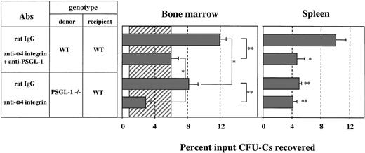
PSGL-1 is a functional E-selectin ligand on HPCs. Bone marrow nucleated cells were harvested from WT control mice and treated with either rat IgG or PS/2 and 4RA10 to block both α4 integrin and the segment of PSGL-1 that interacts with P-selectin. The homing activity remaining after PS/2 and 4RA10 treatment (shaded area) is mediated by E-selectin and its ligands. To evaluate the contribution of PSGL-1 as an E-selectin ligand, PSGL-1 –/– BMNCs were concomitantly transplanted into lethally irradiated WT recipients. Homing of PSGL-1–/– HPCs was further reduced when α4 integrin was blocked, suggesting that PSGL-1 contributes to HPC homing as an E-selectin ligand (*P < .05, **P < .01). Lodgment of HPCs in spleen was significantly reduced when PSGL-1 was inhibited or absent (*P < .05, **P < .01 compared with rat IgG control group). n = 6 to 7 mice per group.
PSGL-1 is a functional E-selectin ligand on HPCs. Bone marrow nucleated cells were harvested from WT control mice and treated with either rat IgG or PS/2 and 4RA10 to block both α4 integrin and the segment of PSGL-1 that interacts with P-selectin. The homing activity remaining after PS/2 and 4RA10 treatment (shaded area) is mediated by E-selectin and its ligands. To evaluate the contribution of PSGL-1 as an E-selectin ligand, PSGL-1 –/– BMNCs were concomitantly transplanted into lethally irradiated WT recipients. Homing of PSGL-1–/– HPCs was further reduced when α4 integrin was blocked, suggesting that PSGL-1 contributes to HPC homing as an E-selectin ligand (*P < .05, **P < .01). Lodgment of HPCs in spleen was significantly reduced when PSGL-1 was inhibited or absent (*P < .05, **P < .01 compared with rat IgG control group). n = 6 to 7 mice per group.
Isolation of cells and CFU-C assay
Blood was harvested by retro-orbital sampling of mice anesthetized with tribromoethanol and collected in polypropylene tubes containing ethylenediaminetetraacetic acid (EDTA). Mononuclear cells were obtained by underlaying 400 μL blood with Lympholyte M (Cedarlane Laboratories, Hornby, ON, Canada) and by centrifugation at room temperature, 280g for 30 minutes. Cells were washed twice in RPMI. BM cells were harvested by aseptically flushing femora of each animal in RPMI using a 21-gauge needle. A single-cell suspension was obtained by gently aspirating several times using the same needle and syringe. The volume of each cell suspension was determined with a graduated pipette. A fraction (25% vol/vol) of a femur was transferred for CFU-C assay. Splenocytes were extracted by homogenizing the spleen using stainless steel blades. Single-cell suspension was ensured by multiple aspirations through a 21-gauge needle, and the suspension volume was measured with a graduated pipette. A fraction (5% vol/vol) of the spleen homogenate was transferred for CFU-C assay. For CFU-C assays, hematopoietic cells were added to 9 vol of Iscove-modified Dulbecco medium containing 0.9% methylcellulose (Nakarai, Japan), 30% fetal bovine serum (StemCell Technologies, Vancouver, BC, Canada), 1% deionized bovine serum albumin (BSA), 10–4 M 2-mercaptoethanol and conditioned medium (20% vol/vol) from WEHI3 cell line (containing interleukin 3 [IL-3]), HM-5 cell line (containing granulocyte macrophage–colony-stimulating factor [GM-CSF]) and BHK/MKL (baby hamster kidney cell line stably transfected with an expression vector containing the cDNA encoding for the secreted form of murine stem cell factor). Cultures were plated in 35 mm culture dishes (StemCell Technologies) and incubated at 37°C in 5% CO2. The numbers of CFU-Cs were scored on day 7 using an inverted microscope.
Competitive reconstitution to assess homing of long-term repopulating stem cells
BM nucleated cells (10 million) from CD45.1 congenic WT mice (donor cells) were treated with 2 μg Abs/106 BMNCs (rat IgG, PS/2, 4RA10, and both PS/2 and 4RA10) and were injected into lethally irradiated (12 Gy, single dose) E–/– mice (CD45.2). Bone marrow cells were harvested 3 hours after injection. After washing with 5 mL RPMI to remove potential contamination of Abs, these BM cells from one femur were injected together with 1 to 2 × 10⁵ CD45.2 WT competitor BM cells into a lethally irradiated (12 Gy, split dose) CD45.2 WT secondary recipient mouse. Blood was harvested monthly from secondary recipient mice, and the expression of CD45.1 and CD45.2 was assessed by flow cytometry. The repopulating units (RUs) were calculated using the following standard formula:34 RU = % (C) / (100-%) where % is the measured percentage of donor cells (CD45.1+ cells) and C is the number of fresh competitor marrow cells per 105. In this calculation, one RU is equivalent to the long-term repopulating stem cell activity of 105 competitor BM cells. Based on this system, the relative stem cell activity was compared among 4 groups of antibody-treated cells after homing to E–/– primary recipient BM.
Flow cytometry
For CD45.1/CD45.2 antigen staining, whole blood (50 μL) was incubated, after blockade of Fc receptors with rat antimouse CD16/CD32 mAb, with FITC-CD45.2 and biotinylated CD45.1 Abs. Blood cells were washed once in phosphate-buffered saline (PBS)/0.03% BSA and stained with PE-streptavidin. Erythrocytes were lysed in 0.8% NH4Cl lysis buffer and the remaining leukocytes were washed twice in PBS/0.03%BSA. Analysis was performed on a FACScan flow cytometer (Becton Dickinson, Mountain View, CA).
Bone marrow intravital microscopy
Six- to 10-week-old nonirradiated or irradiated (12 Gy) C57BL/6 mice were prepared for BM intravital microscopy as described previously.22 Briefly, anesthetized animals were cannulated through the carotid artery to allow injection of cells and Abs, and the frontoparietal skull was exposed for recording of the vasculature of the parietal bone using a fixed-stage custom-designed intravital microscope (MM-40; Nikon, Tokyo, Japan), equipped with a mercury fluorescent lamp and water immersion objectives (Nikon). To assess the role of P- and E-selectin in mediating hematopoietic cell interaction with irradiated endothelium, polymorphonuclear neutrophils (PMNs) were used because previous studies have shown that expression of P- and E-selectin ligands is similar to that of HPCs.22,23 PMNs were purified from the BM of WT or P- and E-selectin double-deficient mice7 using discontinuous Percoll gradients.35 This method reproducibly yields purities over 90% as assessed by Gr-1 antibody fluorescence activated cell sorting (FACS) staining. After lysis of contaminating red blood cells (RBCs) in 0.8% NH4Cl lysis buffer, PMNs were fluorescently labeled with 33 μM carboxyfluorescein succinimidyl ester (CFSE; Molecular Probes, Eugene, OR) for 30 minutes at 6°C and washed twice with RPMI before injection into the carotid artery catheter. Steady-state or irradiated mice prepared for BM intravital microscopy were injected with 60 μg rat IgG (Sigma), rat antimouse P-selectin (clone RB40.34), rat antimouse E-selectin (clone 9A9), or both antiselectin Abs followed by injection of fluorescently labeled PMNs. Images were captured using a SIT camera (Hamamatsu, Japan), a camera-controller (C2400; Hamamatsu), and recorded using a VHS video recorder (SV0-9500MD; Sony, San Jose, CA).
Vessel diameter and cell velocities were measured using a video caliper and sequential single-frame analysis. The maximal velocity (Vmax), which represents the average velocities of free-flowing CFSE-labeled BMNCs or RBCs, was determined for each BM vessel. The mean blood flow velocity (Vmean) was thus calculated: Vmean = Vmax / (2-ϵ2), where ϵ is the ratio of BMNC diameter (about 20 μm) to vessel diameter (Dv). The wall shear rate and critical velocity (Vcrit) were obtained from the following formulas: wall shear rate = 8(Vmean / Dv) and Vcrit = Vmean × ϵ (2–ϵ). Any cell traveling below the Vcrit was considered to be rolling on the vessel wall. Rolling velocities were determined by dividing the rolling distance (in μm) by the rolling duration (in seconds) for each specific event. Cells that remained stationary for at least 5 seconds were considered “arrested” cells.
Statistical analysis
All values are reported as the mean plus or minus SEM. Statistical significance for 2 unpaired groups was assessed by the Student t test for parametric or Mann-Whitney U test for nonparametric comparisons.
Results
PSGL-1 plays a role as a P-selectin ligand in HPC homing to BM but does not cooperate with α4 integrin
To evaluate the function of the PSGL-1/P-selectin pathway alone or in combination with α4 integrin in HPC homing, we used a rat monoclonal antibody (4RA10) generated against the functional N-terminal portion of the PSGL-1 glycoprotein, which specifically blocks the interaction of PSGL-1 with P-selectin.31,32 BMNCs were harvested from WT mice and divided into 4 groups according to their Ab treatment: control rat IgG, anti-α4 integrin, anti–PSGL-1, or both anti-α4 integrin and anti–PSGL-1. As shown in Figure 1A, the percentage of injected HPCs that homed in the BM was 12.6 ± 1.2% in the rat IgG control group. Consistent with previous reports,12,18 we found that anti–α4 integrin treatment of donor BMNCs reduced homing of HPCs by 55% compared with the rat IgG group (P < .01). Interestingly, anti–PSGL-1 treatment significantly reduced homing of HPCs by 33% (P < .05). These reductions were not due to the effects of the Abs on the growth or survival of progenitors since CFU-C counts and colony size from Ab-treated donor cells were similar among the 4 Ab treatment groups (Table 1). Surprisingly, homing was not further reduced when both α4 integrin and PSGL-1 were simultaneously blocked. These results suggest that α4 integrin and PSGL-1 play a partial role and likely act at the same step in HPC homing to the BM, and that even in the absence of 2 major adhesion pathways, approximately 40% of HPCs could still home to the BM.
Effects of antibodies on colony formation in methylcellulose cultures
Antibody . | Number of CFU-Cs per 105 BM nucleated cells . |
|---|---|
| Rat IgG | 258 ± 19 |
| Anti-α4 integrin (PS/2) | 253 ± 20 |
| Anti-PSGL-1 (4RA10) | 257 ± 18 |
| Anti-α4 integrin + anti-PSGL-1 | 243 ± 18 |
Antibody . | Number of CFU-Cs per 105 BM nucleated cells . |
|---|---|
| Rat IgG | 258 ± 19 |
| Anti-α4 integrin (PS/2) | 253 ± 20 |
| Anti-PSGL-1 (4RA10) | 257 ± 18 |
| Anti-α4 integrin + anti-PSGL-1 | 243 ± 18 |
Wild-type donor bone marrow cells were incubated with indicated antibodies (2 μg/106 nucleated cells) for 3 hours and plated for CFU-C assays. Antibody treatment did not alter colony formation. Data are shown as means ± SEMs; n = 7.
E-selectin ligands cooperate with α4 integrin to recruit HPCs into BM
To evaluate whether the remaining homing activity was mediated by E-selectin and its ligands, the same 4 groups of Ab-treated WT BM cells were injected into lethally irradiated E–/– mice. As shown in Figure 1B, the percentage of HPCs homed in BM of the control rat IgG group was similar between WT and E–/– recipients, indicating that E-selectin deficiency itself does not affect HPC homing. Anti–PSGL-1 treatment produced a partial reduction in E–/– recipients similar to that observed in WT recipients (P < .01). This suggests that, in sharp contrast to mature myeloid cells,7,8 P- and E-selectins and their ligands do not cooperate in HPC homing to the BM. Strikingly, anti–α4 integrin treatment in the absence of E-selectin reduced HPC homing to BM by more than 90% (Figure 1B; P < .01). In these experiments, there was a trend toward reduced splenic lodgment in WT recipient mice that received anti–PSGL-1–treated BM cells (Figure 1A, P = .25), and the reduction was statistically significant in the E–/– recipient group (45% reduction, Figure 1B, P < .01), suggesting that PSGL-1 is involved in progenitor lodgment into the spleen. The numbers of circulating CFU-Cs 3 hours after injection were significantly higher in the group treated with both mAbs compared with the control IgG group in both WT and E–/– recipients (Table 2).
Number of circulating CFU-Cs/mL blood 3 hours after injection
Antibody . | WT recipient . | E-/- recipient . |
|---|---|---|
| Rat IgG | 18.8 ± 5.2 | 8.0 ± 0.9 |
| Anti-α4 integrin | 27.9 ± 2.2 | 20.0 ± 3.1* |
| Anti-PSGL-1 | 17.5 ± 3.0 | 16.4 ± 2.6† |
| Anti-α4 integrin + anti-PSGL-1 | 38.9 ± 3.9† | 50.5 ± 17.4† |
Antibody . | WT recipient . | E-/- recipient . |
|---|---|---|
| Rat IgG | 18.8 ± 5.2 | 8.0 ± 0.9 |
| Anti-α4 integrin | 27.9 ± 2.2 | 20.0 ± 3.1* |
| Anti-PSGL-1 | 17.5 ± 3.0 | 16.4 ± 2.6† |
| Anti-α4 integrin + anti-PSGL-1 | 38.9 ± 3.9† | 50.5 ± 17.4† |
Lethally irradiated wild-type (WT) or E-selectin-deficient mice (E-/-) were injected with antibody-treated WT donor BM cells. CFU-Cs were determined from the recipient blood 3 hours after injection. The vast majority of CFU-Cs are rapidly cleared from the circulation but the clearance is significantly delayed when α4 integrin and/or selectins/ligands are inhibited. n = 5 to 6 mice per group.
P < .01 compared with rat IgG control group.
P < .05 compared with rat IgG control group.
E-selectin ligands cooperate with α4 integrin to recruit long-term repopulating stem cells into BM
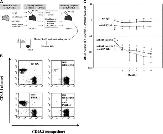
To assess the role of α4 integrin and E-selectin ligands in the homing of long-term repopulating stem cells, we used a competitive reconstitution assay (Figure 2A). BMNCs from CD45.1 congenic WT mice (donor cells) were preincubated with control or adhesion-blocking antibodies and injected into lethally irradiated E–/– mice whose leukocytes express the CD45.2 antigen. Bone marrow cells were harvested 3 hours after injection, and injected together with CD45.2 WT fresh competitor BM cells into lethally irradiated CD45.2 WT secondary recipient mice. Blood was harvested monthly from secondary recipient mice, and the expression of CD45.1 and CD45.2 was assessed by flow cytometry to monitor the contribution of homed donor and competitor cells. Figure 2B demonstrates representative FACS dot plots 3 months after secondary transplantation. Similar dot plots were observed up to 6 months after secondary transplantation. The left upper quadrant of each panel shows CD45.1+ leukocytes which were derived from donor repopulating stem cells homed in BM of E–/– primary recipient mice. Many leukocytes were derived from homed donor stem cells in the control IgG and anti–PSGL-1–treated groups. However, very few leukocytes were derived from CD45.1 donor cells treated with anti–α4 integrin or both anti–α4 integrin and anti–PSGL-1 in the E–/– recipients. Figure 2C depicts RUs between one and 6 months after transplantation. RUs were drastically reduced when the functions of both α4 integrin and E-selectin were absent (P < .05 at each time point throughout 6 months, more than 99% reduction compared with control rat IgG group 6 months after transplantation). There was a slight but significant reduction in RUs one month after transplantation in the anti–PSGL-1–treated group (P = .045), but this difference was no longer significant in the subsequent analyses (months 2 to 6). Analysis by forward and side scatter characteristics revealed that all leukocyte subsets (PMNs, monocytes, and lymphocytes) engrafted and that the RU levels were similar in each leukocyte subset (data not shown). These data suggest that the combination of E-selectin ligands and α4 integrin represents major homing receptors on hematopoietic stem cells.
Competitive reconstitution assay to assess hematopoietic stem cell homing. (A) Schematic representation of competitive repopulation experiment. BM nucleated cells (10 million) from CD45.1 congenic WT mice were treated with rat IgG, anti–α4 integrin, anti–PSGL-1, or both anti–α4 integrin and anti–PSGL-1, and injected into lethally irradiated (12 Gy, single dose) E–/– mice (CD45.2). Femoral bone marrow cells were harvested 3 hours after injection. Bone marrow cells contained in one femur were injected together with 1 to 2 × 10⁵ CD45.2 WT competitor BM cells into a lethally irradiated (12 Gy, split dose) CD45.2 WT secondary recipient mouse. Blood was harvested monthly from secondary recipient mice, and the expression of CD45.1 and CD45.2 was assessed by flow cytometry to determine the repopulating unit (RU). (B) Representative FACS dot plots 3 months after secondary transplantation. The left upper quadrant of each panel shows CD45.1+ leukocytes which were derived from long-term repopulating stem cells homed in the BM of E–/– primary recipient mice. Many CD45.1+ leukocytes derived from homed repopulating cells can be observed in control rat IgG and anti–PSGL-1–treated groups. However, in all mice from the groups treated with anti–α4 integrin or both anti–α4 integrin and anti–PSGL-1, very few leukocytes were derived from cells homed in BM of E–/– first recipients. Similar results were found up to 6 months after secondary transplantation. (C) RU levels following secondary transplantation. RUs were drastically reduced when α4 integrin was inhibited in the absence of E-selectin. n = 4 to 7 mice at one month and n = 3 to 6 at 6 months after secondary transplantation. Data are pooled from 3 independent experiments. *P < .05 for anti–α4 integrin and anti–α4 integrin plus anti–PSGL-1 groups compared with either IgG control or anti–PSGL-1 groups. #P < .05 compared with IgG control.
Competitive reconstitution assay to assess hematopoietic stem cell homing. (A) Schematic representation of competitive repopulation experiment. BM nucleated cells (10 million) from CD45.1 congenic WT mice were treated with rat IgG, anti–α4 integrin, anti–PSGL-1, or both anti–α4 integrin and anti–PSGL-1, and injected into lethally irradiated (12 Gy, single dose) E–/– mice (CD45.2). Femoral bone marrow cells were harvested 3 hours after injection. Bone marrow cells contained in one femur were injected together with 1 to 2 × 10⁵ CD45.2 WT competitor BM cells into a lethally irradiated (12 Gy, split dose) CD45.2 WT secondary recipient mouse. Blood was harvested monthly from secondary recipient mice, and the expression of CD45.1 and CD45.2 was assessed by flow cytometry to determine the repopulating unit (RU). (B) Representative FACS dot plots 3 months after secondary transplantation. The left upper quadrant of each panel shows CD45.1+ leukocytes which were derived from long-term repopulating stem cells homed in the BM of E–/– primary recipient mice. Many CD45.1+ leukocytes derived from homed repopulating cells can be observed in control rat IgG and anti–PSGL-1–treated groups. However, in all mice from the groups treated with anti–α4 integrin or both anti–α4 integrin and anti–PSGL-1, very few leukocytes were derived from cells homed in BM of E–/– first recipients. Similar results were found up to 6 months after secondary transplantation. (C) RU levels following secondary transplantation. RUs were drastically reduced when α4 integrin was inhibited in the absence of E-selectin. n = 4 to 7 mice at one month and n = 3 to 6 at 6 months after secondary transplantation. Data are pooled from 3 independent experiments. *P < .05 for anti–α4 integrin and anti–α4 integrin plus anti–PSGL-1 groups compared with either IgG control or anti–PSGL-1 groups. #P < .05 compared with IgG control.
Endothelial selectins are functional in the bone marrow microvasculature after total body irradiation
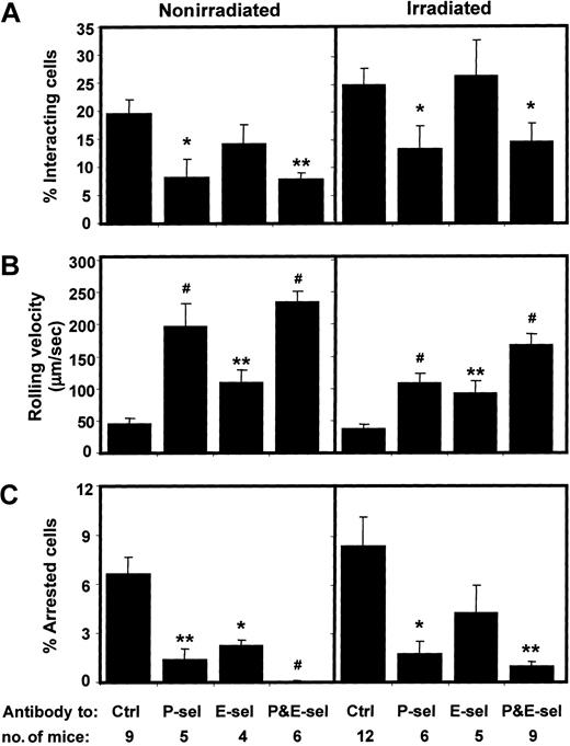
A recent study suggested that the interactions of HPCs with bone marrow microvessels were selectin-independent as early as 3 hours after total body irradiation (TBI), owing to either a down-regulation of endothelial selectins after irradiation or the expression of other compensatory pathways.36 Since this timetable coincides with the present and previous homing studies,12,14,18,37 we further evaluated the expression (and function) of endothelial selectins in irradiated and steady-state BM microcirculation using intravital microscopy. We chose to study the interactions of PMNs with the bone marrow endothelium since we wanted to evaluate selectin function in BM microvessels, and PMNs can be isolated in numbers sufficient to observe their interactions in the BM of a live mouse recipient. Moreover, previous work indicated that mature BM myeloid cells express levels of P- and E-selectin ligands similar to those of immature HPCs.22,23 BM-derived PMNs were purified using Percoll discontinuous gradients, fluorescently labeled and injected via the carotid artery of either steady-state or irradiated mice that were preinjected with control or antiselectin antibodies. Venules in the irradiated groups were recorded 3 to 4 hours after a single 12 Gy dose. In contrast to the study of Mazo et al,36 we observed a significant reduction in the rolling fraction in mice that received anti–P-selectin treatment (Figure 3A) and a significant increase in rolling velocities in mice that received anti–P-selectin and/or anti–E-selectin treatment (Figure 3B), whether the animals received or did not receive prior irradiation. More importantly, there was a dramatic reduction in the number of cell arrests, particularly when mice were treated with both anti–P- and –E-selectin antibodies (Figure 3C). No significant differences in the hemodynamic values were found among these different groups (Table 3). Thus, these results confirm that both endothelial selectins are indeed expressed in BM microvessels 3 to 4 hours after irradiation and can mediate the recruitment of hematopoietic cells.
Endothelial selectins are expressed and functional in the BM microvasculature after irradiation. Fluorescently labeled PMNs obtained from mouse BM were injected into C57BL/6 mice before (nonirradiated, left panels) or 3 hours after lethal irradiation (irradiated, right panels). Mice had been treated with control rat IgG (Ctrl), anti–P-selectin (P-sel), anti–E-selectin (E-sel), or both antiselectin antibodies (P&E-sel). (A) The fractions of cells interacting with the BM microvasculature, (B) rolling velocities, and (C) the fractions of cells arrested on the BM microvasculature were determined by analysis of video recordings from fluorescence intravital microscopy experiments. The number of mice per group is indicated. *P < .05; **P < .01; #P < .001.
Endothelial selectins are expressed and functional in the BM microvasculature after irradiation. Fluorescently labeled PMNs obtained from mouse BM were injected into C57BL/6 mice before (nonirradiated, left panels) or 3 hours after lethal irradiation (irradiated, right panels). Mice had been treated with control rat IgG (Ctrl), anti–P-selectin (P-sel), anti–E-selectin (E-sel), or both antiselectin antibodies (P&E-sel). (A) The fractions of cells interacting with the BM microvasculature, (B) rolling velocities, and (C) the fractions of cells arrested on the BM microvasculature were determined by analysis of video recordings from fluorescence intravital microscopy experiments. The number of mice per group is indicated. *P < .05; **P < .01; #P < .001.
Hemodynamic characteristics of BM microvessels
. | Nonirradiated . | Irradiated . | P . |
|---|---|---|---|
| Vessel diameter (μm) | 42.3 ± 2 | 45.7 ± 4 | .39 |
| V max (μm/s) | 1788.5 ± 193 | 1771.8 ± 181 | .95 |
| WSR (s-1) | 212.7 ± 29 | 191.8 ± 23 | .60 |
. | Nonirradiated . | Irradiated . | P . |
|---|---|---|---|
| Vessel diameter (μm) | 42.3 ± 2 | 45.7 ± 4 | .39 |
| V max (μm/s) | 1788.5 ± 193 | 1771.8 ± 181 | .95 |
| WSR (s-1) | 212.7 ± 29 | 191.8 ± 23 | .60 |
The velocity of free-flowing cells was determined in BM microvessels of irradiated (3 hours after a single 12 Gy dose) or nonirradiated mice after the injection of fluorescently labeled mononuclear and red blood cells obtained from the BM of donor mice. Vessel diameters were measured using a video caliper, and mean blood velocity (Vmax) and wall shear rates (WSRs) were calculated as described.22 Data shown are means ± SEMs from 32 (nonirradiated) and 21 (irradiated) vessels. P values given compare the different parameters between irradiated and nonirradiated groups.
PSGL-1 is a physiologic ligand for E-selectin on HPCs and contributes to homing to BM
The identity of E-selectin ligand(s) expressed on HPCs is unknown. PSGL-1 represents a prime candidate because it is expressed on HPCs at high levels and studies have shown that leukocyte PSGL-1 is involved in E-selectin–mediated leukocyteendothelial interactions.26-29 To test whether PSGL-1 on HPCs is a functional ligand for E-selectin in vivo, we took advantage of PSGL-1–deficient mice whose HPCs are lacking the whole PSGL-1 molecule and the 4RA10 antibody which knocks out only the P-selectin–binding segment of PSGL-1. As shown in Figure 4 (and consistent with results using the pure C57BL/6 donor in Figure 1), treatment of backcrossed WT control BMNCs with both 4RA10 and PS/2 also reduced HPC homing by approximately 50%. Since the remaining homing activity is mediated by E-selectin and its ligands, the role of PSGL-1 as an E-selectin ligand was assayed using PSGL-1–/– BMNCs treated with anti–α4 integrin antibody (PS/2). Homing of PS/2-treated PSGL-1–/– HPCs was further reduced (by 76%, Figure 4), suggesting that PSGL-1 is a functional E-selectin ligand on HPCs. Interestingly, in agreement with data shown in Figure 1, splenic lodgment of HPCs was also reduced when PSGL-1 was absent or inhibited using antibody treatment (Figure 4), suggesting that the PSGL-1/P-selectin pathway indeed plays a significant role in HPC lodgment in the spleen. Treatment of PSGL-1–/– BMNCs with PS/2 antibody did not alter the growth or survival of progenitors (CFU-Cs: 242 ± 25 and 268 ± 9 per 105 BMNCs for rat IgG and PS/2 treated groups, respectively; n = 4; P = .37).
Discussion
Recent data indicate that the adhesion receptors that mediate HPC homing to the BM are not specific to immature blood cells but that, in fact, the adhesion pathways for HPC homing were similar to those used by mature leukocytes to migrate into inflammatory sites. The cooperative function observed in the present study between E-selectin ligands and α4 integrin in hematopoietic progenitor/stem cell homing underscores a major difference between the adhesion mechanisms of mature myeloid cells and immature hematopoietic cells. Previous studies have shown that endothelial selectins and their ligands cooperate for the recruitment of mature leukocytes into inflammatory organs. Function inhibition of, or deficiency in, both P- and E-selectins indeed produce much greater defects in leukocyte rolling and adhesion than singly deficient mice.7-10 In sharp contrast, we show here that E-selectin ligands on murine hematopoietic progenitor/stem cells preferentially cooperate with α4 integrin rather than P-selectin ligands. This unexpected finding raises several interesting points about possible mechanisms at work.
One possibility to account for the limited role of the PSGL-1/P-selectin pathway might be that PSGL-1 isn't fully functional on hematopoietic progenitor/stem cells or that its expression density may be too low to play a more significant role in homing. We have recently shown, for example, that a large subset of cord blood–derived CD34+ cells express an immature (not binding P-selectin) form of PSGL-1 due to incomplete posttranslational modifications.22 However, several facts argue against this possibility, including (1) the density of functional P- and E-selectin ligands on adult human CD34pos CD38neg BM cells is as high as that of mature leukocytes,22,23 (2) murine15 and human22 progenitors can roll on P-selectin in a manner similar to that of leukocytes, and (3) anti–PSGL-1 treatment or PSGL-1 deficiency can partially block progenitor homing (Figures 1 and 4). We have previously described that the initial interactions of human CD34+ cells in bone marrow microvessels of nonobese diabetic–severe combined immunodeficiency mice were largely dependent on endothelial selectins and that VCAM-1 played a modest role.22 Whether selectin ligands exert a more prominent function on human HPCs or whether the difference in selectin function between human and mouse HPCs is due to differential affinities of adhesion receptors (mouse selectins with human ligands and mouse VCAM-1 with human α4β1) across species remains to be determined.
The lack of additive effect of α4 integrin and PSGL-1 might indicate that these 2 receptors act at the same step in the adhesion cascade leading to HPC recruitment. Owing to its rapid on/off association rates, the role of the interaction of PSGL-1 with P-selectin has been limited to the initial rolling step. α4 integrin, however, appears to operate at multiple levels in the recruitment cascade, including rolling, firm adhesion, and migration on extracellular matrix.15,38,39 Thus, when expressed at high density, α4 integrin may partially compensate for the absence or inhibition of PSGL-1. Indeed, the expression level of α4 integrin is higher on HSCs than on total BM cells.40 In contrast to the other selectins, E-selectin appears to be particularly important for the slow rolling of leukocytes that results in long transit times which are essential for efficient leukocyte adhesion in response to local chemotactic stimuli.41 It is possible that E-selectin and its ligands exert similar functions in the interactions of HPCs with the bone marrow vasculature. It is interesting that the inactivation of CD18 and α4 integrin functions37 produced inhibition of HPC homing similar to that of E-selectin and α4 integrin (present study). Thus, this study and the present data suggest similar functions of CD18 and E-selectin ligands on HPCs.
It is also possible that the ligation of E-selectin ligands, but not P-selectin ligands, may produce “cross-talk” signals to α4 integrin that up-regulate integrin affinity and/or avidity, and thus enhance progenitor/stem cell homing. The engagement of either P-selectin or E-selectin with their ligands can transduce signals that activate β2 integrins in leukocytes.42,43 P-selectin binding to monocytes (via PSGL-1) has also been shown to enhance monocyte binding to soluble VCAM-1, presumably by up-regulating α4β1 integrin function.44 Whether a similar phenomenon can occur for β1 integrins on HPCs/HSCs is unknown.
An alternative explanation for the absence of cooperation between P- and E-selectin ligands on HPCs might be that α4 integrin simply represents a natural adhesion partner for E-selectin ligands even in cells that also express P-selectin ligands. Although this possibility may seem counterintuitive, it is notable that PMNs, whose rolling interactions are largely mediated by both endothelial selectins and their ligands, express little α4 integrin. The synergy observed between E-selectin ligands and α4 integrin pathways for hematopoietic progenitor/stem cell adhesion suggests that similar adhesion mechanisms may also be operative in immune responses in which effector cells harbor functional E-selectin ligands and α4 integrin. Indeed, it was recently shown that these adhesion receptors were especially important for lymphocyte infiltration to dermal inflammation.45 It is interesting that the interactions of PSGL-1 expressed on HPCs with P-selectin expressed in the splenic microcirculation significantly contributed to the lodgment of HPCs in the spleen. This feature was not previously noted in our prior homing studies in P/E–/– mice likely because their splenic enlargement may have masked a lodgment defect.14 Progenitors have long been known to form colonies in the spleen (CFU-S),46 a process that requires β1 integrins.47 But the role, if any, of this phenomenon in bone marrow transplantation is not understood. Indeed, HPC homing and engraftment proceeds normally in splenectomized mice (Y.K. and P.S.F., unpublished results, November 2000). It is possible that such P-selectin–mediated splenic trafficking might play a role in certain immune responses.
The dramatic contribution of E-selectin and its ligands in progenitor/stem cell homing to BM clearly indicates that selectins are expressed in the BM microvasculature after irradiation. We confirmed that this was the case by analyzing selectin-dependent leukocyte behavior in the bone marrow using intravital microscopy. The discrepancy between these results and those of Mazo et al36 may be due in part to the lower shear rates in BM microvessels (40 s–1-60 s–1 in Mazo et al36 vs about 200 s–1 in this study) since a minimal shear threshold is also needed for optimal selectin-mediated interactions.48 Perhaps a more likely possibility is the difference in hematopoietic cells used in each study. Since we were interested in evaluating selectins expressed on BM endothelium, not selectin ligands expressed on HPCs, purified PMNs were used for intravital studies instead of fetal liver (FL) cells. FL cells, however, likely express significantly lower cell surface densities of selectin ligands than adult-derived HPCs (or PMNs). Lower levels of PSGL-1 glycoprotein expression on FL cells was reported,36 and the selectin binding avidity of these cells could even be lower if the remaining PSGL-1 molecules were not appropriately modified in the fetal environment. We recently found, for example, that PSGL-1 is expressed in a nonfunctional form (due to immature posttranslational modifications) in a large subset (about 30%) of human cord blood–derived, but not adult-derived BM or G-CSF–mobilized, CD34+ cells.22 It is possible that cells with lower selectin ligand densities may be more sensitive to slight changes in selectin expression in the BM and that such mild alterations may not have been detected by cells expressing higher ligand densities like adult BM–derived HPCs or PMNs. Although we cannot exclude or confirm the possibility that a lethal dose of irradiation might alter selectin expression in the BM, our results indicate that such potential changes do not significantly impact the in vivo behavior of hematopoietic cells that express high densities of functional selectin ligands.
We have demonstrated herein that PSGL-1 is a physiologic ligand for E-selectin which can contribute to HPC homing to BM. We used PSGL-1–deficient mice and an antibody that blocks the interaction with P-selectin to specifically evaluate the contribution of PSGL-1 as an E-selectin ligand. Our results indicate that, when α4 integrin function is inhibited, PSGL-1 contributes to approximately 60% of the E-selectin–mediated homing activity in the bone marrow (Figure 4). It has been shown that mature leukocytes use PSGL-1 to tether to immobilized E-selectin, but PSGL-1 is not needed to support subsequent rolling.29 It will be interesting to evaluate whether PSGL-1 on HPCs/HSCs has similar functions. The corollary of these observations is that other functional ligands for E-selectin must exist on HPCs. A CD44 isoform expressed on human CD34+ cells was recently proposed to bind E-selectin in vitro.24 The fact that homing of CD44-deficient HPCs is as efficient as that of WT animals25 is consistent with this possibility since our data (Figure 1) show that E-selectin deficiency itself does not alter HPC homing. Another candidate is E-selectin ligand-1 (ESL-1), a glycoprotein shown to bind E-selectin in vitro.49 Further work is needed to further characterize functional E-selectin ligands both on mature and immature hematopoietic cells.
In summary, we have identified PSGL-1 as a major E-selectin ligand on HPCs. We have also demonstrated that, in the absence of α4 integrin function, E-selectin ligands expressed on HPCs and long-term repopulating stem cells play significant roles in homing to the bone marrow. The cooperative function of E-selectin and α4 integrin is reminiscent of that previously described for E-selectin and P-selectin ligands on mature leukocytes.7,8,10 These observations underscore a major difference between mature and immature hematopoietic cells in the adhesion mechanisms used to adhere under flow. The partial, but highly significant, contribution of PSGL-1 as an E-selectin ligand suggests the presence of other functional ligands on HPCs and underlines the multiplicity of adhesive pathways operating during progenitor homing.
Prepublished online as Blood First Edition Paper, May 22, 2003; DOI 10.1182/blood-2003-04-1212.
Supported by National Institutes of Health grants R01 DK56638 (P.S.F.) and HL51926 (B.F.).
The publication costs of this article were defrayed in part by page charge payment. Therefore, and solely to indicate this fact, this article is hereby marked “advertisement” in accordance with 18 U.S.C. section 1734.
We would like to thank Linnea Weiss, Anna Peired, and Pegah Jenab for their excellent technical assistance, and Dr Barry Wolitzky for providing the anti–E-selectin antibody.


This feature is available to Subscribers Only
Sign In or Create an Account Close Modal