Abstract
The Steel factor (SF) and its receptor c-Kit play a critical role for various cell types at different levels in the hematopoietic hierarchy. Whether similar or distinct signaling pathways are used upon c-Kit activation in different cell types within the hematopoietic hierarchy is not known. To study c-Kit signaling pathways in the hematopoietic system we have compared c-Kit downstream signaling events in SF-dependent hematopoietic stem cell (HSC)–like cell lines to those of mast cells. Both Erk and protein kinase B (PKB)/Akt are activated by ligand-induced activation of the c-Kit receptor in the HSC-like cell lines. Surprisingly, phosphoinositide-3 (PI-3) kinase inhibitors block not only PKB/Akt activation but also activation of Raf and Erk. SF-induced activation of Ras is not affected by inhibition of PI-3 kinase. In mast cells and other more committed hematopoietic precursors, the activation of Erk by SF is not PI-3 kinase dependent. Our results suggest that a molecular signaling switch occurs during differentiation in the hematopoietic system whereby immature hematopoietic progenitor/stem cells use a PI-3 kinase–sensitive pathway in the activation of both Erk and PKB/Akt, which is then switched upon differentiation to the more commonly described PI-3 kinase–independent mitogen-activated protein (MAP) kinase pathway.
Introduction
The c-Kit gene encodes a 145-kDa transmembrane protein that is a member of the receptor tyrosine kinase subclass III family that includes the platelet-derived growth factor receptor (PDGF-R), macrophage colony-stimulating factor receptor (M-CSF-R or c-fms), and the fms-like tyrosine kinase-3/fetal liver kinase-2 (Flt-3/Flt-2).1 Attenuating mutations in the c-Kit locus (white spotted, or W mutants) lead to a lethal anemia, hematopoietic stem cell (HSC) defects, mast cell deficiency, as well as a series of nonhematologic defects in mice.2-8 The ligand for c-Kit, Steel factor (SF), which is also known as kit ligand, stem cell factor, and mast cell growth factor, exists both as a secreted and membrane-bound form where the latter appears to be most important for biologic activity in vivo.9 Mice deficient in SF expression (Steel or Sl mutants) show similar defects as W mice including in utero lethality due to severe anemia.2 The mast cell deficiency, together with the observation that the entire hematopoietic progenitor/stem cell pool in viable W and Sl mutants is compromised,2,10,11 suggests that c-Kit signaling is important at many different levels in the hematopoietic hierarchy.
Binding of SF to the c-Kit receptor induces dimerization and autophosphorylation of the receptor on tyrosine residues, thereby creating docking sites for cytoplasmic signaling molecules containing Src homology 2 (SH2) domains.7,12,13 As a result of c-Kit activation by SF, proteins of the Ras/Erk and the phosphoinositide-3 (PI-3) kinase pathways are subsequently activated. The mechanism by which these pathways are activated in distinct types of hematopoietic cells remains to be clarified. Thus, elucidation of the c-Kit signaling pathway in HSCs is essential for the understanding of the maintenance of the normal hematopoietic system. However, HSCs are difficult to access experimentally due to their low abundance in hematopoietic tissues. A common approach to study hematopoietic progenitor cells has been to analyze immortalized cell lines at different levels in the hematopoietic hierarchy. Few, if any, of the established cell lines reveal growth characteristics in vitro similar to normal HSCs, suggesting that the characteristics of existing cell lines only partly overlap with those of their normal counterparts.14-18 Thus, development of more accurate model systems is necessary to increase our understanding of the signal transduction pathways responsible for the physiological stimuli that the SF/c-Kit activation exerts on HSCs in vivo.
We have recently generated immortalized HSC-like cell lines by expressing the LIM (lin11, isl-1, and mec3)–homeobox gene Lhx2 in hematopoietic progenitor cells derived from embryonic stem (ES) cells differentiated in vitro19,20 or expression in HSCs derived from adult bone marrow (BM).21 These HSC-like cell lines are dependent on SF for their maintenance in an immature state and share many properties in vitro and in vivo with normal HSCs.19-21 Such characteristics make these HSC-like cell lines unique when compared with previously described cell lines and therefore provide a valuable model system to study intracellular signaling in immature hematopoietic progenitor/stem cells.
To analyze c-Kit signaling in different cell types in the hematopoietic system, we have compared c-Kit signaling in the HSC-like cell lines to that of more committed hematopoietic cells. A transient activation of both Erk and protein kinase B (PKB)/Akt (from here on denoted PKB) can be observed in the HSC-like cell lines upon ligand-induced activation of c-Kit. Surprisingly, we observed that PI-3 kinase inhibitors block phosphorylation not only of PKB, but also Raf-1 and Erk, while not affecting the activation of Ras and upstream signaling molecules, such as Shc and Grb2. Thus, activation of the c-Kit receptor in HSC-like cell lines leads to a PI-3 kinase–dependent Raf/MEK/Erk activation. Analyses of the c-Kit signaling pathway in more committed hematopoietic cells revealed that PI-3 kinase inhibitors do not prevent Erk activation in these cell types. These results suggest that a molecular signaling switch in the c-Kit pathway occurs during differentiation from immature hematopoietic progenitor/stem cells to more committed cell types.
Materials and methods
Generation and maintenance of the HSC-like cell lines
Hematopoietic progenitor cell (HPC) and BM-HPC lines were generated by expressing Lhx2 in hematopoietic progenitor/stem cells derived from ES cells differentiated in vitro or in hematopoietic progenitor/stem cells derived from adult BM, as previously described.19,21 The characteristics of these cells lines in vitro and in vivo have been thoroughly investigated previously.19-21 The cell lines were maintained in Iscove modified Dulbecco media (IMDM; Gibco, Grand Island, NY) supplemented with 5% fetal calf serum (FCS; Integro, Dordrecht, the Netherlands), 1.5 × 10⁻⁴ M monothioglycerol (MTG; Sigma, St Louis, MO), and 100 ng/mL recombinant murine Steel factor (SF; R&D Systems, Minneapolis, MN) at a cell density between 5 × 10⁵ to 4 × 10⁶/mL.
Generation of bone marrow mast cells
Total BM cells from the femurs of 10-week-old C57BL/6-cast mice were harvested and cultured in IMDM supplemented with 1.5 × 10⁻⁴ M MTG, 10% FCS, and 100 ng/mL SF. Nonadherent cells were expanded for 2 to 3 weeks. Biochemical analyses were carried out on expanded cell populations where more than 95% of the cells show mast cell morphology based on May-Grünwald Giemsa (MERCK, Darmstadt, Germany) staining of cytospun cells.
Generation of mast cells from ES cells differentiated in vitro
ES cells transduced with control murine stem cell virus (MSCV) vector or vector containing Lhx2 cDNA (MSCV-Lhx2) were generated, maintained, and differentiated as previously described.19 Briefly, ES cells were maintained in Dulbecco modified Eagle medium (DMEM; Gibco) supplemented with leukemia inhibitory factor (LIF; R&D Systems), 15% FCS (Boehringer Mannheim, Mannheim, Germany), and 1.5 × 10⁻⁴ M MTG. Two days prior to differentiation, the ES cells were passaged into IMDM supplemented as the DMEM. ES cells were trypsinized, and a single-cell suspension was prepared and cultured on suspension culture Petri dishes (Corning 25060-60; Corning, NY) in IMDM supplemented with 15% FCS (Integro), 4.5 × 10⁻⁴ M MTG, and 50 μg/mL ascorbic acid (Sigma). After 6 days of differentiation, embryoid bodies (EBs) were collected and resuspended in trypsin-EDTA (trypsin–ethylenediaminetetraacetic acid) solution and incubated for 3 minutes. Two milliliters of FCS were added, and the cells were gently passaged 2 to 3 times through a 20-gauge needle; 10 mL IMDM was added, and the cells were spun down. The cells were resuspended in the same medium as used for expanding mast cells from the BM, and nonadherent cells were expanded for 2 to 3 weeks. Biochemical analyses were carried out on expanded cell populations where more than 95% of the cells show mast cell morphology based on May-Grünvald Giemsa staining of cytospun cells.
Generation of mast cells and mixed myeloid cells from the HPC line
The HPC line was cultured for 1 week in IMDM supplemented with interleukin-3 (IL-3) (1% vol/vol conditioned media from X63 Ag8-653 myeloma cells transfected with murine IL-3 cDNA)22 to induce differentiation of HPCs as previously described.19 To obtain cell populations containing either mast cells or mixed myeloid cells, the cells were plated in IMDM containing 1% methylcellulose (Fluka, Buchs, Switzerland) and supplemented with l-glutamine, 5% FCS, and 100 ng/mL SF. After 10 days individual colonies were picked from the methylcellulose cultures and transferred to IMDM supplemented with 5% FCS and IL-3. After an additional 7 days each individually expanded clone was split into IMDM supplemented with either IL-3 or SF and cultured for an additional 7 to 10 days. The cells cultured in SF differentiated mainly into mast cells, and biochemical analyses were only carried out in cell populations where more than 95% of the cells were mast cells based on May-Grünwald Giemsa staining of cytospun cells. The population of cells cultured in IL-3 that was subjected to biochemical analyses was mostly composed of myeloiderythroid cells at various stages of differentiation, where 90% of the cells were of the neutrophil and monocyte/macrophages lineages, and 10% of the cells were of erythroid (erythroblasts) lineage.
Antibodies, GST fusion proteins
Anti–c-Kit antibody was a kind gift from Dr L. Rönnstrand, Malmö.23 Anti-Ras (no. R02120) and anti-Erk antibodies were from Transduction Laboratories (no. 610123; Lexington, KY). The antibodies phospho-p42/44 mitogen-activated protein kinase (MAPK), phospho-PKB (Ser473), pan-Akt, and phospho–mitogen-activated protein kinase/ERK kinase 1/2 (phospho-MEK1/2) (Ser217/221) were obtained from Cell Signaling Technology (Beverly, MA). LY294002, PD98059, and wortmannin were from Calbiochem (San Diego, CA) and PP1 from Alexis (Lausen, Switzerland). Glutathione S-transferase fusion protein encompassing the Ras-binding domain of Raf-1 (GST-Raf-RBD) has been described and used according to Henriksson et al.24
Cell lysis, immunoprecipitation, in vitro Raf kinase assay, and Western blotting
The cells were starved overnight (in IMDM without FCS and SF) and subsequently stimulated with SF (50 ng/mL or as indicated in the figures) for various times at 37° C. Thereafter, the cells were washed in ice-cold phosphate-buffered saline (PBS) and lysed on ice in lysis buffer (1% [vol/vol] Triton X-100, 1 mM EDTA, 100 mM NaCl, 50 mM Tris [tris(hydroxymethyl)aminomethane]–HCl [pH 7.5], 1 mM EGTA [ethylene glycol tetraacetic acid], 12 mM MgCl, 1 mM phenylmethylsulfonyl fluoride [PMSF] supplemented with protease inhibitors [10 μg/mL aprotinin, pepstatin, and leupeptin]). Lysates were cleared by centrifugation at 15 000g for 10 minutes at 4° C. Similar amounts of lysate were incubated with primary antibody or GST-Raf-RBD fusion proteins for 1 hour and with protein G–agarose or glutathione-agarose (Pharmacia, Uppsala, Sweden) for a further 30 minutes. After 4 washes in lysis buffer, samples were boiled in sodium dodecyl sulfate–polyacrylamide gel electrophoresis (SDS-PAGE) sample buffer and analyzed by immunoblotting.25,26 Raf-1 kinase assays were performed using the Raf-1 immunoprecipitation kinase cascade assay kit from Upstate Biotechnology (Lake Placid, NY) with some modifications. Lysates from 60 × 106 cells were used for each Raf-1 immunoprecipitation. Serum-starved cells were incubated 30 minutes with 20 μM LY294002 prior to 5 minutes of stimulation with 50 ng/mL SF.
Results
Erk and PKB are transiently activated upon stimulation of an HSC-like cell line with SF
SF binding and subsequent transient tyrosine phosphorylation of the c-Kit receptor on hematopoietic cells activates distinct signaling cascades and mediates different effects on cell survival, proliferation, differentiation, and effector function. These signaling pathways include the Ras/Raf/Erk cascade27 and the PI-3 kinase/PKB pathway.28 Because cell lines generated by expressing Lhx2 in hematopoietic progenitor/stem cells, denoted hematopoietic progenitor cell (HPC) lines, share many characteristics with normal HSCs,20 the initial signaling events downstream the c-Kit receptor were investigated in the HPC lines.
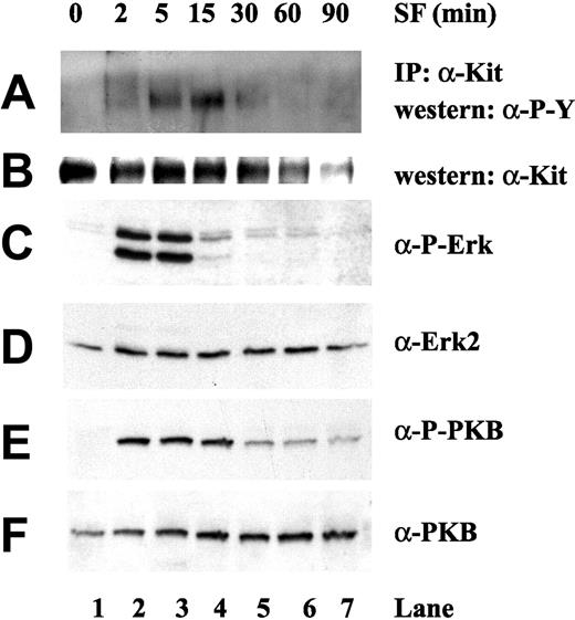
We observe that the c-Kit receptor is transiently tyrosine phosphorylated upon ligation with the SF (Figure 1A). The intrinsic autophosphorylation activity of the c-Kit receptor can be observed after 2 minutes of stimulation with SF and was not observed in nonstimulated cells (Figure 1A). We detected an activation of the c-Kit receptor up to 15 minutes after stimulation with SF. After 15 minutes, however, the transient tyrosine phosphorylation of the c-Kit receptor declines, and after 90 minutes of stimulation with SF, tyrosine phosphorylation of c-Kit could no longer be observed (Figure 1A, lane 7). The membrane was stripped and reprobed with anti–c-Kit receptor antibodies, thus confirming that the band of 145 kDa was the c-Kit receptor (Figure 1B). Furthermore, immunoblotting with anti–c-Kit antibodies revealed that c-Kit receptors were down-regulated over time, suggesting an internalization process upon ligation, which has been described and reported previously in response to SF.29,30
Steel factor activation of c-Kit receptor, Erk, and PKB. HPCs were harvested after stimulation with SF (50 ng/mL) for various times, as indicated. c-Kit protein was immunoprecipitated and immunoprecipitates (A-B) or whole cell lysates (20 μg per lane) (C-F) analyzed by SDS-PAGE, followed by immunoblotting with the indicated antibodies. (A) Immunoblotting with antiphosphotyrosine antibody PY100. (B) Membrane from panel A was stripped and reprobed with anti–c-Kit antibody. (C) Immunoblotting with phospho-specific antibody toward Erk. (D) Membrane from panel C was stripped and reprobed with anti-Erk2 antibody. (E) Immunoblotting with phospho-specific antibody toward PKB. (F) Membrane from panel E was stripped and reprobed with antipan-PKB antibody.
Steel factor activation of c-Kit receptor, Erk, and PKB. HPCs were harvested after stimulation with SF (50 ng/mL) for various times, as indicated. c-Kit protein was immunoprecipitated and immunoprecipitates (A-B) or whole cell lysates (20 μg per lane) (C-F) analyzed by SDS-PAGE, followed by immunoblotting with the indicated antibodies. (A) Immunoblotting with antiphosphotyrosine antibody PY100. (B) Membrane from panel A was stripped and reprobed with anti–c-Kit antibody. (C) Immunoblotting with phospho-specific antibody toward Erk. (D) Membrane from panel C was stripped and reprobed with anti-Erk2 antibody. (E) Immunoblotting with phospho-specific antibody toward PKB. (F) Membrane from panel E was stripped and reprobed with antipan-PKB antibody.
The downstream endogenous targets of c-Kit signaling, such as the Erk and PKB kinases, were analyzed using phospho-specific antibodies, which are a sensitive readout for any changes in activity status. Phosphorylation of both Erk1/2 and PKB was observed upon SF stimulation (Figure 1C,E). The membranes were stripped and reprobed with anti-Erk and PKB antibodies as a control for equal loading of the gel (Figure 1D,F). Thus, as expected, both Erk and PKB are rapidly and transiently activated upon stimulation with SF in this HSC-like cell line.
PI-3 kinase is required for both SF-induced PKB activation and SF-induced Erk activation
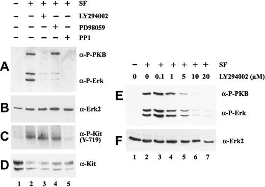
To further investigate SF activation of PKB and the Ras/Raf/Erk cascade, we used the inhibitors LY294002, PD98059, and PP1, which inhibit PI-3 kinase, MEK, and tyrosine kinases, respectively. HPC lines were treated with or without inhibitors for 30 minutes prior to stimulation with SF for 5 minutes (Figure 2). To our surprise we observed that PI-3 kinase inhibitors blocked not only the phosphorylation of PKB but also the phosphorylation of Erk with high efficiency (Figure 2A, compare lane 3 with lane 2). Conversely, PKB was not inhibited when cells were pretreated with the MEK inhibitor PD98059, although Erk phosphorylation/activation was efficiently blocked (Figure 2A, lane 4), confirming that the treatment was effective. As expected and described previously, the tyrosine kinase inhibitor PP1 inhibited both PKB and Erk activation (Figure 2A, lane 5).31 This was further investigated using a phospho-specific antibody towards phosphorylated tyrosine 719 on the c-Kit receptor. The phosphorylation of tyrosine 719 on the c-Kit receptor upon stimulation with SF was inhibited by pretreatment with PP1 but not by pretreatment with the inhibitors LY294002 and PD98059 (Figure 2C, compare lanes 2, 3, and 4 with lanes 1 and 5). Membranes were stripped and reprobed with both anti–c-Kit and anti-Erk as a control for equal loading of the gel (Figure 2B,D).
PI-3 kinase inhibition affects both PKB and Erk activation upon SF stimulation. Serum-starved HPCs were incubated 30 minutes with 20 μM LY294002 (lane 3), 10 μM PD98059 (lane 4), or 10 μM PP1 (lane 5), as indicated (+) prior to 5 minutes of stimulation with 50 ng/mL SF (lanes 2-5, panels A-D). Cell lysates (20 μg per lane) were subjected to SDS-PAGE and then immunoblotted with antiphospho-specific Erk and PKB (A), anti-Erk (B), antiphospho-specific Y-719 c-Kit (C), and anti–c-Kit (D) antibodies. Dose-dependent inhibition of PKB and Erk by LY294002 (E). Cell lysates (20 μg protein per lane) were subjected to SDS-PAGE and then immunoblotted with antiphospho-specific Erk and PKB (E) and anti-Erk (F). In panels E-F, serum-starved cells were incubated 30 minutes with 0.1 μM (lane 3), 1 μM (lane 4), 5 μM (lane 5), 10 μM (lane 6), and 20 μM (lane 7) LY294002, as indicated prior to 5 minutes of stimulation with 50 ng/mL SF (lanes 2-7).
PI-3 kinase inhibition affects both PKB and Erk activation upon SF stimulation. Serum-starved HPCs were incubated 30 minutes with 20 μM LY294002 (lane 3), 10 μM PD98059 (lane 4), or 10 μM PP1 (lane 5), as indicated (+) prior to 5 minutes of stimulation with 50 ng/mL SF (lanes 2-5, panels A-D). Cell lysates (20 μg per lane) were subjected to SDS-PAGE and then immunoblotted with antiphospho-specific Erk and PKB (A), anti-Erk (B), antiphospho-specific Y-719 c-Kit (C), and anti–c-Kit (D) antibodies. Dose-dependent inhibition of PKB and Erk by LY294002 (E). Cell lysates (20 μg protein per lane) were subjected to SDS-PAGE and then immunoblotted with antiphospho-specific Erk and PKB (E) and anti-Erk (F). In panels E-F, serum-starved cells were incubated 30 minutes with 0.1 μM (lane 3), 1 μM (lane 4), 5 μM (lane 5), 10 μM (lane 6), and 20 μM (lane 7) LY294002, as indicated prior to 5 minutes of stimulation with 50 ng/mL SF (lanes 2-7).
We then asked if the inhibition of PKB and Erk by LY294002 displays a similar range of dose dependence, which would be an important validation of PI-3 kinase–dependent Erk activation. Pretreatment of HPCs with increasing dose of LY294002, from 0.1 μM to 20 μM, indicates a comparable inhibition on both PBK and Erk in response to LY294002 (Figure 2E).
Our observations demonstrate that tyrosine kinase activities are required for activation of both Erk and PKB in HPC lines. Furthermore, the PI-3 kinase inhibitor LY294002 visibly inhibits both Erk and PKB phosphorylation, clearly showing that PI-3 kinase is required not only for SF-induced PKB activation but also for SF-induced Erk activation.
PI-3 kinase–dependent Erk activation is specific for immature hematopoietic progenitor/stem cells
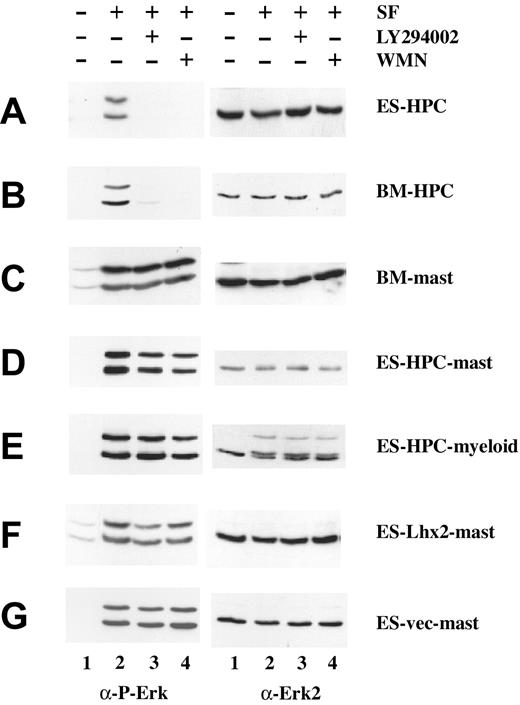
To evaluate if this PI-3 kinase–dependent Erk activation was specific for our hematopoietic precursor cell line, we employed an SF-dependent HSC-like cell line generated by expressing Lhx2 in HSCs derived from adult bone marrow (BM).21 This cell line, denoted bone marrow–derived (BM)–HPC line, also showed a PI-3 kinase–dependent Erk activation, because both LY294002 and wortmannin inhibited SF-induced Erk phosphorylation (Figure 3B), in a manner similar to HPCs (Figure 3A). Thus, Erk activation is PI-3 kinase dependent in immature HSC-like cell lines irrespective of the ontogenic origin of such cell lines.
Analysis of Erk activation in immature and differentiated hematopoietic cells. Serum-starved cells were incubated 30 minutes with 20 μM LY294002 (lane 3) or 50 nM wortmannin (lane 4), as indicated (+) prior to 5 minutes of stimulation with 50 ng/mL SF (lanes 2 to 4). Cell lysates (20 μg per lane) were subjected to SDS-PAGE and then immunoblotted with antiphospho-specific Erk (left panels) and then stripped and reprobed with anti-Erk (right panels), as indicated. Cells analyzed were as follows: (A) ES-HPCs; (B) bone marrow–derived HPCs (BM-HPC); (C) mast cells differentiated from bone marrow cells (BM-mast); (D) mast cells obtained from differentiated HPCs (ES-HPC-mast); (E) myeloid-committed cells obtained from differentiated HPCs (ES-HPC-myeloid); (F) mast cells generated from Lhx2-transduced ES cells (ES-Lhx2-mast); and (G) mast cells generated from vector-transduced ES cells (ES-vec-mast).
Analysis of Erk activation in immature and differentiated hematopoietic cells. Serum-starved cells were incubated 30 minutes with 20 μM LY294002 (lane 3) or 50 nM wortmannin (lane 4), as indicated (+) prior to 5 minutes of stimulation with 50 ng/mL SF (lanes 2 to 4). Cell lysates (20 μg per lane) were subjected to SDS-PAGE and then immunoblotted with antiphospho-specific Erk (left panels) and then stripped and reprobed with anti-Erk (right panels), as indicated. Cells analyzed were as follows: (A) ES-HPCs; (B) bone marrow–derived HPCs (BM-HPC); (C) mast cells differentiated from bone marrow cells (BM-mast); (D) mast cells obtained from differentiated HPCs (ES-HPC-mast); (E) myeloid-committed cells obtained from differentiated HPCs (ES-HPC-myeloid); (F) mast cells generated from Lhx2-transduced ES cells (ES-Lhx2-mast); and (G) mast cells generated from vector-transduced ES cells (ES-vec-mast).
PI-3 kinase is not required for Erk activation in differentiated hematopoietic stem cells
Many studies in different cell lines have shown that the activation of Erk kinase is PI-3 kinase independent. However, is this valid for the hematopoietic system?
To address this we investigated c-Kit signaling in differentiated hematopoietic cells derived from different sources. Initially we analyzed c-Kit signaling in mast cells because it is relatively easy to generate a homogenous population of mast cells in large quantities by culturing BM cells in the presence of SF (see “Materials and methods”). In contrast to the HSC-like cell lines, SF-induced Erk activation was not inhibited by LY294002 or wortmannin in mast cells derived from adult BM (Figure 3C). We have previously reported that the HPC line can differentiate into various myeloid lineages including mast cells using specific cytokines, and the differentiated progeny of the HPC lines maintains Lhx2 expression.19 To exclude that PI-3 kinase–dependent Erk activation is unique to cells immortalized by Lhx2 and/or cells expressing Lhx2, c-Kit signaling was analyzed in differentiated progeny generated from the HPC line. Mast cells and more myeloid committed cells obtained from HPCs in culture (see “Materials and methods”) were starved and treated with LY294002 or wortmannin for 30 minutes prior to stimulation with SF. We observed that neither LY294002 nor wortmannin was able to block Erk phosphorylation in the differentiated cells derived from the HPC line (Figure 3D-E, compare with A). Thus, PI-3 kinase activity is required for c-Kit–dependent Erk phosphorylation in immature hematopoietic progenitor/stem cells but not in more committed hematopoietic cells. However, all cell populations analyzed showed the expected PI-3 kinase–dependent activation of PKB (data not shown). These data suggest that a molecular switch of signaling activities from the c-Kit receptor occurs upon differentiation within the hematopoietic system.
Because the HPC lines were generated by differentiating ES cells transduced with Lhx2 cDNA, we wanted to exclude that Lhx2 expression during the early events of hematopoietic commitment might influence c-Kit signaling. To address this issue, we generated mast cells by in vitro differentiation of ES cells transduced with Lhx2 or an empty vector (see “Materials and methods”). Mast cells generated from the Lhx2-transduced ES cells expressed Lhx2 whereas mast cells generated from the control ES cells did not (data not shown). Both of these mast cell populations showed a PI-3 kinase–independent Erk activation, because LY294002 did not inhibit Erk phosphorylation upon SF stimulation (Figure 3F-G), indicating that Lhx2 expression does not influence c-Kit signaling. These results further support the idea that PI-3 kinase–dependent Erk activation in hematopoietic cells is restricted to immature hematopoietic progenitor/stem cells.
LY294002 inhibits SF-induced MEK phosphorylation but not Ras activation
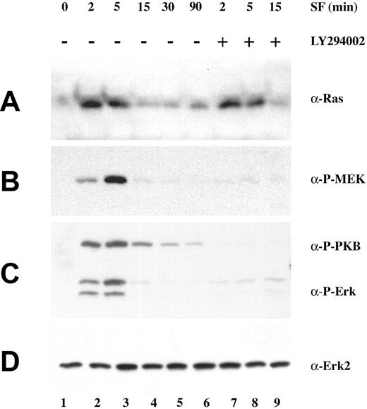
The observation that PI3-K inhibitors were able to abrogate not only PKB, but also Erk phosphorylation, raised the question of whether Ras activation or signaling proteins upstream of Ras were affected by PI-3 kinase inhibition in the HPC line. To investigate this further, HPCs were treated with LY294002 for various times before SF stimulation. Lysates were incubated either with GST-Raf-RBD fusion protein immobilized on glutathione-Sepharose or immunoprecipitated with anti-Shc antibodies. After washing, bound proteins were eluted and subjected to SDS-PAGE analysis followed by immunoblotting for Ras, MEK, PKB, and Erk (Figure 4). From this analysis we also observed that pretreatment of the HPCs with LY294002 followed by stimulation with SF did not affect the phosphorylation of Shc; neither was the Shc-Grb2 interaction upon SF stimulation affected (Supplementary Figure 1; at the Blood website, see the Supplemental Figures link at the top of the online article). Moreover, the ability of Ras to interact with downstream targets upon stimulation with SF, such as the Ras-binding domain of Raf-1, was not impaired on pretreatment of the HPCs with LY294002 (Figure 4A, compare lanes 7 to 9 with lanes 2 to 6). In contrast, MEK phosphorylation was totally blocked by the inhibitor LY294002 (Figure 4B, compare lanes 7 to 9 with lanes 2 to 6), suggesting that a signal between PI-3 kinase and MEK is critical for Erk activation in the HPC line. After 5 minutes of stimulation with SF, Erk, MEK, and PKB were still blocked by LY294002, and this was also observed after stimulation for 15 minutes (Figure 4B-C), suggesting that the block of MEK and Erk activation is not the result of a delayed activation signal. Taken together, these results show that PI-3 kinase does not appear to act upstream or at the level of Ras. Instead, our results indicate that PI-3 kinase plays a critical role in Erk activation at some point between Ras and MEK in the HPC line.
The PI-3 kinase inhibitor, LY294002, blocks phosphorylation of Erk and MEK but not Ras–Raf-1 interaction. HPCs were harvested after stimulation with SF (50 ng/mL) for the indicated times (lanes 2 to 9). Prior to SF stimulation, serum-starved HPCs were incubated 30 minutes with 20 μM LY294002 (lanes 7 to 9). Lysates were subjected to affinity precipitation with GST-Raf-RBD prior to detection of activated Ras proteins by immunoblotting with anti-Ras antibodies (A). Cell lysates were subjected to SDS-PAGE and then immunoblotted with phospho-specific anti-MEK (B), phospho-specific anti-Erk and PKB (C), and anti-Erk2 antibodies (D).
The PI-3 kinase inhibitor, LY294002, blocks phosphorylation of Erk and MEK but not Ras–Raf-1 interaction. HPCs were harvested after stimulation with SF (50 ng/mL) for the indicated times (lanes 2 to 9). Prior to SF stimulation, serum-starved HPCs were incubated 30 minutes with 20 μM LY294002 (lanes 7 to 9). Lysates were subjected to affinity precipitation with GST-Raf-RBD prior to detection of activated Ras proteins by immunoblotting with anti-Ras antibodies (A). Cell lysates were subjected to SDS-PAGE and then immunoblotted with phospho-specific anti-MEK (B), phospho-specific anti-Erk and PKB (C), and anti-Erk2 antibodies (D).
PI-3 kinase activity is necessary for Raf activation in the HPC line
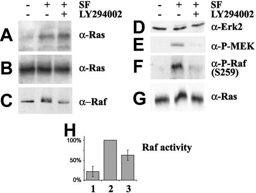
Because PI-3 kinase activity does not appear to play an important role upstream of Ras, but LY294002 is able to completely block MEK phosphorylation, we analyzed Raf-1 activity in the HPC line (Figure 5). To test whether PI-3 kinase activity affects Raf phosphorylation, HPCs were starved and treated with LY294002 before SF stimulation. Lysates were incubated either with GST-Raf-RBD fusion protein, as above, or immunoprecipitated with anti-Raf antibodies. After washing, bound proteins were eluted and subjected to SDS-PAGE analysis followed by immunoblot analysis. Similar to the results shown in Figure 4, the ability of Ras to interact with downstream targets upon stimulation with SF, such as the Ras-binding domain of Raf-1, was not impaired on pretreatment of the HPCs with LY294002 (Figure 5A). A clear Raf-1 shift was observed by SDS-PAGE in HPC cells stimulated with SF, indicating that Raf-1 becomes phosphorylated (Figure 5C, compare lane 1 with lane 2). This shift was not observed in starved HPCs or in SF-stimulated HPCs pretreated with LY294002 (Figure 5C, lanes 1 and 3). To further analyze Raf activation, starved HPC lysates were probed with phospho-specific antibodies toward phosphorylated amino acid 259 on Raf-1 (Figure 5F). We observed phosphorylation of Raf-1 on amino acid 259 upon stimulation with SF, which was blocked when the cells were pretreated with LY294002 prior to SF stimulation (Figure 5F, lane 3). Because the phosphorylation status of Raf kinase is not necessarily an equivalent to enzymatic activity, we immunoprecipitated Raf-1 from starved cells, from cells stimulated with SF, and from cells pretreated with LY294002 prior to stimulation with SF. Immunoprecipitates were then used to perform in vitro kinase assays to evaluate the enzymatic activity of Raf (Figure 5H). Activation of Raf kinase activity was observed upon stimulation with SF, as compared with the basal level of Raf activation observed in starved HPC cells. However, the activation of Raf in response to SF was lowered dramatically upon pretreatment with LY294002 (Figure 5H, lane 3). Taken together, these data indicate that PI-3 kinase activity is necessary for the efficient phosphorylation and activation of Raf-1 upon stimulation with SF in HPCs.
PI-3 kinase activity is required for activation of Raf. Effect of SF (50 ng/mL) stimulation on Raf-1 phosphorylation (lanes 2 and 3). HPCs were pretreated with 20 μM LY294002 (lane 3) for 30 minutes. (A) Lysates were precipitated with GST-Raf-RBD and immunoblotted with anti-Ras antibody. (B) Control immunoblot analysis with anti-Ras. (C) Lysates were immunoprecipitated with anti–Raf-1 antibodies and immunoblotted with anti–Raf-1 antibodies. (D) Control immunoblot analysis of lysates with anti-Erk2. (E) Antiphospho-MEK analysis of the lysates. (F) Antiphospho-specific (S259) Raf-1 immunoblot analysis of lysates. (G) Anti-Ras immunoblot analysis of lysates. (H) In vitro Raf kinase activity. Values (SEM) are calculated from 3 independent experiments performed as described in “Materials and methods.”
PI-3 kinase activity is required for activation of Raf. Effect of SF (50 ng/mL) stimulation on Raf-1 phosphorylation (lanes 2 and 3). HPCs were pretreated with 20 μM LY294002 (lane 3) for 30 minutes. (A) Lysates were precipitated with GST-Raf-RBD and immunoblotted with anti-Ras antibody. (B) Control immunoblot analysis with anti-Ras. (C) Lysates were immunoprecipitated with anti–Raf-1 antibodies and immunoblotted with anti–Raf-1 antibodies. (D) Control immunoblot analysis of lysates with anti-Erk2. (E) Antiphospho-MEK analysis of the lysates. (F) Antiphospho-specific (S259) Raf-1 immunoblot analysis of lysates. (G) Anti-Ras immunoblot analysis of lysates. (H) In vitro Raf kinase activity. Values (SEM) are calculated from 3 independent experiments performed as described in “Materials and methods.”
Inhibition of PI-3 kinase in HPCs induces an apoptotic response
Clarification of the importance of SF-induced PI-3 kinase activation for both Erk and PKB phosphorylation in the HPC line is important for our understanding of c-Kit signaling, but it does not reveal whether these signaling pathways are involved in the survival and/or proliferation of these cells. To test if these signals are important for HPCs in survival and proliferation, we monitored viability and proliferation of the cells with and without inhibitors. Firstly, we observed that SF is absolutely necessary for the HPC line to proliferate, because upon withdrawal of SF no cells survived (Pinto do O et al19 and data not shown). Secondly, treatment of HPCs with 20 μM LY294002 inhibited proliferation profoundly, and fewer cells were observed per milliliter in our assay after 24 hours (Figure 6A). Treatment with the MEK inhibitor PD98059 showed that this pathway is absolutely necessary for proliferation, because cell number increased by only 0.5 × 10⁶ cells compared with the control, where cell number increased by 2.0 × 10⁶ cells after 24 hours (Figure 6A). Thirdly, upon treatment with LY294002, more than 60% of the cells were dead after 24 hours; this was not observed in the control or cells treated with the MEK inhibitor PD98059 (Figure 6B). These observations clearly indicate that PI-3 kinase is important in HPC physiology.
SF-induced HPC line cellular viability and proliferation requires PI-3 kinase activity in vivo. (A) HPCs were cultured in the presence of SF without inhibitors (♦), SF with 20 μM LY294002 (▴), or SF with PD98059 (▪). (B) HPCs were cultured in the presence of SF without inhibitors (♦), SF with 20 μM LY294002 (▴), or SF with PD98059 (▪). Cells were stained with 0.4% trypan blue (Gibco) and examined under visible light for the ability to exclude trypan blue. Each time point represents 2 independent experiments performed in triplicate (SEM).
SF-induced HPC line cellular viability and proliferation requires PI-3 kinase activity in vivo. (A) HPCs were cultured in the presence of SF without inhibitors (♦), SF with 20 μM LY294002 (▴), or SF with PD98059 (▪). (B) HPCs were cultured in the presence of SF without inhibitors (♦), SF with 20 μM LY294002 (▴), or SF with PD98059 (▪). Cells were stained with 0.4% trypan blue (Gibco) and examined under visible light for the ability to exclude trypan blue. Each time point represents 2 independent experiments performed in triplicate (SEM).
Discussion
Clarification of the signaling events downstream of the c-Kit receptor in hematopoietic progenitor/stem cells is important for the understanding of stem cell physiology. In this study we have used novel SF-dependent multipotent HPC lines that share many characteristics with normal HSCs. This HPC line is dependent on SF for its maintenance in an immature state and shares many characteristics in vitro with normal HSCs, such as responses to specific cytokines, interactions with stromal cells, transcription factor expression, and cell surface marker expression.19-21 Upon ligation of SF we show that the c-Kit receptor is transiently tyrosine phosphorylated and down-regulated at the protein level, which is in agreement with recent reports that phosphorylation of the c-Kit receptor is followed by an internalization of the active growth factor receptor.32-35 In addition, we also observed complex formation of a Grb2-Shc complex upon SF stimulation, which further correlated with Ras/Erk/PKB activation. More detailed analysis of Erk and PKB activation upon stimulation with SF was carried out using the PI-3 kinase inhibitors, LY294002 and wortmannin, and the MEK inhibitor, PD98059. PD98059 blocks the SF stimulation of Erk proteins as expected; however, we were surprised to find that the inhibitor LY294002 and wortmannin clearly inhibit not only PKB phosphorylation but also Erk phosphorylation in HSCs. This is important because it indicates that PI-3 kinase is required not only for SF-induced PKB activation but also for Erk activation in these cells. Furthermore, our analysis indicates that PI-3 kinase activity in HPC lines is necessary for the activation of Raf-1, because LY294002 inhibits both the activation of Raf as well as phosphorylation of serine 259 on Raf-1.
While the existing general model for Raf and Erk kinase activation does not reveal an obvious role for PI-3 kinase in hematopoietic cells, a few reports have documented inhibition of Erk kinase phosphorylation/activation by inhibitors of PI-3 kinase in other systems.36-39 Conversely, it is interesting that overexpression of some activated forms of p110 have been reported to stimulate the Ras/MAP kinase pathway.38,40-43 Differentiation stage–specific inhibition of Erk by PI-3 kinase and PKB has been reported in differentiated myotubes but not in their myoblast precursors, indicating an importance for PI-3 kinase modulation during differentiation.44,45 Constitutive PKB expression (via phosphorylation of Raf-1), inhibition of Erk pathway by inhibitors, or dominant negative Raf-1 can all promote differentiation and myotube formation, whereas enforced Raf-1 activity blocks differentiation in the precursor myoblast stage.46,47 The mechanisms involved in the activation of Erk by PI-3 kinase in these systems are not fully understood. However, regulation of the crosstalk between these pathways may vary with cell stage specificity in a temporal and spatial manner. Interestingly, in Xenopus laevis oocytes, which may be thought of as an immature derived cell, it has been suggested that PI-3 kinase is able to stimulate Ras.41 However, in HPC lines, the LY294002 inhibitor clearly does not block Ras activation or activation of signaling molecules upstream of Ras. We also noted that inhibition of the activation by SF was not a result of a delayed activation signal from the receptor or due to excessive concentrations of LY294002, because 200 ng/mL SF was clearly inhibited by 20 μM LY294002 (200 ng/mL SF is 4-fold higher required amount of SF) (data not shown). From this we conclude that the PI-3 kinase does not appear to play a role on/at the level of Ras or upstream of Ras in the SF/c-Kit cascade but that PI-3 kinase drives Raf/MEK/Erk activation through the modulation of the activation of Raf by an as yet uncharacterized mechanism.
Presence of SF is absolutely necessary for the HPC line to survive. Similar to withdrawal of SF, we show that addition of LY294002 to growing HPC lines decreases the proliferation and survival rate dramatically. This correlates with cleavage of the caspase-3 death protein (data not shown) and indicates that PI-3 kinase plays an important and physiologically relevant role in HPC physiology and survival. Conversely, PD98059, the MEK inhibitor, did not affect the survival of HPCs dramatically but did have a marked effect on proliferation rate. Furthermore, recent studies have shown that the key signaling pathway responsible for maintaining the viability of erythroid cells is via PKB, which is activated by both erythropoietin and SF.48,49
A valid concern regarding the HPC line and the observed PI-3 kinase–dependent Erk activation is the Lhx2 gene, with which the HPC line is transformed. However, we observed that mast cells, derived from ES cells differentiated in vitro, regardless of transduction by Lhx2, lose their PI-3 kinase–dependent Erk activation. Thus, we conclude that Lhx2 itself plays no obvious role in the PI-3 kinase–dependent Erk activation we have observed. Similar to our results in differentiated ES cells, neither differentiated myeloid nor mast HPC lines or mast cells of bone marrow origin show PI-3 kinase–dependent Erk activation. Thus, during the differentiation process a molecular signaling switch appears to occur between c-Kit and Erk. Putative explanations for these results could be that either the amount of c-Kit receptors on the cell surface changes during the differentiation process or that different splice variants of the c-Kit receptor50 are differentially expressed during the differentiation process. However, there is no correlation between the amount of receptor expressed by the cells and the signaling pathway used (Supplementary Figure 2); nor is there any change in the expression of the 2 described splice variants of the receptor during differentiation (data not shown).
In summary, we show that both Erk and PKB are transiently activated by SF-induced ligation of the c-Kit receptor in hematopoietic stem cells. Surprisingly, PI-3 kinase inhibitors block both phosphorylation of PKB, MEK, and Raf-1 but not the activation of Ras and upstream signaling molecules, such as Shc and Grb2. These results show that, in addition to Ras activation, a PI-3 kinase–dependent pathway is essential for SF-induced Erk activation in hematopoietic stem cells. Indeed, activation of Ras in the absence of PI-3 kinase activity in these cells is insufficient for Erk activation.
Differentiation of these hematopoietic stem cells results in a signaling switch from PI-3 kinase–dependent to PI-3 kinase–independent Erk activation. This molecular signaling switch appears to be due to alternate crosstalk between the PI-3 kinase and Erk pathways at the level of Raf, together with the cellular background and/or stage of differentiation. Taken together, our results suggest that PI-3 kinase/PKB signaling plays an important and complex role in the maintenance of hematopoietic progenitor cells in the multipotent state.
Prepublished online as Blood First Edition Paper, March 2, 2004; DOI 10.1182/blood-2003-07-2554.
Supported by the Swedish Cancer Society, Swedish Natural Science Research Council, and Lions Cancer Fund, Umeå, Sweden.
E.W. and C.E.E. contributed equally to this study.
The online version of the article contains a data supplement.
The publication costs of this article were defrayed in part by page charge payment. Therefore, and solely to indicate this fact, this article is hereby marked “advertisement” in accordance with 18 U.S.C. section 1734.
We thank Dr Lars Rönnstrand, Malmö, Sweden, for the generous gift of the c-Kit antibody.


This feature is available to Subscribers Only
Sign In or Create an Account Close Modal