Abstract
To investigate the role of constitutively active internal tandem duplication (ITD) mutants of the Fms-like tyrosine kinase 3 (Flt3) receptor in leukemogenesis, we introduced the Flt3-ITD, W51, into human cord blood CD34+ cells and evaluated their phenotype in diverse hematopoietic assays. W51 expression resulted in a strong proliferative advantage and enhanced erythropoiesis as determined by immunophenotyping, colony assays, and molecular analyses. In MS-5 stromal cocultures, numerous early cobblestone areas (CAs) were generated within 10 to 14 days. Such W51-associated early CAs disappeared by 4 weeks, yet retained self-renewal properties as demonstrated by generation of secondary and tertiary CAs upon replating. This phenotype appears related to the expression of W51 since it was abolished by exposure to the FLT3 inhibitor, AG1295, but not to the c-kit inhibitor PD16. Wild-type Flt3–overexpressing CD34+ cells exposed to high levels of its physiologic ligand did not produce early CAs, highlighting differences in intracellular signaling between wild-type Flt3 and W51. W51-associated signal transducer and activator of transcription 5 (Stat5) activation plays a major role in this phenotype, although additional downstream targets of W51 may be relevant. Flt3-ITD+ acute myeloid leukemia (AML) blasts from patients invariably generated early AG1295-sensitive CAs in MS-5 cocultures, further validating the phenotype observed in transduced CD34+ cells.
Introduction
Human Fms-like tyrosine kinase 3 (Flt3) is a class III tyrosine kinase receptor expressed in early human hematopoietic stem cells (HSCs).1,2 In the mouse, selective Flt3 expression is observed in common lymphoid progenitors, whereas multilineage repopulating HSCs lack Flt3 in the steady state,3 and up-regulation of its expression in Lin– Sca1+ c-kit+ HSCs leads to loss of self-renewal capacity, further highlighting its restricted role in murine hematopoiesis.4 In contrast, nearly all human multilineage-reconstituting HSCs in cord blood express Flt3 and are responsive to its ligand (FL) that ensures survival of these cells.5 High Flt3 levels, and receptor phosphorylation, are found in many hematologic malignancies including almost all acute myeloid leukemias (AMLs) of all French-American-British (FAB) subtypes, many cases of myelodysplastic syndrome (MDS), T-cell acute lymphoblastic leukemia (t-ALL), and chronic myelogenous leukemia (CML).6-8 This led to the discovery of activating mutations of Flt3, which represent the most common mutation in AML (accounting for up to 35% of the cases), span all FAB subtypes, and confer a poor prognosis with increased risk of relapse.9-11 The most common mutation involves an in-frame internal tandem duplication (ITD) of the sequence encoding the juxtamembrane domain of Flt3.12 Additional Flt3-ITDs vary in location within the juxtamembrane domain and in length. The further search for mutations that could confer constitutive Flt3 activation led to the identification of a point mutation in the codon for a specific, highly conserved aspartate residue, Asp835, present in as many as 7% of patients with AML.13,14
Flt3-ITD mutants dimerize in a ligand-independent manner and display constitutive activation of the tyrosine kinase domain.15,16 Flt3-ITDs can form heterocomplexes with wild-type Flt3 (wtFlt3), resulting in tyrosine phosphorylation of both molecules.15 Expression of Flt3-ITDs confers cytokine-independent growth on Ba/F3 and 32D cells, and results in a lethal oligoclonal myeloproliferative disorder in a retroviral murine bone marrow transplant model.16,17 In contrast, overexpression of wtFlt3 even in the presence of FL does not replace the interleukin-3 (IL-3) growth requirement in factor-dependent cell lines, suggesting a unique biologic feature of the Flt3-ITD. Several groups have demonstrated qualitative differences in signaling by wtFlt3 and ITD mutants. Mizuki et al showed that Flt3-ITDs signal through the mitogen-activated protein kinase (MAPK) and AKT pathways very weakly relative to activated wtFlt3 in murine Flt3-ITD+ cell lines and ITD+ AML blasts.18 Constitutive signal transducer and activator of transcription 5a/b (Stat5a/b) activation was induced by all Flt3-ITDs studied, whereas Stat5 phosphorylation by the wtFlt3 is minimal and does not translate into enhanced DNA binding in response to FL, suggesting that Stat5 may be a critical downstream target of the Flt3-ITDs.18
The Asp835 Flt3 mutation, like the ITDs, has also been shown to constitutively activate the tyrosine kinase domain and confer cytokine independence in murine cell lines such as 32D-Cl.3.14 The mechanism that leads to activation of the receptor remains unclear, but a current hypothesis suggests that this mutation triggers the activating loop within the cytoplasmic domain of the receptor into an active conformation and stabilizes it in the activated state.19 However, no clear prognostic significance is demonstrated in patients with AML harboring this mutation, whereas the Flt3-ITDs are clearly associated with an adverse prognostic impact and increased risk of relapse.9,10 These findings have prompted the development of Flt3 inhibitors including SU11248, CT53518, and AG1295, which have shown promise for targeted therapy.20-23
The species-specific differences identified in functional studies of Flt3 highlight the difficulties in extrapolating results of murine studies to the human hematopoiesis. Hence, we established a retroviral transduction model to address the role of Flt3-ITDs in the most relevant target—the primary human CD34+ cell. Enforced expression of the Flt3-ITD in human CD34+ cells resulted in accelerated self-renewal and proliferation, and development of early (days 10-14) cobblestone areas (CAs) in MS-5 stromal cocultures, a system where cobblestone formation by control cells was observed only at 5 weeks. Early CAs appeared to be a direct consequence of Flt3-ITD expression since they were undetectable in cells overexpressing wtFlt3 exposed to high levels of FL. Furthermore, the nonadherent cells generated in W51 cocultures appeared enriched in progenitors (particularly erythroid) as determined by colony assays and phenotypic analyses. Cocultures of primary AML blasts with Flt3 mutations fully confirmed the early CA finding. The in vitro phenotype described in this paper may have potentially important diagnostic and clinical implications for AML.
Materials and methods
Retroviral vectors and constructs
A murine stem cell virus (MSCV)–based retroviral expression vector (MIGR1) was used for all experiments. The MIGR1 plasmid (kindly provided by Dr Warren Pear, The University of Pennsylvania, Philadelphia) contains a multicloning site upstream of an internal ribosomal entry site 2 (IRES2) element and the coding sequence for the enhanced green fluorescent protein (EGFP) downstream of the latter. MIGR1 constructs carrying the cDNAs encoding human wtFlt3 and the W51 and W78 Flt3-ITDs17 were kindly provided by Dr Gary Gilliland (Harvard Institute of Medicine, Boston, MA). To generate stable, high-titer retroviral packaging cell lines, H29 cells were transiently transfected with 10 μg vector DNA by calcium-phosphate precipitation. After 72 hours, the H29 supernatant, containing vesicular stomatitis virus (VSV)–pseudotyped retrovirus, was used for cross-transduction of PG13 packaging cells, in the presence of polybrene (8 μg/mL; Sigma, St Louis, MO). High-titer retrovirus-producing PG13 subclones were selected by EGFP+ cell sorting.
Retroviral transduction protocol
Human cord blood–derived CD34+ cells isolated as detailed were prestimulated for 48 hours in serum-free Quality Biological serum-free–60 medium (Quality Biological, Gaithersburg, MD) supplemented with c-Kit ligand (KL, 100 ng/mL), Flt3 ligand (FL, 100 ng/mL), and thrombopoietin (Tpo, 100 ng/mL). KL and Tpo were provided by Peprotech (Rocky Hill, NJ); and FL, by Imclone Systems (New York, NY). Retrovirus-containing supernatants were harvested from PG13 clones incubated in QBSF for 8 to 12 hours; supplemented with 100 ng/mL KL, FL, and Tpo, and polybrene (4 μg/mL); filtered through a 0.45-μm filter (Costar, Cambridge, MA); and applied immediately to the CD34+ cells on retronectin-coated 6-well plates (Takara, Otsu, Japan). Every 8 to 12 hours, 3 consecutive transduction rounds were performed.
Cell culture and stromal cell lines
Human umbilical cord blood (CB) was kindly provided by the Cord Blood Bank subdivision of the New York Blood Bank from healthy full-term pregnancies. Human CD34+ cells were selected from the ficoll-separated mononuclear CB cells using the MiniMACS CD34 isolation kit (Miltenyi Biotech, Auburn, CA). Mononuclear cells were separated from peripheral blood and bone marrow of patients with AML and without Flt3 mutations under an institutional review board and ethics committee–approved clinical protocol with informed consent at Memorial Sloan-Kettering Cancer Center (MSKCC). The murine MS-5 stromal cell line was kindly provided by Dr Itoh (Department of Biology, Niigata University, Japan) and grown in α-modified essential medium (α-MEM; Gibco-BRL Life Technologies, Grand Island, NY) supplemented with 10% fetal bovine serum (FBS; Hyclone, Logan, UT). H29 cells were cultured in Dulbecco modified Eagle medium (Gibco-BRL Life Technologies) supplemented with 10% FBS, penicillin streptomycin, and 200 mM glutamine. MS-5 stromal cocultures and long-term culture-initiating cell (LTC-IC) experiments were performed in Gartner medium (α-MEM containing 12.5% FBS, 12.5% horse serum [Hyclone], penicillin and streptomycin, 200 mM glutamine, 57.2 μM β2-mercaptoethanol [Fisher Scientific, Fair Lawn, NJ], and 1 μM hydrocortisone [Sigma]).
Cytokine-driven (Delta) cultures were performed in Iscoves modified Eagle medium (IMDM; Gibco) containing 20% FBS and 100 ng/mL KL, FL, and Tpo. Population doubling (PDL) curves were calculated based on weekly total nucleated cell counts and replating of 1 × 10⁵ cells.
In some experiments, an adenoviral vector expressing human soluble Flt3 ligand (kindly provided by Dr Demeteo, MSKCC) was used for transduction of MS-5 stromal cells for 12 hours in QBSF-60, at a multiplicity of infection (MOI) of 15.
Colony-forming cells (CFCs), cobblestone area–forming cells, LTC-IC, and serial CAFC assays
Colony assays were performed in triplicate in 35-mm plates using 1.2% methylcellulose (Dow Chemical, Waterloo, NY); 30% FBS; 57.2 μM β2-mercaptoethanol; 2 mM glutamine; 0.5 mM hemin (Sigma); 20 ng/mL IL-3, IL-6, granulocyte colony-stimulating factor (G-CSF), and KL; and 6 U/mL erythropoietin (Epo). IL-3, IL-6, and KL were from Peprotech (Rocky Hill, NJ), G-CSF was from Amgen (Thousand Oaks, CA), and Epo was from Ortho Biotech (Bridgetown, NJ). Colonies were scored 14 days after plating.
Cobblestone area–forming cell (CAFC) assays were performed by plating 1 × 10⁴ CB-CD34+ cells on to MS-5 monolayers in T12.5 tissue-culture flasks (Becton Dickinson, Franklin Lanes, NJ) in triplicate. Weekly demidepopulations were performed, with phenotypic analysis of nonadherent cells. CAs were scored through the course of 5 weeks. Cobblestone areas were defined as groups of at least 10 phase-contrast dark cells tightly associated beneath the MS-5 monolayer.
Limiting dilution assays were performed by plating sorted EGFP+ MIGR1– and W51-transduced CD34+ cells in 24-well tissue-culture plates containing MS-5 monolayers, at 5 progressive dilutions with 12 replicate wells per dilution. Additional LTC-IC experiments were performed using sorted EGFP+ CD34+/CD38– and CD34+/CD38+ subsets or patients' blast cells to determine the CAFC frequency.
Secondary CAFC assays were performed by trypsin harvesting day-10 to day-14 adherent cells and replating all cultures, or sorted EGFP+ cells, on fresh MS-5 monolayers.
The Flt3-RTK inhibitor AG1295 (Sigma) was applied to MS-5 cocultures at 10 μM. The c-kit–specific inhibitor PD16 (kindly provided by Dr Bayard Clarkson, Sloan-Kettering Institute, NY) was also applied at 10 μM in specific experiments.
An Olympus (Melville, NY) fluorescent microscope was used to visualize cells at 400 × original magnification; SPOT advanced software was used for acquisitions, and a SPOT II 1.4.0 camera captured images (Diagnostic Instruments, Sterling Heights, MI).
Cell fractionation, Western blots, immunocytochemistry, and morphologic analysis
To prepare nuclear extracts, 3 × 10⁵ sorted EGFP+ MIGR1–, wtFlt3-, and W51-transfected cells were centrifuged and incubated on ice for 15 minutes in an extraction buffer containing 10 mM HEPES (N-2-hydroxyethylpiperazine-N′-2-ethanesulfonic acid, pH 7.9), 10 mM KCl, 0.1 mM EDTA (ethylenediaminetetraacetic acid), 0.1 mM EGTA (ethylene glycol tetraacetic acid), 1 mM dithiothreitol (DTT), and 1:100 protease inhibitor cocktail. Nonidet P-40 (NP-40, 1.2%) was then added. Nuclear pellets and postnuclear supernatants were obtained by centrifugation (10 000g, 30-60 seconds) and used for immunoblot evaluations. Both nuclear pellets and postnuclear supernatants were boiled for 5 minutes in Laemmli sample buffer prior to separation on 4% to 20% gradient sodium dodecyl sulfate (SDS)–acrylamide gels. Proteins were transferred to nitrocellulose filters (Millipore, Billerica, Spain) in Tris (tris(hydroxymethyl)aminomethane)–glycine buffer/20% methanol at 8 V for 1.5 hours using the BioRAD (Hercules, CA) electroblotter. Membranes were blocked overnight in phosphate-buffered saline (PBS) containing 4% nonfat milk prior to incubation with antibodies. The enhanced chemiluminescence detection method was used according to the manufacturer's instructions (Amersham Pharmacia Biotech, Piscataway, NJ). Antibodies (all from Santa Cruz Biotechnology, Santa Cruz, CA) to Flt3 (C20) and Stat5 (L20) were used at a dilution of 1:1000, and the secondary horseradish peroxidase–conjugated rabbit antimouse antibodies, at a 1:200 dilution.
Cell cytospins were fixed with 4% paraformaldehyde, permeabilized in PBS 0.1% Tween-20, and stained with antibodies (1:100). Secondary cyanin 3–conjugated antibodies (Jackson Immuno-research, West Grove, PA) were used at 1:200 dilution for visualization using a Zeiss inverted fluorescent microscope (Carl Zeiss, Heidelberg, Germany). Max-Grünwald-Giemsa staining was also performed on cytospins for morphologic evaluation.
RT-PCR analysis
Reverse-transcription–polymerase chain reaction (RT-PCR) was performed using total RNA isolated from 0.5 to 1.0 × 10⁶ sorted cells by the RNAeasy kit (Qiagen, Valencia, CA) following the manufacturer's protocol. The Qiagen OneStep RT-PCR kit was used, involving reverse transcription of 2 μg RNA with OneStep RT-PCR Enzyme mix and Omniscript and Sensiscript RTs, and amplification of the cDNA with primers and HotStarTaq DNA-polymerase as indicated. Negative controls contained the same amounts of RNA but no RT. Aliquots (20 μL) were run on 2% agarose gels.
Flow cytometry
Phycoerythrin-conjugated antibodies were from Pharmingen (San Diego, CA). Cells were incubated with the relevant antibodies at 4°C for 1 hour, washed once with PBS/2% FCS, and analyzed on a FACScalibur (Becton Dickinson). The data were acquired and analyzed with the CellQuest (Becton Dickinson) software. All cell sortings were performed using MoFlo (Cytomation, Denver, CO).
Results
Retroviral transduction of human CB-CD34+ cells
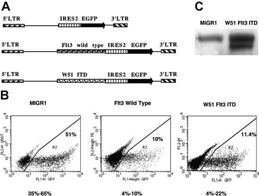
Purified human CB-CD34+ cells were prestimulated into cell cycle and then transduced with bicistronic retroviral constructs carrying the cDNAs for the Flt3-ITDs (W51 and, in some experiments, W78), wtFlt3, or no transgene upstream of an IRES element and the EGFP reporter gene (Figure 1A). Gene transfer efficiencies were determined by measuring the EGFP fluorescence 24 hours after the final transduction round. In multiple independent experiments, mean transduction efficiencies of 15 ± 5%, 15 ± 5%, and 45 ± 10% were obtained from W51, wtFlt3, and control (MIGR1) viral supernatants, respectively (Figure 1B). Expression of the W51 Flt3-ITD in transduced cells was confirmed by Western blot, in comparison with MIGR1-transduced cells (Figure 1C).
Enforced expression of wild-type Flt3-RTK and W51 Flt3-ITD in human CB-CD34+ cells. (A) Map of MIGR1, wtFlt3, and W51 Flt3-ITD retroviruses used in this study. (B) Transduction efficiencies were determined by EGFP expression using flow cytometry analysis. Purified CB-CD34+ cells were prestimulated for 48 hours in QBSF-60 supplemented with KL, FL, and Tpo (100 ng/mL) and then subjected to 3 transduction rounds as described in “Materials and methods.” (C) Immunoblot of whole-cell protein extracts from transduced and sorted CB-CD34+ cells with anti-Flt3 antibody (Ab). LTR indicates long terminal repeat.
Enforced expression of wild-type Flt3-RTK and W51 Flt3-ITD in human CB-CD34+ cells. (A) Map of MIGR1, wtFlt3, and W51 Flt3-ITD retroviruses used in this study. (B) Transduction efficiencies were determined by EGFP expression using flow cytometry analysis. Purified CB-CD34+ cells were prestimulated for 48 hours in QBSF-60 supplemented with KL, FL, and Tpo (100 ng/mL) and then subjected to 3 transduction rounds as described in “Materials and methods.” (C) Immunoblot of whole-cell protein extracts from transduced and sorted CB-CD34+ cells with anti-Flt3 antibody (Ab). LTR indicates long terminal repeat.
Selective in vitro proliferative advantage of W51-expressing cells and induction of erythropoiesis
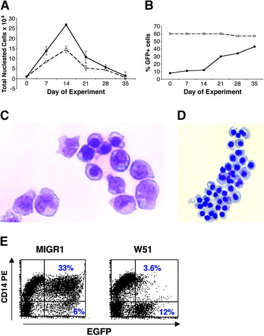
CB-CD34+ cells transduced with W51, wtFlt3, or MIGR1 were cocultured on a monolayer of the MS-5 stromal cell line and demidepopulated weekly for evaluation. In nonsorted experiments, W51-expressing cells demonstrated a proliferative advantage over the course of 5 weeks as determined by the increase of the EGFP+ nonadherent progeny from 10% to more than 50% by week 5, whereas the MIGR1 EGFP+ population remained constant (Figure 2A-B). A similar proliferative advantage was observed in cocultures with other stromal cell lines capable of supporting human hematopoiesis including AGM-S2 and OP-9 (not shown).
Proliferative advantage of CB-CD34+ cells expressing the W51 Flt3-ITD in stromal coculture and liquid cultures. CB-CD34+ cells were transduced with MIGR1 or W51 and evaluated in stromal MS-5 coculture in Gartner media. Cells were demipopulated weekly for counting (A) and FACS analyses (B). (A) Absolute expansion of nonsorted, nonadherent cells. (B) Percentage of EGFP+ cells in the nonadherent fraction. (C) Cytoprep of nonadherent cells from week-2 MS-5 coculture of W51-transduced cells reveals primitive myelomonocytic and erythroid blasts. (D) Clusters of erythoblasts from the same cocultures. (E) Comparative FACS analysis of CD14 expression in nonadherent, week-2 MS-5 coculture of W51+ and MIGR1 cells highlights a drastic reduction in CD14 expression in Flt3-ITD–expressing cell cultures.
Proliferative advantage of CB-CD34+ cells expressing the W51 Flt3-ITD in stromal coculture and liquid cultures. CB-CD34+ cells were transduced with MIGR1 or W51 and evaluated in stromal MS-5 coculture in Gartner media. Cells were demipopulated weekly for counting (A) and FACS analyses (B). (A) Absolute expansion of nonsorted, nonadherent cells. (B) Percentage of EGFP+ cells in the nonadherent fraction. (C) Cytoprep of nonadherent cells from week-2 MS-5 coculture of W51-transduced cells reveals primitive myelomonocytic and erythroid blasts. (D) Clusters of erythoblasts from the same cocultures. (E) Comparative FACS analysis of CD14 expression in nonadherent, week-2 MS-5 coculture of W51+ and MIGR1 cells highlights a drastic reduction in CD14 expression in Flt3-ITD–expressing cell cultures.
Cytokine-driven (Delta) cultures (not shown) confirmed the proliferative advantage of W51+ cells, with a persistent increase in the EGFP+ population up to 2 weeks, in contrast to control cultures where the EGFP+ fraction declined through the culture period. However, cytokine-independent growth could not be demonstrated for the W51-expressing cells.
The expanding W51+ population in stromal cocultures and in cytokine-driven cultures consisted predominantly of immature myelo-monocytic blasts and erythroblasts at various stages of differentiation, as demonstrated by immunophenotype and cytospin morphology (Figure 2C-D). Flow cytometry revealed a dramatic reduction in the monocytic marker, CD14, and an increase in erythroid markers CD71bright and glycophorin A within the first 2 weeks of coculture in comparison with controls, which accumulated CD14+ monocytes and macrophages (Figure 2E; Table 1).
Immunophenotype of EGFP+ adherent and nonadherent cells from week-2 MS-5 coculture of transduced CB-CD34+ cells in percentages
. | Adherent cells . | . | Nonadherent cells . | . | ||
|---|---|---|---|---|---|---|
. | MIGR1 . | W51 (Flt3-ITD) . | MIGR1 . | W51 (Flt3-ITD) . | ||
| CD14 | 74 | 11.5 | 70 | 12 | ||
| CD15 | 8 | 5 | 20 | 41 | ||
| CD19 | 0 | 0 | 0 | 0 | ||
| CD33 | 64 | 84 | 78 | 53 | ||
| CD34 | 2 | 0.5 | 2 | 4 | ||
| CD41 | 0 | 0 | 0 | 0 | ||
| CD71 | 18 | 35 | 14 | 35 | ||
| GlyA | 4 | 80 | 16 | 26 | ||
| CD135 | 2 | 0.5 | 2 | 11 | ||
The data represent the average of 2 to 5 independent experiments.
GlyA indicates glycophorin A.
The total progenitor expansion of W51+ cells in MS-5 cocultures was significantly greater than that of MIGR1+ cells throughout the course of 4 weeks as enumerated by weekly colony assays, indicating a proliferative advantage of these progenitors (Figure 3A). Immediately following transduction, the W51+ population contained significantly more burst-forming units–erythroid (BFU-Es, 198 vs 116/1000 cells), colony-forming units–macrophage (CFU-Ms, 14 vs 6/1000 cells), and CFUs–granulocyte macrophage (CFU-GMs, 44 vs 35/1000 cells) than control MIGR1 cells (Figure 3B). Thus, W51 expression induced the expansion of both myeloid and, more pronouncedly, erythroid progenitors (Figure 3C). Accordingly, RT-PCR analysis of freshly transduced, purified W51-expressing cells showed a significant increase in the expression of the β-globin transcript compared with control cells (Figure 3D).
Progenitor expansion in W51-transduced CD34+ cells cultures. Colony assays of sorted MIGR1- and W51-transduced CD34+ cells. Week-0 cells were plated after transduction and sorting. (A) Total colonies were enumerated in triplicate at each time point. (B) The lineage distribution of colonies (CFU–granulocyte-erythrocyte-megakaryocyte-macrophage [CFU-GEMM], CFU-GM, CFU-M, BFU-E) from week-0 sorted cells. The data shown are representative of 3 separate experiments. (C) Representative BFU-Es generated from the week-0 colony assay of sorted W51+ cells (C, 1). Note the terminally differentiated erythrocytes (C, 2) within the BFU-Es. (D) RT-PCR evaluation of the α-globin mRNA in EGFP+ sorted cells expressing W51 Flt3-ITD and MIGR1 control. Total RNA was isolated and used as described in “Materials and methods” for RT-PCR.
Progenitor expansion in W51-transduced CD34+ cells cultures. Colony assays of sorted MIGR1- and W51-transduced CD34+ cells. Week-0 cells were plated after transduction and sorting. (A) Total colonies were enumerated in triplicate at each time point. (B) The lineage distribution of colonies (CFU–granulocyte-erythrocyte-megakaryocyte-macrophage [CFU-GEMM], CFU-GM, CFU-M, BFU-E) from week-0 sorted cells. The data shown are representative of 3 separate experiments. (C) Representative BFU-Es generated from the week-0 colony assay of sorted W51+ cells (C, 1). Note the terminally differentiated erythrocytes (C, 2) within the BFU-Es. (D) RT-PCR evaluation of the α-globin mRNA in EGFP+ sorted cells expressing W51 Flt3-ITD and MIGR1 control. Total RNA was isolated and used as described in “Materials and methods” for RT-PCR.
W51-expressing CD34+ cells specifically generate early cobblestone areas with self-renewing capacity
The week-5 to week-6 cobblestone area–forming cell (CAFC) assays on MS-5 stromal cells classically enumerate primitive hematopoietic stem cells that migrate, dwell, and proliferate below the stroma. Early (week 2) cobblestone areas, believed to arise from a committed progenitor, are also observed in MS-5 cocultures of CB-CD34+ cells, but rapidly decline in frequency following the cytokine exposure required for the transductions (Figure 4A).
Day-12 cobblestone areas in MS-5 cocultures of W51+ cells. Nonsorted, transduced CB-CD34+ cells expressing W51 were plated onto a monolayer of the MS-5 stroma. The photograph shows a representative early cobblestone area (CA). All early CAs were strongly EGFP+ (A). (B) The frequency of early week-2 CA formation depends upon the interval of cytokine prestimulation of CB-CD34+ cells. CB-CD34+ cells were prestimulated for either 0, 1, 2, 3, 4, and 5 days and plated (1 × 10⁴/T12.5 in triplicate) onto an MS-5 monolayer, with enumeration of week-2 early CAs.
Day-12 cobblestone areas in MS-5 cocultures of W51+ cells. Nonsorted, transduced CB-CD34+ cells expressing W51 were plated onto a monolayer of the MS-5 stroma. The photograph shows a representative early cobblestone area (CA). All early CAs were strongly EGFP+ (A). (B) The frequency of early week-2 CA formation depends upon the interval of cytokine prestimulation of CB-CD34+ cells. CB-CD34+ cells were prestimulated for either 0, 1, 2, 3, 4, and 5 days and plated (1 × 10⁴/T12.5 in triplicate) onto an MS-5 monolayer, with enumeration of week-2 early CAs.
Cocultures of W51+ cells showed a dramatic development of early (day 10-14) EGFP+ CAs (Figure 4B), which were absent in control cell cocultures, with a frequency of 1.8% to 4% as determined by limiting dilution CAFC assays of sorted EGFP+ cells on MS-5 stroma, in independent experiments (Table 2). These early CAs were no longer detected by week 4 of coculture, whereas MIGR1 control cells gave rise to classic week-5 CAs at a frequency 4-fold lower than the early W51+ CAs (Table 3). Initially, in addition to W51, we also investigated the effect of enforced expression of the W78 Flt3-ITD. W78-expressing CD34+ cells generated abundant early CAs (Table 2) with a profile indistinguishable from that induced by W51. Hence in the development of the study, we chose to pursue the study of the latter mutation.
Two representative experiments demonstrating early CAs in MS-5 coculture in triplicate T12.5 flasks
. | Experiment A . | . | Experiment B . | . | ||
|---|---|---|---|---|---|---|
. | Day 7 . | Day 14 . | Day 11 . | Day 18 . | ||
| Control | 0 | 0 | 0 | 0 | ||
| MIGR1 | 0 | 0 | 0 | 0 | ||
| wtFlt3 | 0 | 0 | 0 | 0 | ||
| W51 ITD | 29 ± 3 | 200+ | 48 ± 7 | 200+ | ||
| W78 ITD | 10 ± 1 | 200+ | 37 ± 10 | 200+ | ||
CA frequencies determined by limiting dilution assays
Limiting dilution CAFC assay, sorted GFP* . | Experiment 1, % . | Experiment 2, % . |
|---|---|---|
| W51 Flt3-ITD, d 10 | 4 | 1.8* |
| W51 Flt3-ITD, wk 5 | < 0.1 | < 0.1 |
| MIGR1 control, wk 5 | 1.0 | 0.5 |
Limiting dilution CAFC assay, sorted GFP* . | Experiment 1, % . | Experiment 2, % . |
|---|---|---|
| W51 Flt3-ITD, d 10 | 4 | 1.8* |
| W51 Flt3-ITD, wk 5 | < 0.1 | < 0.1 |
| MIGR1 control, wk 5 | 1.0 | 0.5 |
The secondary CAFC frequency ≥ 1/cobblestone area.
Early CAs isolated by flow cytometry from the stromal monolayer consisted predominantly of myelo-monocytic blasts and erythroblasts at various stages of differentiation. W51+ early CAs contained more glycophorin A+ and CD71bright cells compared with week-2 adherent cells from MIGR1 cocultures (Table 1), mirroring the progenitor population identified in the nonadherent fraction. In contrast, the week-5 CA cells harvested from MIGR1 cocultures contained exclusively myeloid blasts but no erythroid cells. Upon replating on fresh MS-5, purified W51 early CA cells generated secondary and tertiary early CAs with a replating efficiency of 1 or more. These, like the parental CAs, displayed a life span of less than 4 weeks.
Sorting of stem cell–enriched CD34+/CD38–/low and progenitor-enriched CD34+/CD38+ subsets from W51-transduced cells revealed that both fractions contained early CAFCs and generated immature myeloid and erythroid progenitors in MS-5 cocultures. The CD34+/CD38–/low fraction, however, displayed a 5-fold greater CAFC frequency than the CD34+/CD38+ counterpart (7% vs 1.5%). Moreover, only the CD34+/CD38– subset had self-renewal capacity and was able to develop secondary and tertiary CAs upon passage.
To address the specificity of the W51-dependent early CA formation, we compared MS-5 cocultures of CB-CD34+ cells transduced with wtFlt3, W51, or MigR1 in the presence of high levels of FL. Only the W51+ cocultures generated early CAFCs (data not shown), suggesting that their formation involves specific signaling events downstream of the mutated, constitutively active Flt3 receptor.
Flt3-ITD–mediated early CA formation is abolished by the Flt3-specific inhibitor tyrphostin (AG1295), but not by the c-kit–specific inhibitor PD16
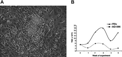
Previous studies have shown that tyrphostin is an effective Flt3 tyrosine kinase inhibitor in hematopoietic cells. Therefore, we tested whether exposure to tyrophostin affected the phenotype observed in W51-transduced cells. As a control, to rule out possible toxicity, CB-CD34+ cells transduced with a constitutively activated mutant of Stat5, a downstream target of the Flt3-ITD, that also develop early CAs (Schuringa et al submitted) were treated with AG1295. Administration of 10 μM AG1295 to MS-5 cocultures of W51-transduced cells every 48 hours (a) inhibited the expansion of W51-transduced cells in coculture (Figure 5A); and (b) resulted in a 7-fold reduction in early CAs compared with nonexposed cocultures (Figure 5B). Application of 10 μM AG1295 to W51+ cocultures on day 14 caused the dramatic disappearance of all CAs within 72 hours (not shown). As expected, treatment with AG1295 did not affect the expansion or the early CA formation of CD34+ cells expressing constitutively active Stat5 (Figure 5A-B).
Early CAs in MS-5 coculture are mediated by the Flt3-ITD. Transduced CB-CD34+ cells expressing the MIGR1 control, W51 Flt3-ITD, and a constitutively activated Stat5 mutant (Stat5A1*6) were plated in triplicate MS-5 cocultures, with weekly evaluation of nonadherent cell expansion in control cultures (A) and in cultures supplemented with 10 μM AG1295 (Flt3-RTK inhibitor) every 48 hours. Day-10 CAs were enumerated for each set of cultures (B). Parallel MS-5 coculture experiments were also performed with W51 Flt3-ITD cells in the presence of 10 μMof the c-kit RTK specific inhibitor, PD16, demonstrating no effect on early CAs (C). TNC indicates total nucleated cell.
Early CAs in MS-5 coculture are mediated by the Flt3-ITD. Transduced CB-CD34+ cells expressing the MIGR1 control, W51 Flt3-ITD, and a constitutively activated Stat5 mutant (Stat5A1*6) were plated in triplicate MS-5 cocultures, with weekly evaluation of nonadherent cell expansion in control cultures (A) and in cultures supplemented with 10 μM AG1295 (Flt3-RTK inhibitor) every 48 hours. Day-10 CAs were enumerated for each set of cultures (B). Parallel MS-5 coculture experiments were also performed with W51 Flt3-ITD cells in the presence of 10 μMof the c-kit RTK specific inhibitor, PD16, demonstrating no effect on early CAs (C). TNC indicates total nucleated cell.
As an additional control, the c-kit–specific tyrosine kinase inhibitor, PD16, was applied to W51+ cocultures at 10 μM. No effect on progenitor expansion and CA formation could be discerned, confirming the specific role of Flt3-ITD in the generation of early CAs (Figure 5C).
Stat5 is a downstream target of the Flt3-ITD
Stat5 activation is likely to play a key role in the development of early CAs by Flt3-ITD+ cells as evidenced by the abundance of early CAs in CD34+ cells expressing a constitutively active mutant of Stat5.24 Western blots and immunocytochemistry revealed a strong increase in nuclear localization of Stat5 in both wtFlt3 and W51+ CD34+ cells (Figure 6) in the presence of Flt3 ligand.
Nuclear localization of Stat5 in wtFlt3- and W51-transduced CD34+ cells in the presence of FL. Immunoblots of nuclear and cytoplasmic extracts of transduced and sorted W51 Flt3-ITD and wtFlt3 CB-CD34+ cells were stained with anti-Stat5 Ab, as detailed in “Materials and methods.”
Nuclear localization of Stat5 in wtFlt3- and W51-transduced CD34+ cells in the presence of FL. Immunoblots of nuclear and cytoplasmic extracts of transduced and sorted W51 Flt3-ITD and wtFlt3 CB-CD34+ cells were stained with anti-Stat5 Ab, as detailed in “Materials and methods.”
In preliminary experiments (not shown), CD34+ cells cotransduced with W51 and a dominant-negative form of Stat5 (Y694F) were unable to form early CAs. This suggests that Stat5 activation is essential for the phenotype observed in W51-expressing cells.
Primary Flt3-ITD+ AML blasts generate early CAs
Bone marrow or peripheral blood mononuclear cells from AML patients (4 with Flt3-ITD+, 1 harboring the Flt3-Asp835 mutation, and 5 clinically matched Flt3-ITD–) were evaluated comparatively in MS-5 cocultures. Immunophenotypically, all samples contained predominantly myeloid blasts with few CD14+ cells. The Flt3-ITD+ and the Flt3-Asp835+ cells generated large numbers of CAs within 10 days that persisted for 5 weeks, whereas the cells from the Flt3-ITD– patients did not generate early CAs, but only low numbers of week-5 CAs (Table 4). Secondary CAs developed upon passage of the early Flt3-ITD+ CAs, confirming the self-renewing properties of these cells consistent with the phenotype of W51-transduced CB-CD34+ cells.
Day-10 to day-14 CA formation in Flt3-ITD+ and Flt3-ITD- AML blasts
AML patient . | Blast cells . | Day-10 to day-14 CAs, input 5 × 10⁵ cells . | Day-35 CAFCs . |
|---|---|---|---|
| Flt3-ITD+ | |||
| Patient 1 | PB | 98 ± 5 | 200+ |
| Patient 2 | BM | 109 ± 14 | 200+ |
| Patient 3 | BM | 200+ | 200+ |
| Patient 4 | BM | 200+ | 200+ |
| Asp 835+ | |||
| Patient 1 | PB | 84 ± 3 | 200+ |
| Flt3-ITD- | |||
| Patient 1 | BM | 0 | 25 ± 1 |
| Patient 2 | PB | 0 | 19 ± 2 |
| Patient 3 | BM | 0 | 12 ± 2 |
| Patient 4 | BM | 0 | 28 ± 4 |
| Patient 5 | BM | 0 | 21 ± 3 |
PB indicates peripheral blood; BM, bone marrow.
Evaluation of one representative Flt3-ITD+ sample (patient no. 3) by LTC-IC limiting dilution revealed an early CAFC frequency of approximately 1:50 000 cells and a 30-fold higher week-5 CAFC frequency (Figure 7). Administration of AG1295 every 48 hours resulted in virtually complete abolishment of the early CAs and an associated decline in total cell expansion (Figure 7). This faithfully mirrored the phenotype identified in Flt3-ITD–transduced CB-CD34+ cells.
Generation of early, tyrphostin-sensitive CAs in MS-5 cocultures of Flt3-ITD+ AML blasts. Day-10 to day-14 CAs were enumerated in the cocultures of AML blasts harboring Flt3-ITDs, from patient no. 3 (A). The addition of 10 μM AG1295 (Flt3-RTK inhibitor) every 48 hours prevented the formation of early CAs, with an associated decline in nonadherent cell expansion in MS-5 stromal coculture (B).
Generation of early, tyrphostin-sensitive CAs in MS-5 cocultures of Flt3-ITD+ AML blasts. Day-10 to day-14 CAs were enumerated in the cocultures of AML blasts harboring Flt3-ITDs, from patient no. 3 (A). The addition of 10 μM AG1295 (Flt3-RTK inhibitor) every 48 hours prevented the formation of early CAs, with an associated decline in nonadherent cell expansion in MS-5 stromal coculture (B).
Discussion
The establishment of a retroviral transduction model to enforce expression of the Flt3-ITD, W51, in human CD34+ cells has allowed us to investigate the role of this constitutively active mutant receptor in primary, nonleukemic human hematopoietic stem and progenitor cells. Our results show that W51 expression confers selective growth advantage and accelerated self-renewal on CD34+ cells, thereby enhancing the generation of myeloid and especially erythroid progenitors in MS-5 stromal cocultures. One striking feature of the W51-expressing CD34+ cells is the development of abundant early cobblestone areas in MS-5 cocultures. The expression of constitutively active Flt3-ITD appears essential for the generation and maintenance of such phenotype, since exposure to the Flt3 inhibitor, AG1295 (but not to the c-kit–specific inhibitor PD16), not only prevents their development but also induces rapid disappearance of pre-existing CAs (Figure 5; data not shown). Classically, the week-5 to week-6 CAs that develop in stromal or Dexter-type cultures are believed to derive from primitive hematopoietic cells, representative of stem cells capable of long-term repopulation in nonobese diabetic–severe combined immunodeficient (NOD/SCID) mice and referred to as SCID-repopulating units (SRUs). In contrast, earlier-developing CAs have been attributed to a more committed pool of short-term repopulating hematopoietic progenitors based on their enhanced cycling status and sensitivity to the S-phase–specific antimetabolite 5-fluorouracil (5-FU).25 Breems et al observed day-10 to day-14 CAs in cocultures of CB-CD34+ cells with the FBMD-1 stromal cell line. These early CAs disappeared upon exposure to 5-FU, whereas the development of week-5 CA was not affected.25 Although this suggests that the more primitive stem cells were not in S-phase, it did not address the biologic significance of early CAFCs.
In our retroviral gene transfer model, CB-CD34+ cells are activated into cell cycle by the stimulatory cytokines KL, FL, and Tpo prior to transduction, and the early CAs are drastically reduced or virtually absent in cells transduced with void vector. We have previously detected week-2 CAs in MS-5 cocultures, using fresh CB-CD34+, or G-CSF–mobilized PB-CD34+ cells, at a frequency of 2% and 1%, respectively. We believe that these represent committed progenitors, possibly equivalent to the common myeloid progenitor (CMP).26 The fact that these early CAs were not detected in MIGR1-transduced CD34+ cells is most likely a consequence of the 4 to 5 days of cytokine stimulation that induces proliferation and possibly alters their mobility, homing ability, or responsiveness to proliferative signals from the MS-5 stroma. In addition, cytokines may induce differentiation of immature CMPs to more committed progenitors, unable to migrate beneath the stroma. The delayed initiation of stem cell proliferation (after 48-72 hours of cytokine treatment) would in turn delay CMP replacement, with consequent depletion of this compartment. Consistent with these hypotheses, a time-course stimulation of CB-CD34+ cells with KL, FL, and Tpo for 0 to 5 days prior to plating in MS-5 cocultures revealed a sharp decline in early CA formation by day 3 (Figure 4A).
It is likely that the high frequency of early CAs with extensive replating potential—a specific feature of Flt3-ITD–transduced CD34+ cells, and in particular of the more primitive CD34+/CD38–/low subset—may reflect accelerated and persisting activation of cell cycle in the immature CAFC population. This would also explain the exhaustion of these early CAs within 4 weeks of coculture unless replated on fresh stroma. Expression of the Flt3-ITD in the CD34+ population was observed in both the CD38+ and CD38–/low compartments, but the ability to form early CAs that could be serially passaged was confined to the latter fraction, depleted of progenitors and enriched for stem cells. This observation further supports our view that the early CAs derive from transduced stem cells.
Differences in activation of intracellular signaling pathways between wild-type Flt3 and the Flt3-ITD have been suggested by cell-line studies. Hayakawa et al16 demonstrated constitutive activation of MAPK and Stat5 in Flt3-ITD–transfected 32D and Ba/F3 cells, and the absence of MAPK activation in wtFlt3-transfected counterpart, a finding validated by the study of many ITD+ AMLs. Further Ba/F3 studies demonstrate constitutive Stat5 activation only in Flt3-ITD–transduced Ba/F3 but not wtFlt3-transduced cells exposed to saturating levels of FL (20 ng/mL).27 These findings were consistent with a recent report showing selective induction of STAT-responsive genes in Flt3-ITD–transformed 32Dcl3 cells.28 Hence, the absence of early CAs in MS-5 cocultures of wtFlt3-transduced cells continually exposed to high levels of its physiologic ligand further highlights the specific effects of the Flt3-ITD mediated by downstream effectors.
Our study delineates a role for Stat5 activation in the development of early CAs by W51-expressing cells. A strong increase in nuclear localization of Stat5 was observed in sorted, W51-transduced CD34+ cells (Figure 2) as well as in the Flt3-ITD+ patients' blasts. Expression of a constitutively active variant of Stat5A in CB-CD34+ cells produces a phenotype that mirrors in many aspects the one described here.24 In preliminary experiments, suppression of Stat5 activity by coexpression of a dominant-negative Stat5 (Y694F) in W51+ cells completely abolished the generation of early CAs. Taken together, these data strongly suggest that continual signal transduction through Stat5 results in enhanced proliferation and self-renewal of immature hematopoietic cells expressing Flt3-ITDs. CD34+ cells transduced with wtFlt3 exposed to high levels of FL also show significant Stat5 activation, as demonstrated by its nuclear localization (Figure 6), but do not display an early CA phenotype. Different hypotheses may explain this observation: persistence of the ITD mutant receptor at the cell surface, accumulation of Stat5, inhibition of modulatory pathways, and finally activation of a different spectrum of downstream signaling mechanisms by FLT3-ITDs. Analyses of the transcript and protein profiles in W51-expressing cells are being carried out and will help identify molecules that may contribute to the generation of the phenotype of Flt3-ITD–expressing cells.
The early CA formation observed in W51-transduced CD34+ cells was validated by cocultures of AML blast cells expressing Flt3-ITDs or the Asp835 Flt3 mutation.
Rombouts et al29 reported that Flt3-ITD+ AML blasts had a reduced proliferative ability in FBMD-1 stromal coculture relative to AML blasts not bearing Flt3 mutations. In addition, high levels of early CAs were found in both AML categories and fell progressively to zero by 6 weeks.29,30 This contrasted with our results obtained using MS-5 stroma, where early CAs were formed only by primary AML Flt3-ITD+ blasts or Flt3-ITD–transduced normal CD34+ cells. This is most likely the consequence of differences between MS-5 cells and the stromal cell line used by Rombouts et al and/or of the addition of IL-3 and G-CSF to stromal cocultures in the Rombouts et al studies.
The fact that early CAs are a distinctive and specific feature of AML blasts bearing mutations of the Flt3 gene, and that the CAs are susceptible to an anti–Flt3-RTK agent, further validates our findings in primary normal CD34+ cells, suggesting that the biologic features observed are at least in large part conserved in AML blasts. The diagnostic and therapeutic implications are significant. For instance, the early CA phenotype may provide a reliable quantitative assay for leukemic stem cells and be used to determine their responsiveness to Flt3-ITD inhibitors.
In summary, the experimental in vitro evidence described in this paper provides clues to the role of the Flt3-ITDs in leukemogenesis. Constitutive activation of Flt3 appears to endow primitive hematopoietic cells with considerable proliferative advantage, accelerate CA formation, stimulate progenitor expansion, and induce preferential erythroid differentiation. The clinical relevance of the early CAs is highlighted by its specific reproduction in primary AML blasts harboring Flt3 mutations. Expression of Flt3-ITD does not render normal CD34+ cells cytokine independent, nor does it induce any obvious features resembling malignant transformation, in agreement with the observation of Kelly et al in an in vivo murine system where the single “hit” with W51 resulted in a lethal myeloproliferation but not in frank AML, whereas coexpression of promyelocytic leukemia/retinoic acid receptor α (PML-RARα) caused acute leukemia.31 The enhanced proliferation resulting from Flt3-ITD expression may therefore induce expansion of stem cells that may in turn acquire additional mutations and eventually develop full-blown acute myeloid leukemia. The in vitro model and data presented here will prove valuable in the dissection of the molecular mechanism of action of Flt3-ITDs in primary human stem/progenitor cells and in the study of its cooperation with other oncogenes in the pathogenesis of myeloid leukemias, and may provide a useful system for the evaluation of novel therapeutic agents.
Prepublished online as Blood First Edition Paper, July 8, 2004; DOI 10.1182/blood-2003-12-4445.
K.Y.C. was supported by a Charles A. Dana Foundation Fellowship and National Institutes of Health grant no. CA-09512 (Ruth L. Kirschstein National Research Service Award); J.J.S., by a grant from the European Molecular Biology Organization (ALTF-412-2001); G.M., by a grant from the Italian Ministry of University and Research (PRIN); and M.A.S.M., by P01 CA 59350, R01 HL 61401, and the Leukemia and Lymphoma Society Specialized Centers for Research (SCOR) grants.
The publication costs of this article were defrayed in part by page charge payment. Therefore, and solely to indicate this fact, this article is hereby marked “advertisement” in accordance with 18 U.S.C. section 1734.
The authors would like to acknowledge Diane Domingo, Patrick Anderson, and Jan Hendrikx for excellent flow cytometry assistance; Katja Weisel for providing AGM S2; Agnes Viale (Genomics Core Facility, MSKCC) for microarray analyses; and Dianna Ngok for assistance with cord blood. We gratefully acknowledge M. Heaney, MD, PhD, for providing the AML samples.


![Figure 3. Progenitor expansion in W51-transduced CD34+ cells cultures. Colony assays of sorted MIGR1- and W51-transduced CD34+ cells. Week-0 cells were plated after transduction and sorting. (A) Total colonies were enumerated in triplicate at each time point. (B) The lineage distribution of colonies (CFU–granulocyte-erythrocyte-megakaryocyte-macrophage [CFU-GEMM], CFU-GM, CFU-M, BFU-E) from week-0 sorted cells. The data shown are representative of 3 separate experiments. (C) Representative BFU-Es generated from the week-0 colony assay of sorted W51+ cells (C, 1). Note the terminally differentiated erythrocytes (C, 2) within the BFU-Es. (D) RT-PCR evaluation of the α-globin mRNA in EGFP+ sorted cells expressing W51 Flt3-ITD and MIGR1 control. Total RNA was isolated and used as described in “Materials and methods” for RT-PCR.](https://ash.silverchair-cdn.com/ash/content_public/journal/blood/105/1/10.1182_blood-2003-12-4445/6/m_zh80010571890003.jpeg?Expires=1765891909&Signature=I1o499jtFEMMadyzdZgU4qnz7hEfJv3SV-pzdzZTn1UZwUEXvsqYo~D1OkvnQB6Lga2PwTWTQLslOXdk7qqKCi62YH7JJcd9GTVKf6wSosI2m5WhSUyY2pvd62vTDPz7jAb-RoNN1tHLGVupjtQaxhuF2oJeB9iO0GJeRC0MyBdUyfFDnpXPRQpQBKTue2cdlRVOTihx~93rRxYfaq4rRa7Ue2TFzNyZsGXjxO0CkRcmtfgY6l-ndHiMOE~6RkdyFYY7Spgr6b4XJBUVSIoXoPAOozBEYlwls2B69BiVQ5MysjFU73hbTEqEBrT9jJSbVFXQArAlTU4gAnLgHv8oEA__&Key-Pair-Id=APKAIE5G5CRDK6RD3PGA)


This feature is available to Subscribers Only
Sign In or Create an Account Close Modal