Abstract
Although the mechanisms by which all-trans-retinoic acid (RA) regulates gene transcription are well understood, very little is known on the signaling events regulating RA-dependent initiation of mRNA translation. We examined whether the mammalian target of rapamycin (mTOR)/p70 S6 kinase pathway is activated by RA. RA treatment of sensitive cell lines resulted in phosphorylation/activation of mTOR and downstream induction of p70 S6 kinase activity. Such phosphorylation/activation of p70 S6 kinase was inducible in primary acute promyelocytic leukemia (APL) blasts and RA-sensitive NB-4 cells, but was defective in an NB-4 variant cell line (NB-4.007/6) that is resistant to the biologic effects of RA. The RA-dependent activation of p70 S6 kinase was also phosphatidylinositol 3′ kinase (PI3′K)-dependent, and resulted in downstream phosphorylation of the S6 ribosomal protein on Ser235/236 and Ser240/244, events important for initiation of translation for mRNAs with oligopyrimidine tracts in their 5′ untranslated region. RA treatment of leukemia cells also resulted in an mTOR-mediated phosphorylation of the 4E-BP1 repressor of mRNA translation, to induce its deactivation and dissociation from the eukaryotic initiation factor-4E (eIF-4E) complex. Altogether, these findings provide evidence for the existence of a novel RA-activated cellular pathway that regulates cap-dependent translation, and strongly suggest that this cascade plays a role in the induction of retinoid responses in APL cells. (Blood. 2005;105:1669-1677)
Introduction
All-trans-retinoic acid (RA)1 acts as a potent inducer of cellular differentiation and growth arrest of neoplastic cells in vitro and in vivo.1-8 A very important biologic activity of RA is its ability to induce differentiation of acute promyelocytic leukemia (APL) cells.5,9-11 This activity of RA has led to its introduction in the clinical management of APL and has dramatically changed the outcome of patients suffering from this historically fatal form of acute leukemia.5 It is now well established that, in addition to APL cells, all-trans-retinoic acid and other retinoids inhibit cell proliferation or induce programmed cell death of malignant cells of diverse origin.12-19
Retinoids induce their activities by binding to specific nuclear receptors, the retinoic acid receptors (RARs).20-23 Two families of retinoid receptors exist: RARs (types α, β, γ), which are activated in response to either all-trans-retinoic acid or 9-cis-retinoic acid; and RXRs (types α, β, γ), which are selectively activated by 9-cis-retinoic acid.20-23 RARs and RXRs form homo- or heterodimers that bind to specific response elements present in the promoters of target genes, called retinoic acid response elements (RAREs). Such binding of RA-nuclear receptor complexes to RAREs results in initiation of transcription of genes for protein products that mediate RA-dependent differentiation and growth inhibition, including p21WAF, C/EBPβ, and HoxA1.20-23
In addition to the induction of formation of RAR/RXR complexes, retinoids induce activation of other cellular pathways that appear to play roles in the generation of their effects on target cells. Members of the src-family of kinases (Lyn and Fgr) are activated during RA-induced cell differentiation,24 whereas the guanine exchange factor Vav24 and the CrkL adaptor protein25 are phosphorylated in an RA-dependent manner and are engaged in RA-cellular pathways. Other mechanisms via which retinoids induce their biologic effects on malignant cells include suppression of AP-1 activity,26,27 modulation of histone acetylation,28 and up-regulation of transforming growth factor β 2 (TGF-β2) and insulin-like growth factor binding protein-3 (IGFBP-3) expression.29 Recent studies have also suggested that RA induces an early phosphorylation of Stat1 on serine727 and tyrosine 701, and that such phosphorylation is required for RA-dependent induction of cell-cycle G0/G1 arrest and inhibition of cell proliferation.30,31
The p70 S6 kinase was originally identified as a kinase that regulates serine phosphorylation of the 40S ribosomal S6 protein (rpS6).32-36 This kinase plays important roles in various cellular functions, such as regulation of cell-cycle progression, cell survival, and initiation of mRNA translation via phosphorylation of rpS6.32-42 There is also accumulating evidence that mammalian target of rapamycin (mTOR) and the phosphatidylinositol 3′ kinase (PI3′K)/PDK1 pathway regulate engagement of the p70 S6K,37-44 to generate signals ultimately required for mRNA translation.
The cellular pathways activated by retinoids to regulate mRNA translation for protein products that mediate their biologic effects are not known. In the present study we determined whether the p70 S6 kinase is activated by all-trans-retinoic acid. Our data demonstrate that the p70 S6 kinase is rapidly phosphorylated and activated during treatment of the RA-sensitive NB-4 acute promyelocytic leukemia cell line. Such activation appears to require both PI3′K and mTOR activation, while it is defective in an NB-4 RA-resistant variant cell line. We also demonstrate that RA induces the phosphorylation of the S6 ribosomal protein on Ser235/236 and Ser240/244, strongly suggesting that activation of the p70 S6 kinase plays an important role in the generation of signals for mRNA translation. In other studies we establish that the translational repressor 4E-BP1 is phosphorylated in an RA-dependent manner, resulting in its deactivation and dissociation from the eIF4E complex to allow cap-dependent translation.
Materials and methods
Cells lines and reagents
The all-trans-retinoic acid-sensitive human acute promyelocytic leukemia NB-4 and the resistant variant NB-4.007/6 (NB-4R) cell lines45,46 were grown in RPMI 1640 supplemented with 10% fetal bovine serum and antibiotics. MCF-7 cells were grown in Dulbecco modified Eagle medium supplemented with 10% fetal bovine serum and antibiotics. Antibodies against the phosphorylated forms of p70 S6 kinase, mTOR, and 4EBP-1 were obtained from Cell Signaling Technology (Beverly, MA). The mTOR inhibitor, rapamycin, and the PI3′K inhibitor, LY294002, were obtained from Calbiochem (La Jolla, CA). Peripheral blood mononuclear cells were isolated from the peripheral blood of a patient with acute promyelocytic leukemia, after obtaining informed consent approved by the institutional review board of Northwestern University.
Cell lysis and immunoblotting
Cells were treated with all-trans-retinoic acid (final concentration 1 μM), for the indicated times, and lysed in phosphorylation lysis buffer, as previously described.47,48 Immunoprecipitations and immunoblotting, using an enhanced chemiluminescence (ECL) method, were performed as previously described.48 In the studies in which MCF-7 cells were used, the cells were incubated for 24 hours in medium containing 2% to 5% fetal calf serum (FCS), and RA was subsequently added in the cultures for the indicated time periods. In the experiments in which pharmacologic inhibitors of tacrolimus (FK506)-binding protein-rapamycin-associated protein (FRAP)/mTOR or the PI3′K were used, the cells were incubated in the presence of RA for indicated time periods, and treated with inhibitors for 2.5 hours prior to lysis in phosphorylation lysis buffer.
p70 S6 kinase assays
Assays to detect RA-dependent activation of the p70 S6 kinase were performed as previously described.25,49 Briefly, cells were lysed in phosphorylation lysis buffer and cell lysates were immunoprecipitated with an antibody against p70 S6 kinase or control nonimmune rabbit immunoglobulin (RIgG). In vitro kinase assays were performed using a synthetic peptide substrate (AKRRRLSSLRA), and p70 S6 kinase activity was measured using an S6 kinase assay kit (Upstate Biotechnology, Lake Placid, NY), according to the manufacturer's instructions. Values were calculated by subtracting nonspecific activity detected in RIgG immunoprecipitates, from kinase activity detected in anti-p70 S6K immunoprecipitates.
Luciferase reporter assays
Luciferase assays were performed as previously described.50 Briefly, MCF-7 cells were transfected with a β-galactosidase expression vector and a RARE-luciferase plasmid,51 using the superfect transfection reagent as per the manufacturer's recommended procedure (Qiagen, Valencia, CA). Forty-eight hours after transfection, triplicate cultures were either left untreated or treated with RA for 16 hours in the presence or absence of rapamycin (20 nM), as indicated. The cells were then washed twice with cold phosphate-buffered saline, and after cell lysis, luciferase activities were measured using the protocol of the manufacturer (Promega, Madison, WI). The measured luciferase activities were normalized for β-galactosidase activity for each sample.
Cell proliferation assays
Flow cytometric analysis
Flow cytometric studies were performed as in our previous studies.45,50 Briefly, NB-4 cells were treated with RA for the indicated times, and cell differentiation was determined by staining with the anti-CD11b monoclonal antibody. The anti-CD11b monoclonal antibody and a matched isotype control were purchased from Becton Dickinson (San Jose, CA).
Results
We initially examined whether treatment of RA-responsive cell lines with all-trans-retinoic acid results in phosphorylation and activation of the p70 S6 kinase. Studies were performed with the acute promyelocytic leukemia NB-4 cell line,45,50 in which RA induces cell differentiation and suppresses growth, and with the breast carcinoma MCF-7 cell line, which is sensitive to the growth inhibitory effects of RA.52,53 Cells were incubated with RA for different time periods, and after cell lysis in phosphorylation lysis buffer, total cell lysates were analyzed by sodium dodecyl sulfate-polyacrylamide gel electrophoresis (SDS-PAGE) and immunoblotted with an antibody against the phosphorylated form of p70 S6 kinase on threonine 421 and serine 424. RA treatment resulted in strong phosphorylation of p70 S6 kinase in both NB-4 (Figure 1A) and MCF-7 (Figure 1C) cells, whereas there was no increase in the amounts of p70 S6 kinase protein detected after RA treatment (Figure 1B,D). Such RA-dependent phosphorylation of p70 S6 kinase was dose dependent, being detectable with RA concentrations as low as 0.1 μM, with maximum phosphorylation being detectable at 1 μM (Figure 1E-F). It was also time dependent, with the signal occurring as early as after 2 hours of treatment of NB-4 cells and maximizing at 12 to 24 hours (Figure 1G,H). Thus, RA induces rapid and sustained phosphorylation of p70 S6 kinase in RA-responsive cell lines, suggesting that this kinase may be playing a role in the generation of the biologic effects of RA.
All-trans-retinoic acid induces phosphorylation of the p70 S6 kinase. (A) NB-4 cells were treated with RA (1 μM) for the indicated times. Equal amounts of total cell lysates were analyzed by SDS-PAGE and immunoblotted with an antibody against the phosphorylated/activated form of the p70 S6 kinase on threonine 421 and serine 424. (B) The blot shown in panel A was stripped and reprobed with an anti-p70 S6 kinase antibody, to control for protein loading. (C) MCF-7 cells were treated with RA (1 μM) for the indicated times. Equal amounts of total cell lysates were analyzed by SDS-PAGE and immunoblotted with an antibody against the phosphorylated/activated form of the p70 S6 kinase on threonine 421 and serine 424. (D) The blot shown in panel C was stripped and reprobed with an anti-p70 S6 kinase antibody, to control for protein loading. (E) NB-4 cells were treated for 48 hours with the indicated doses of RA. Equal amounts of total cell lysates were analyzed by SDS-PAGE and immunoblotted with an antibody against the phosphorylated/activated form of the p70 S6 kinase on threonine 421 and serine 424. (F) The blot shown in panel E was stripped and reprobed with an anti-p70 S6 kinase antibody, to control for protein loading. (G) NB-4 cells were treated with RA for the indicated times. Equal amounts of total cell lysates were analyzed by SDS-PAGE and immunoblotted with an antibody against the phosphorylated/activated form of the p70 S6 kinase on threonine 421 and serine 424. (H) The blot shown in panel G was stripped and reprobed with an anti-p70 S6 kinase antibody, to control for protein loading. (I) RA-dependent induction of cell differentiation. NB-4 cells were incubated with RA for the indicated times in hours. The cells were subsequently stained with an anti-CD11b monoclonal antibody and analyzed by flow cytometry. (J) NB-4 cells were treated with RA for the indicated times. Equal amounts of total cell lysates were analyzed by SDS-PAGE and immunoblotted with an antibody against the phosphorylated/activated form of the p70 S6K. (K) The blot shown in panel J was stripped and reprobed with an anti-p70 S6K antibody.
All-trans-retinoic acid induces phosphorylation of the p70 S6 kinase. (A) NB-4 cells were treated with RA (1 μM) for the indicated times. Equal amounts of total cell lysates were analyzed by SDS-PAGE and immunoblotted with an antibody against the phosphorylated/activated form of the p70 S6 kinase on threonine 421 and serine 424. (B) The blot shown in panel A was stripped and reprobed with an anti-p70 S6 kinase antibody, to control for protein loading. (C) MCF-7 cells were treated with RA (1 μM) for the indicated times. Equal amounts of total cell lysates were analyzed by SDS-PAGE and immunoblotted with an antibody against the phosphorylated/activated form of the p70 S6 kinase on threonine 421 and serine 424. (D) The blot shown in panel C was stripped and reprobed with an anti-p70 S6 kinase antibody, to control for protein loading. (E) NB-4 cells were treated for 48 hours with the indicated doses of RA. Equal amounts of total cell lysates were analyzed by SDS-PAGE and immunoblotted with an antibody against the phosphorylated/activated form of the p70 S6 kinase on threonine 421 and serine 424. (F) The blot shown in panel E was stripped and reprobed with an anti-p70 S6 kinase antibody, to control for protein loading. (G) NB-4 cells were treated with RA for the indicated times. Equal amounts of total cell lysates were analyzed by SDS-PAGE and immunoblotted with an antibody against the phosphorylated/activated form of the p70 S6 kinase on threonine 421 and serine 424. (H) The blot shown in panel G was stripped and reprobed with an anti-p70 S6 kinase antibody, to control for protein loading. (I) RA-dependent induction of cell differentiation. NB-4 cells were incubated with RA for the indicated times in hours. The cells were subsequently stained with an anti-CD11b monoclonal antibody and analyzed by flow cytometry. (J) NB-4 cells were treated with RA for the indicated times. Equal amounts of total cell lysates were analyzed by SDS-PAGE and immunoblotted with an antibody against the phosphorylated/activated form of the p70 S6K. (K) The blot shown in panel J was stripped and reprobed with an anti-p70 S6K antibody.
In subsequent experiments, we considered the possibility that the RA-inducible phosphorylation of p70 S6 kinase may not be a primary RA-induced cellular event but reflect cellular changes during differentiation of NB-4 cells by RA. We first examined the time course of RA-induced cellular differentiation of NB-4 cells, as reflected by the expression of the CD11b myeloid marker on the surface of cells. RA-induced differentiation of NB-4 cells did not occur until 48 hours of treatment of cells with RA (Figure 1I), whereas the phosphorylation of p70 S6 kinase was detectable as soon as 2 hours after treatment of cells with RA (Figure 1G and H). Similarly, the induction of antiproliferative responses by RA on NB-4 cells, measured by MTT assays, was not detectable until after approximately 48 hours of treatment of cells with RA (data not shown). Moreover, in very short time-course experiments we found that RA can induce phosphorylation of p70 S6 kinase within 10 minutes of RA treatment of the cells, firmly establishing that such phopshorylation is an early RA-induced signaling event (Figure 1J-K). Thus, the phosphorylation of p70 S6 kinase by RA is an early event that precedes induction of differentiation and growth inhibition of acute promyelocytic leukemia cells.
We subsequently determined whether RA-dependent phosphorylation of p70 S6 kinase occurs in an NB-4 variant cell line, NB-4.007/6, which is refractory to RA-dependent cell differentiation and growth inhibition, due to constitutive degradation of PML-RARα.45,46 When NB-4 and NB-4.007/6 cells were analyzed in parallel, phosphorylation of p70 S6 kinase in response to RA treatment was only detectable in NB-4 cells, but not in NB-4.007/6, despite the fact that similar amounts of protein were expressed in both cell lines (Figure 2A-B). In parallel studies, we determined whether phosphorylation of p70 S6 kinase results in functional activation of its kinase domain. NB-4 cells were incubated in the presence or absence of RA, and after immunoprecipitation of cell lysates with an anti-p70S6K antibody, immunoprecipitates were subjected to in vitro kinase assays.25,49 RA treatment resulted in activation of the catalytic domain of p70S6K, whereas such an activation was not detectable in NB-4.007/6 cells (Figure 2C). RA-inducible phosphorylation/activation of p70 S6 kinase was also detected in primary leukemia cells isolated from the peripheral blood of a patient with acute promyelocytic leukemia (Figure 2D-E), suggesting that engagement of this kinase in RA signaling occurs under physiologically relevant conditions.
RA induces phosphorylation of p70 S6 kinase in NB-4 cells and primary APL blasts, but not in the RA-resistant NB-4.007/6 (NB-4R) cell line. (A) NB-4 or NB-4.007/6 cells were treated with RA (1 μM) for the indicated times. Equal amounts of total cell lysates were analyzed by SDS-PAGE and immunoblotted with an antibody against the phosphorylated/activated form of p70 S6 kinase on threonine 421 and serine 424. (B) The blot shown in panel A was stripped and reprobed with an anti-p70 S6 kinase antibody, to control for protein loading. (C) RA activates the kinase domain of p70 S6 kinase in NB-4, but not NB-4R, cells. NB-4 and NB-4R cells were treated with RA for 48 hours. The cells were subsequently lysed, and equal amounts of protein were immunoprecipitated with an anti-p70 S6 kinase antibody or nonimmune rabbit immunoglobulin (RIgG). In vitro kinase assays to detect p70 S6K activity were subsequently carried out on the immunoprecipitates. Kinase activity is expressed as CPM (counts per minute) after normalizing for nonspecific activity present in RIgG immunoprecipitates. Means plus or minus the standard error (SE) of 2 independent experiments are shown. (D) Isolated peripheral blood mononuclear cells from a patient with acute promyelocytic leukemia were treated with RA (1 μM) for the indicated times. Equal amounts of total cell lysates were analyzed by SDS-PAGE and immunoblotted with an antibody against the phosphorylated/activated form of p70 S6 kinase on threonine 421 and serine 424. (E) The blot shown in panel D was stripped and reprobed with an anti-tubulin antibody, to control for protein loading.
RA induces phosphorylation of p70 S6 kinase in NB-4 cells and primary APL blasts, but not in the RA-resistant NB-4.007/6 (NB-4R) cell line. (A) NB-4 or NB-4.007/6 cells were treated with RA (1 μM) for the indicated times. Equal amounts of total cell lysates were analyzed by SDS-PAGE and immunoblotted with an antibody against the phosphorylated/activated form of p70 S6 kinase on threonine 421 and serine 424. (B) The blot shown in panel A was stripped and reprobed with an anti-p70 S6 kinase antibody, to control for protein loading. (C) RA activates the kinase domain of p70 S6 kinase in NB-4, but not NB-4R, cells. NB-4 and NB-4R cells were treated with RA for 48 hours. The cells were subsequently lysed, and equal amounts of protein were immunoprecipitated with an anti-p70 S6 kinase antibody or nonimmune rabbit immunoglobulin (RIgG). In vitro kinase assays to detect p70 S6K activity were subsequently carried out on the immunoprecipitates. Kinase activity is expressed as CPM (counts per minute) after normalizing for nonspecific activity present in RIgG immunoprecipitates. Means plus or minus the standard error (SE) of 2 independent experiments are shown. (D) Isolated peripheral blood mononuclear cells from a patient with acute promyelocytic leukemia were treated with RA (1 μM) for the indicated times. Equal amounts of total cell lysates were analyzed by SDS-PAGE and immunoblotted with an antibody against the phosphorylated/activated form of p70 S6 kinase on threonine 421 and serine 424. (E) The blot shown in panel D was stripped and reprobed with an anti-tubulin antibody, to control for protein loading.
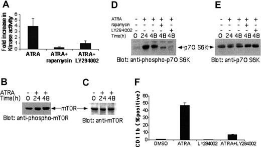
We subsequently sought to identify upstream regulatory signals that mediate RA-dependent activation of the p70 S6 kinase. The activation of the kinase domain of p70 S6 kinase was blocked by pretreatment of cells with the mTOR inhibitor rapamycin, as well as by the PI3′K inhibitor LY294002, suggesting that RA-dependent induction of p70 S6 kinase activity is mTOR and PI3′K dependent (Figure 3A). Consistent with this, in experiments in which the phosphorylation/activation of mTOR was examined in lysates from RA-treated cells, we detected RA-dependent phosphorylation of serine 2448 in mTOR (Figure 3B-C), suggesting that mTOR is activated in an RA-dependent manner to phosphorylate the p70S6 kinase. Moreover, the RA-inducible phosphorylation of p70 S6 kinase on Thr421/Ser424 was inhibited by concomitant treatment of cells with rapamycin or LY294002 (Figure 3D-E). In other studies, we sought to examine whether activation of the PI3′K pathway by RA is required for RA-inducible differentiation of APL cells. NB-4 cells were incubated with or without RA, in the presence or absence LY294002, and the induction of RA-dependent differentiation was assessed by flow cytometry using the anti-CD11b monoclonal antibody. As expected, RA treatment resulted in an increase in the expression of CD11b in NB-4 cells, reflecting granulocytic differentiation (Figure 3F). Concomitant treatment with LY294002 reversed such increase in the expression of CD11b, indicating that activation of the PI3′K is essential for differentiation of NB-4 cells (Figure 3F).
Activation of the kinase domain of the p70 S6 kinase by RA is FRAP/mTOR- and PI3′K-dependent, and PI3′K activity is required for differentiation of APL cells. (A) NB-4 cells were treated with RA for 48 hours. Two and a half hours prior to cell lysis, the indicated inhibitors were added in the cultures. The cells were subsequently lysed, and immunoprecipitated with an anti-p70 S6 kinase antibody or nonimmune rabbit immunoglobulin (RIgG). In vitro kinase assays to detect p70 S6K activity were subsequently carried out on the immunoprecipitates. The data are expressed as fold increase over untreated controls, and represent the mean plus or minus SE of 2 independent experiments. (B) NB-4 cells were incubated with RA for the indicated times. Total cell lysates were resolved by SDS-PAGE and immunoblotted with an antibody against the phosphorylated form of mTOR on serine 2448. (C) The blot shown in panel B was subsequently stripped and reprobed with an anti-mTOR antibody, to control for protein loading (right panel). (D) NB-4 cells were treated with RA for the indicated times. Two and half hours prior to cell lysis, rapamycin (20 nM) or LY294002 (50 μM) were added in the cultures, as indicated. Equal amounts of total cell lysates were analyzed by SDS-PAGE and immunoblotted with an antibody against the phosphorylated/activated form of the p70 S6 kinase on threonine 421 and serine 424. (E) The blot shown in panel D was stripped and reprobed with an anti-p70 S6 kinase antibody, to control for protein loading. (F) NB-4 cells were incubated with RA, in the presence or absence of the PI3′K inhibitor LY294002. The cells were subsequently stained with an anti-CD11b monoclonal antibody and analyzed by flow cytometry. The data represent mean ± SE of 3 experiments.
Activation of the kinase domain of the p70 S6 kinase by RA is FRAP/mTOR- and PI3′K-dependent, and PI3′K activity is required for differentiation of APL cells. (A) NB-4 cells were treated with RA for 48 hours. Two and a half hours prior to cell lysis, the indicated inhibitors were added in the cultures. The cells were subsequently lysed, and immunoprecipitated with an anti-p70 S6 kinase antibody or nonimmune rabbit immunoglobulin (RIgG). In vitro kinase assays to detect p70 S6K activity were subsequently carried out on the immunoprecipitates. The data are expressed as fold increase over untreated controls, and represent the mean plus or minus SE of 2 independent experiments. (B) NB-4 cells were incubated with RA for the indicated times. Total cell lysates were resolved by SDS-PAGE and immunoblotted with an antibody against the phosphorylated form of mTOR on serine 2448. (C) The blot shown in panel B was subsequently stripped and reprobed with an anti-mTOR antibody, to control for protein loading (right panel). (D) NB-4 cells were treated with RA for the indicated times. Two and half hours prior to cell lysis, rapamycin (20 nM) or LY294002 (50 μM) were added in the cultures, as indicated. Equal amounts of total cell lysates were analyzed by SDS-PAGE and immunoblotted with an antibody against the phosphorylated/activated form of the p70 S6 kinase on threonine 421 and serine 424. (E) The blot shown in panel D was stripped and reprobed with an anti-p70 S6 kinase antibody, to control for protein loading. (F) NB-4 cells were incubated with RA, in the presence or absence of the PI3′K inhibitor LY294002. The cells were subsequently stained with an anti-CD11b monoclonal antibody and analyzed by flow cytometry. The data represent mean ± SE of 3 experiments.
In further studies, we sought to define the precise functional contribution of RA-dependent activation of p70 S6 kinase in the generation of RA-responses. A target for p70 S6 kinase activity is the S6 ribosomal protein (rpS6), phosphorylation of which is essential for initiation of mRNA translation of proteins with oligopyrimidine tracts in the 5′ untranslated region. We initially examined whether RA induces phosphorylation of the S6 ribosomal protein. NB-4 cells were incubated for different times with RA, and cell lysates were resolved by SDS-PAGE and immunoblotted with antibodies against the phosphorylated forms of S6 ribosomal protein on Ser240/244 or Ser 235/236. Treatment with RA resulted in strong phosphorylation on both Ser 240/244 (Figure 4A-B) and Ser 235/236 (Figure 4C-D,G-H). Treatment of HL60 cells with RA also resulted in phosphorylation of the S6 ribosomal protein on Ser 240/244 and Ser 235/236 (Figure 4E-F and data not shown). The phosphorylation of both sites was blocked when NB-4 cells were preincubated with either rapamycin or LY294002 (Figure 4I-J and data not shown), indicating that such phosphorylation occurs in a p70 S6 kinase-dependent manner, downstream of mTOR and the PI3′K. To further understand the functional relevance of such RA-inducible phosphorylation/activation of rpS6, the induction of phosphorylation of this protein was analyzed in RA-resistant NB-4 cells (NB-4.007/6). As shown in Figure 5, phosphorylation of rpS6 in response to RA was only detectable in NB-4 cells but not in NB-4.007/6 cells (Figure 5A,D). Interestingly, the amounts of S6 ribosomal protein were also downregulated in this NB-4 resistant variant (Figure 5B-C,E-F).
RA-induced phosphorylation of the S6 ribosomal protein (rpS6). (A) NB-4 cells were incubated with RA (1 μM) for the indicated times. Equal amounts of total cell lysates were analyzed by SDS-PAGE and immunoblotted with an antibody against the phosphorylated form of rpS6 on Ser240/244. (B) The blot shown in panel A was stripped and reprobed with an anti-rpS6 antibody, to control for protein loading. (C) NB-4 cells were incubated with RA (1 μM) for the indicated times. Equal amounts of total cell lysates were analyzed by SDS-PAGE and immunoblotted with an antibody against the phosphorylated form of rpS6 on Ser235/236. (D) The blot shown in panel C was stripped and reprobed with an anti-rpS6 antibody, to control for protein loading. (E) HL60 cells were incubated with RA (1 μM) for 48 hours. Equal amounts of total cell lysates were analyzed by SDS-PAGE and immunoblotted with an antibody against the phosphorylated form of rpS6 on Ser240/244. (F) The blot shown in panel E was stripped and reprobed with an anti-rpS6 antibody, to control for protein loading. (G) NB-4 cells were incubated with RA (1 μM) for indicated times. Equal amounts of total cell lysates were analyzed by SDS-PAGE and immunoblotted with an antibody against the phosphorylated form of rpS6 on Ser235/236. (H) The blot shown in panel G was stripped and reprobed with an anti-rpS6 antibody, to control for protein loading. (I) NB-4 cells were incubated with RA for indicated times and 2.5 hours prior to cell lysis indicated inhibitors were added in the cultures. Equal amounts of total cell lysates were analyzed by SDS-PAGE and immunoblotted with an antibody against the phosphorylated form of rpS6 on Ser240/244. (J) The blot shown in panel I was stripped and reprobed with an anti-rpS6 antibody, to control for protein loading.
RA-induced phosphorylation of the S6 ribosomal protein (rpS6). (A) NB-4 cells were incubated with RA (1 μM) for the indicated times. Equal amounts of total cell lysates were analyzed by SDS-PAGE and immunoblotted with an antibody against the phosphorylated form of rpS6 on Ser240/244. (B) The blot shown in panel A was stripped and reprobed with an anti-rpS6 antibody, to control for protein loading. (C) NB-4 cells were incubated with RA (1 μM) for the indicated times. Equal amounts of total cell lysates were analyzed by SDS-PAGE and immunoblotted with an antibody against the phosphorylated form of rpS6 on Ser235/236. (D) The blot shown in panel C was stripped and reprobed with an anti-rpS6 antibody, to control for protein loading. (E) HL60 cells were incubated with RA (1 μM) for 48 hours. Equal amounts of total cell lysates were analyzed by SDS-PAGE and immunoblotted with an antibody against the phosphorylated form of rpS6 on Ser240/244. (F) The blot shown in panel E was stripped and reprobed with an anti-rpS6 antibody, to control for protein loading. (G) NB-4 cells were incubated with RA (1 μM) for indicated times. Equal amounts of total cell lysates were analyzed by SDS-PAGE and immunoblotted with an antibody against the phosphorylated form of rpS6 on Ser235/236. (H) The blot shown in panel G was stripped and reprobed with an anti-rpS6 antibody, to control for protein loading. (I) NB-4 cells were incubated with RA for indicated times and 2.5 hours prior to cell lysis indicated inhibitors were added in the cultures. Equal amounts of total cell lysates were analyzed by SDS-PAGE and immunoblotted with an antibody against the phosphorylated form of rpS6 on Ser240/244. (J) The blot shown in panel I was stripped and reprobed with an anti-rpS6 antibody, to control for protein loading.
RA induces phosphorylation of rpS6 in NB-4 cells, but not in RA-resistant NB-4.007/6 (NB-4R) cells. (A) NB-4 or NB-4.007/6 cells were treated with RA (1 μM) for the indicated times. Equal amounts of total cell lysates were analyzed by SDS-PAGE and immunoblotted with an antibody against the phosphorylated form of the rpS6 on Ser235/236. (B) The blot shown in panel A was stripped and reprobed with an anti-rpS6 antibody. (C) The same blot, shown in panels A and B, was stripped and reprobed with an anti-tubulin antibody. (D) NB-4 or NB-4.007/6 cells were treated with RA (1 μM) for the indicated times. Equal amounts of total cell lysates were analyzed by SDS-PAGE and immunoblotted with an antibody against the phosphorylated form of the rpS6 on Ser240/244. (E) The blot shown in panel D was stripped and reprobed with an anti-rpS6 antibody. (F) The same blot, shown in panels D and E, was stripped and reprobed with an anti-tubulin antibody.
RA induces phosphorylation of rpS6 in NB-4 cells, but not in RA-resistant NB-4.007/6 (NB-4R) cells. (A) NB-4 or NB-4.007/6 cells were treated with RA (1 μM) for the indicated times. Equal amounts of total cell lysates were analyzed by SDS-PAGE and immunoblotted with an antibody against the phosphorylated form of the rpS6 on Ser235/236. (B) The blot shown in panel A was stripped and reprobed with an anti-rpS6 antibody. (C) The same blot, shown in panels A and B, was stripped and reprobed with an anti-tubulin antibody. (D) NB-4 or NB-4.007/6 cells were treated with RA (1 μM) for the indicated times. Equal amounts of total cell lysates were analyzed by SDS-PAGE and immunoblotted with an antibody against the phosphorylated form of the rpS6 on Ser240/244. (E) The blot shown in panel D was stripped and reprobed with an anti-rpS6 antibody. (F) The same blot, shown in panels D and E, was stripped and reprobed with an anti-tubulin antibody.
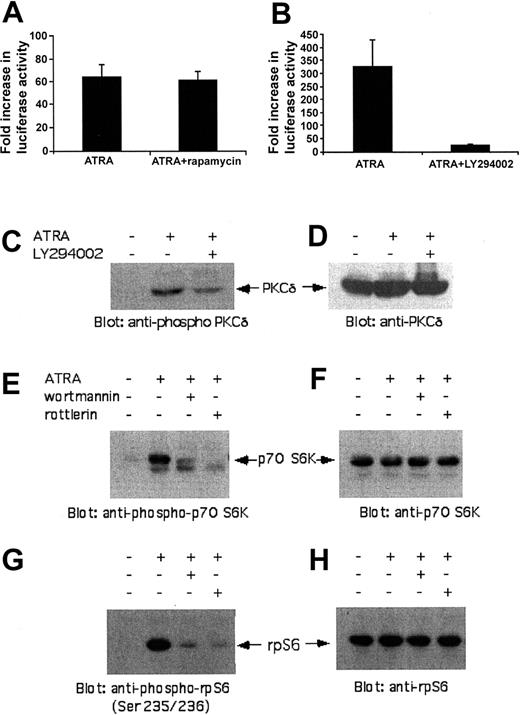
Altogether, these findings suggested that activation of the mTOR/p70 S6K cascade plays an important role in RA-inducible mRNA translation, as demonstrated by the regulation of rpS6 activation. To determine whether other RA-inducible signaling events, unrelated to mRNA translation, are regulated by mTOR and p70 S6K, studies were performed in which the effects of rapamycin on RARE-driven gene transcription were determined. MCF-7 cells were transfected with a plasmid containing a RARE-luciferase construct and treated with RA, in the presence or absence of rapamycin. As expected, RA treatment of cells resulted in a significant increase in RARE-driven luciferase activity (Figure 6A). Treatment of cells with rapamycin had no effects on the induction of such luciferase activity, indicating that the mTOR/p70S6K pathway does not mediate signals for RA-driven gene transcription (Figure 6A). Consistent with this, when chromatin immunoprecipitation (ChIP) assays were performed we found that pretreatment of cells with rapamycin did not affect binding of RARα to RARE elements in NB-4 cells (data not shown). Thus, activation of mTOR and p70 S6K in response to RA treatment appears to selectively regulate signals that target mRNA translation. On the other hand, inhibition of the PI3′K using the LY294002 inhibitor resulted in defective RARE-driven transcription (Figure 6B), indicating that in contrast to mTOR, the PI3′K also regulates signals distinct from p70 S6K that participate in the induction of gene transcription. In previous studies we had shown that PKC-δ is activated in an RA-dependent manner and regulates gene transcription via RAREs.50 To determine whether the regulatory effects of PI3′K on RARE-driven transcription are mediated by PKC-δ, we examined the effects of LY294002 on the RA-inducible phosphorylation/activation of PKC-δ. As shown in Figure 6C-D, treatment of cells with LY294002 blocked the RA-dependent phosphorylation of PKC-δ, strongly suggesting that the PI3′K acts as an upstream effector for the RA-dependent activation of PKC-δ. Thus, the PI3′K pathway controls activation of pathways that are involved both in RA-dependent gene transcription (protein kinase Cδ [PKC-δ]) and initiation of mRNA translation (p70 S6K/S6 ribosomal protein). As PKC-δ has been previously shown to interact with mTOR,54 we examined whether its kinase activity is required for downstream activation of the p70 S6 kinase. Pretreatment of NB-4 cells with the PKC-δ inhibitor rottlerin50 blocked the RA-inducible phosphorylation of the p70 S6 kinase and S6 ribosomal protein (Figure 6E-H), strongly suggesting that this kinase links mTOR to p70 S6 kinase activation.
Binding of RARα to RAREs and RA-dependent gene transcription via RAREs is mTOR independent, but PI3′K sensitive. (A) MCF-7 cells were transfected with a RARE-luciferase construct. Forty-eight hours after transfection, cells were treated for 60 minutes in the presence or absence of rapamycin (20 nM). The cells were then incubated overnight with or without RA (1 μM), in the continuous presence or absence of rapamycin, and luciferase activity was measured. The data are expressed as fold increase in luciferase activity over RA-untreated control samples for each condition, normalized for β-galactosidase activity. Means plus or minus SE values of 3 independent experiments are shown. (B) MCF-7 cells were transfected with a RARE-luciferase construct. Forty-eight hours after transfection, cells were treated for 60 minutes in the presence or absence of LY294002 (50 μM). The cells were then incubated overnight with or without RA (1 μM), in the continuous presence or absence of LY294002, and luciferase activity was measured. The data are expressed as fold increase in luciferase activity over RA-untreated control samples for each condition, normalized for β-galactosidase activity. Means plus or minus SE values of 3 independent experiments are shown. (C) NB-4 cells were incubated with RA in the presence or absence of the PI3′K inhibitor LY294002. Equal amounts of total cell lysates were resolved by SDS-PAGE and immunoblotted with an antibody against the phosphorylated form of PKC-δ at Thr505. (D) The same blot shown in panel C was stripped and reprobed with an anti-PKC-δ antibody, to control for protein loading. (E) NB-4 cells were treated with RA in the presence or absence of the PI3′K inhibitor wortmannin (50 nM) or the PKC-δ inhibitor rottlerin (10 μM). Equal amounts of total cell lysates were resolved by SDS-PAGE and immunoblotted with antibody against the phosphorylated form of p70 S6K. (F) The blot shown in panel E was stripped and reprobed with an anti-p70 S6 kinase antibody. (G) Similar experiment to the one shown in panel E, except that immunoblotting was performed using an anti-phospho-235/236-ribosomal S6 protein (rpS6) antibody. (H) The blot shown in panel G was stripped and reprobed with anti-rpS6 antibody.
Binding of RARα to RAREs and RA-dependent gene transcription via RAREs is mTOR independent, but PI3′K sensitive. (A) MCF-7 cells were transfected with a RARE-luciferase construct. Forty-eight hours after transfection, cells were treated for 60 minutes in the presence or absence of rapamycin (20 nM). The cells were then incubated overnight with or without RA (1 μM), in the continuous presence or absence of rapamycin, and luciferase activity was measured. The data are expressed as fold increase in luciferase activity over RA-untreated control samples for each condition, normalized for β-galactosidase activity. Means plus or minus SE values of 3 independent experiments are shown. (B) MCF-7 cells were transfected with a RARE-luciferase construct. Forty-eight hours after transfection, cells were treated for 60 minutes in the presence or absence of LY294002 (50 μM). The cells were then incubated overnight with or without RA (1 μM), in the continuous presence or absence of LY294002, and luciferase activity was measured. The data are expressed as fold increase in luciferase activity over RA-untreated control samples for each condition, normalized for β-galactosidase activity. Means plus or minus SE values of 3 independent experiments are shown. (C) NB-4 cells were incubated with RA in the presence or absence of the PI3′K inhibitor LY294002. Equal amounts of total cell lysates were resolved by SDS-PAGE and immunoblotted with an antibody against the phosphorylated form of PKC-δ at Thr505. (D) The same blot shown in panel C was stripped and reprobed with an anti-PKC-δ antibody, to control for protein loading. (E) NB-4 cells were treated with RA in the presence or absence of the PI3′K inhibitor wortmannin (50 nM) or the PKC-δ inhibitor rottlerin (10 μM). Equal amounts of total cell lysates were resolved by SDS-PAGE and immunoblotted with antibody against the phosphorylated form of p70 S6K. (F) The blot shown in panel E was stripped and reprobed with an anti-p70 S6 kinase antibody. (G) Similar experiment to the one shown in panel E, except that immunoblotting was performed using an anti-phospho-235/236-ribosomal S6 protein (rpS6) antibody. (H) The blot shown in panel G was stripped and reprobed with anti-rpS6 antibody.
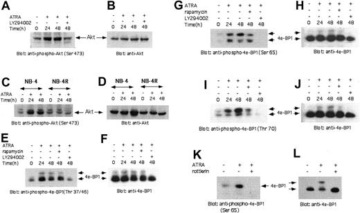
Previous studies have shown that in response to insulin, the 4E-BP1 repressor of mRNA translation is phosphorylated in a PI3′K- and mTOR-dependent manner,55-57 and that such phosphorylation leads to its dissociation from the initiation factor eIF4E, to allow initiation of mRNA translation.57 In addition, the downstream effector of PI3′K, Akt kinase, has been identified as an indirect regulator of 4E-BP1 phosphorylation, via a mechanism that requires FRAP/mTOR activity.56 As phosphorylation of 4E-BP1 constitutes a translational mechanism, distinct from phosphorylation of rpS6, we sought to determine whether RA treatment of NB-4 cells results in phosphorylation/activation of the Akt kinase, and phosphorylation/deactivation of 4E-BP1. In studies in which the phosphorylation status of Akt kinase was evaluated in cell lysates from RA-treated NB-4 cells, we found that Akt was phosphorylated on serine 473 (Figure 7A-B). As expected, such phosphorylation/activation of the Akt kinase was inhibited by concomitant treatment of cells with the PI3′K inhibitor LY294002 (Figure 7A-B). Furthermore, as in the case of p70 S6 kinase, such phosphorylation/activation of Akt was defective in the RA-resistant NB-4 0.007/6 cell line (Figure 7C-D).
RA-dependent phosphorylation/activation of the Akt-kinase and phosphorylation of the 4E-BP1 repressor of mRNA translation. (A) NB-4 cells were incubated with RA for the indicated times. Two and a half hours prior to cell lysis, LY294002 was added to the cultures, as indicated. The cells were subsequently lysed and equal amounts of total cell lysates were resolved by SDS-PAGE and immunoblotted with an antibody against the phosphorylated form of Akt on serine 473. (B) The blot shown in panel A was stripped and reprobed with an anti-Akt antibody, to control for protein loading. (C) NB-4 or NB-4 0.007/6 (NB-4R) cells were incubated with RA for the indicated times. The cells were lysed and equal amounts of total cell lysates were resolved by SDS-PAGE and immunoblotted with an antibody against the phosphorylated form of Akt on serine 473. (D) The blot shown in panel C was stripped and reprobed with an anti-Akt antibody, to control for protein loading. (E-L) NB-4 cells were incubated with RA for the indicated times. Two and a half hours before lysis, the indicated inhibitors were added in the culture. The cells were subsequently lysed, and equal amounts of cell lysates were analyzed by SDS-PAGE and immunoblotted with an anti-phospho-Thr37/46-4E-BP1 antibody (E) or an anti-phospho-Ser65-4E-BP1 antibody (G, K), or an anti-phospho-Thr70-4E-BP1 antibody (I). The same blots were subsequently stripped and reprobed with an anti-4E-BP1 antibody (F, H, J, and L, respectively).
RA-dependent phosphorylation/activation of the Akt-kinase and phosphorylation of the 4E-BP1 repressor of mRNA translation. (A) NB-4 cells were incubated with RA for the indicated times. Two and a half hours prior to cell lysis, LY294002 was added to the cultures, as indicated. The cells were subsequently lysed and equal amounts of total cell lysates were resolved by SDS-PAGE and immunoblotted with an antibody against the phosphorylated form of Akt on serine 473. (B) The blot shown in panel A was stripped and reprobed with an anti-Akt antibody, to control for protein loading. (C) NB-4 or NB-4 0.007/6 (NB-4R) cells were incubated with RA for the indicated times. The cells were lysed and equal amounts of total cell lysates were resolved by SDS-PAGE and immunoblotted with an antibody against the phosphorylated form of Akt on serine 473. (D) The blot shown in panel C was stripped and reprobed with an anti-Akt antibody, to control for protein loading. (E-L) NB-4 cells were incubated with RA for the indicated times. Two and a half hours before lysis, the indicated inhibitors were added in the culture. The cells were subsequently lysed, and equal amounts of cell lysates were analyzed by SDS-PAGE and immunoblotted with an anti-phospho-Thr37/46-4E-BP1 antibody (E) or an anti-phospho-Ser65-4E-BP1 antibody (G, K), or an anti-phospho-Thr70-4E-BP1 antibody (I). The same blots were subsequently stripped and reprobed with an anti-4E-BP1 antibody (F, H, J, and L, respectively).
In parallel studies, we examined whether 4E-BP1 is phosphorylated in an RA-inducible manner in NB-4 cells. As shown in Figure 7, treatment of NB-4 cells with RA resulted in phosphorylation of 4E-BP1 on all sites required for its deactivation and dissociation from 4E-BP1, including Thr37/46 (Figure 7E-F), Ser65 (Figure 7G-H), and Thr70 (Figure 7I-J). Inhibition of PI3′K activity using the LY294002 inhibitor blocked phosphorylation of 4E-BP1 in all sites (Figure 7E,G,I). On the other hand, phosphorylation on Ser65 (Figure 7G) and Thr70 (Figure 7I) was rapamycin-sensitive, whereas phosphorylation on Thr37/46 was not (Figure 7E). The phosphorylation of 4E-BP1 was also PKC-δ-dependent, as evidenced by the inhibition of 4E-BP1 phosphorylation during pretreatment of cells with the PKC-δ inhibitor rottlerin (Figure 7K-L). Thus, in addition to activation of p70S6K and phosphorylation of rpS6, RA induces phosphorylation and deactivation of 4E-BP1, to selectively regulate initiation of cap-dependent mRNA translation.
Discussion
The retinoids have important biologic properties, including their ability to induce differentiation and suppress proliferation of malignant cells in vitro and in vivo.4,6,7,23 These metabolites of vitamin A generate their effects by entering the cells and associating with retinoic acid nuclear receptors. All-trans-retinoic acid activates the family of RARs, whereas cis-retinoic acid activates RARs as well as RXRs.20-22,58,59 The resulting complexes between retinoids and their nuclear receptors bind to specific elements in the promoters of target genes, to initiate gene transcription for protein products that mediate generation of their biologic activities.4,6,7,20-23 Although the identities of RA-induced proteins that account for the generation of distinct biologic effects have not been precisely defined, there are several candidate proteins that may mediate such effects. For instance, regulated by retinoic acid receptors are the genes for p21WAF, C/EBPβ, and HoxA1, all of which encode for protein products that play important roles in regulation of cell growth and myeloid differentiation (reviewed in Lin et al60 ). In addition, retinoids exhibit synergistic activities with interferons, and regulate transcription of the Stat1 gene,61-63 a member of the Stat family of proteins that plays a critical role in interferon-signaling and generation of growth inhibitory responses (reviewed in Platanias and Fish64 and Platanias65 ).
Extensive studies have shown that all-trans-retinoic acid induces cell differentiation of acute promyelocytic leukemia blast cells in vitro and in vivo.1-9 This activity of RA has had a dramatic impact in the natural history of this subtype of acute myelogenous leukemia, and has provided the first model of an effective differentiation therapy for the treatment of hematologic malignancies.1-9 APL is characterized by the t(15;17) chromosomal translocation, which results in the aberrant expression of the abnormal PML-RARα fusion protein.1-7,23 PML-RARα exhibits dominant-negative effects on the function of normal retinoic acid receptors, and suppresses RARα-driven gene transcription.4,6,7,60 A mechanism by which this occurs may be the very-high-binding affinity of PML-RARα for RXR, and also for nuclear corepressors, such as SMRT or N-CoR.4,6,7,60 This results in the recruitment of histone deacetylases (HDACs) in the complex, followed by deacetylation of nuclear histones, chromosomal condensation, and transcriptional repression.4,6,7,60 Such effects are seen in the absence of RA or in the presence of small, physiologically present, concentrations of RA. However, pharmacologic concentrations of RA result in dissociation of corepressor molecules from PML-RARα and restoration of the ability of normal RARα/RXR complexes to regulate transcription.4-7,66,67
Although the mechanisms of transcriptional regulation of RA-responsive genes have been extensively characterized, including the mechanisms of transcription in the presence of PML-RARα, very little is known on the signals required for initiation of mRNA translation by retinoids. In the present report we provide the first evidence for the existence of an RA-activated signaling cascade that regulates initiation of mRNA translation. Our data demonstrate that the mammalian target of rapamycin is phosphorylated/activated in RA-responsive cell lines, and regulates downstream engagement and activation of the p70 S6 kinase. Such RA-dependent activation of p70 S6 kinase may be of importance in the regulation of mRNA translation, as it results in the first step of translation for mRNAs with oligopyrimidine tracts, the downstream phosphorylation of the S6 ribosomal protein. Activation of p70 S6 kinase and phosphorylation of the S6 ribosomal protein is inducible in the APL-derived NB-4 cell line, but not in an RA-resistant NB-4 variant, further demonstrating the functional relevance of this pathway. Our data also establish that activation of mTOR and the PI3′K mediates downstream phosphorylation of 4E-BP1, a translational repressor protein that negatively regulates the function of the eukaryotic initiator of translation eIF4E. Moreover, 4E-BP1 is optimally phosphorylated by RA in all sites required for its deactivation and dissociation from eIF4E. Altogether, these findings strongly suggest that this cascade plays a role in the generation of protein products that mediate the biologic effects of all-trans-retinoic acid and, likely, other retinoids as well.
PI3′K-dependent and mTOR-inducible signaling cascades are generally thought of as pathways that promote cell growth and antiapoptotic responses. This likely reflects the fact that most studies have been focused on the roles of these proteins in growth factor signaling, or in the context of malignant transformation by oncogenes. However, there is accumulating evidence that these pathways have diversified capacities, and are apparently capable of mediating signals for cytokines that suppress cell growth. For instance, recent studies have shown that mTOR and the p70 S6 kinase are activated by interferons,43,44 which are cytokines with growth inhibitory properties, and which exhibit synergistic effects with retinoids.61-63 The regulation of p70 S6 kinase by interferons appears to be regulated by the interferon (IFN)-activated PI3′K pathway,68-70 whereas interferons also induce phosphorylation and deactivation of 4E-BP1.43,44 Other studies have shown that activation of the PI3′K promotes RA-induced differentiation of human neuroblastoma,71 endometrial carcinoma cells,72 and HL-60 leukemia cells.73 In addition, activation of the PI3′K by retinoic acid has been shown to be essential for expression and activation of the tissue transglutaminase,74 an enzyme whose function participates in the regulation of RA-induced cell differentiation.75 Our findings are consistent with such previous reports, and for the first time identify downstream effector mechanisms of the RA-activated PI3′K pathway that directly facilitate initiation of mRNA translation.
It should be pointed out that previous studies have shown that at the late stages of hematopoietic cell differentiation, mRNA translation is decreased,76 whereas a decrease in the protein levels of 4E-BP1 in HL-60 cells during terminal myeloid differentiation has been reported.76 Our findings suggest that the suppression of mRNA translation seen at the late stages of cell differentiation by retinoids likely involves inhibitory signals on pathways distinct from the S6 ribosomal protein and 4E-BP1, as these elements are used in the early stages of RA-induced differentiation. It is possible that certain RA-induced protein products, regulated by rpS6- or cap-dependent translation, exhibit suppressive effects on mRNA translation induced by mitogenic growth factor signaling cascades, but the validity of such an hypothesis remains to be established in future studies. It should be pointed out that our studies have established that the phosphorylation/activation of p70 S6 kinase by RA occurs early, indicating that it is a primary, RA-induced signaling event.
The precise mechanisms and kinases responsible for direct phosphorylation and deactivation of 4E-BP1 downstream of mTOR during RA treatment of cells remain to be determined. Recent studies have shown that PKC-δ, a member of the protein kinase C family of proteins, is activated by all-trans-retinoic acid and is implicated in the regulation of RARE-driven transcription and generation of the effects of RA on acute promyelocytic leukemia cells.50 Interestingly, other studies have shown that PKC-δ interacts with mTOR,54 and acts as a direct serine kinase for 4E-BP1 to initiate cap-dependent mRNA translation in other systems.77 Our studies strongly suggest that PKC-δ may also act as a serine kinase for 4E-BP1 during its activation by RA in APL cells, as evidenced by the inhibition of 4E-BP1 phosphorylation by pharmacologic inhibition of the kinase. Moreover, the study for the first time implicates this kinase in the regulation of additional pathways downstream of mTOR, as the PKC-δ inhibitor rottlerin also inhibits RA-induced phosphorylation of the p70 S6 kinase and the S6 ribosomal protein.
Independently of the precise mechanisms involved, our data establish that cascades downstream of FRAP/mTOR participate in RA signaling. These findings may be of clinical relevance, as mTOR inhibitors, including rapamycin and the related CCI-779 analog, are currently being studied in clinical oncology trials for the treatment of certain tumors.78,79 The basis of such efforts is the ability of these compounds to inhibit growth factor-dependent malignant cell proliferation. However, our findings suggest that mTOR and the p70 S6 kinase may positively regulate the generation of RA responses. Thus, caution should be taken in designing clinical trials combining mTOR inhibitors with retinoids, as it is possible that mTOR inhibitors may block the antitumor properties of retinoids in vivo.
Prepublished online as Blood First Edition Paper, October 7, 2004; DOI 10.1182/blood-2004-06-2078.
Supported by a Merit Review grant from the Department of Veterans Affairs (L.C.P.), by National Institutes of Health grants CA77816 and CA94079 (L.C.P.), and by grant DAMD 17-03-1-0254 from the Department of Defense (L.C.P.).
An Inside Blood analysis of this article appears in the front of this issue.
The publication costs of this article were defrayed in part by page charge payment. Therefore, and solely to indicate this fact, this article is hereby marked “advertisement” in accordance with 18 U.S.C. section 1734.


This feature is available to Subscribers Only
Sign In or Create an Account Close Modal