Abstract
Using 2-dimensional gel electrophoresis (2D-gel) analysis, we show here that cell-cycle entry is associated with a significant increase in p27kip1 phosphorylation in human primary B cells. A similar pattern of increase in p27kip1 phosphorylation was also seen in 2 fast-growing tumor cell lines, Burkitt lymphoma cell line BL40 and breast carcinoma cell line Cal51, where inactive p27kip1 is expressed at high levels. Detailed analysis revealed for the first time that different cyclins and cyclin-dependent kinases (cdk's) interact with distinct posttranslationally modified isoforms of p27kip1 in vivo. Cyclin E but not cyclin A selectively interacts with phosphorylated p27kip1 isoforms, while cyclin D1 and D2 favor unphosphorylated p27kip1 isoforms in vivo. Interestingly, cyclin D3 and cdk4 selectively interact with phosphorylated p27kip1 in BL40 cells. Among all D-type cyclin/cdk4 and cdk6 complexes, cyclin D3/cdk4 is most active in sequestering the inhibitory activity of p27kip1 in vitro in a cyclinE/cdk2 kinase assay. This novel feature of the binding specificity of p27kip1 to cyclins and cdk's in vivo is interpreted in the context of overexpression of cyclin D3 in the presence of high levels of p27kip1 in human B-cell lymphomas with adverse clinical outcome.
Introduction
Cell-cycle progression is controlled by cyclin-dependent kinases (cdk's). The activity of cyclin-dependent kinases is positively and negatively regulated by cyclins and cdk inhibitors, respectively. All cdk inhibitors cause G1 arrest when overexpressed in transfected cells. There are 2 families of cdk inhibitors, the cip/kip and ink4 families. Members of the ink4 family specifically inhibit the activity of cdk4 and cdk6, whereas the cip/kip family share sequence homology and inhibit the kinase activity of various cdk's. In vitro, p21cip1, p27kip1, and p57kip2 can inhibit all cyclin-dependent kinases. However, in vivo, p21cip1 and p27kip1 arrest cells at G1 by inhibiting the kinase activity of cyclin E/cdk2. This growth inhibitory activity of p21cip1 and p27kip1 can be titrated out by D-type cyclins. It was reported previously that p27kip1 only induces cell-cycle arrest when it is in complex with cyclin E/cdk2.1 Thus, a shift from cyclin E/cdk2 to cyclin D2/cdk4 complex compromised the growth inhibitory function of p27kip1.
In several different types of human tumor, the prognostic value of p27kip1 expression level has been well established. Down-regulation of p27kip1 is often associated with an increased degree of malignancy and poor prognosis.2 However, high levels of p27kip1 expression were observed in some fast-growing human tumor cells.3,4 In particular, a good correlation between high-level expression of cyclin D1 and p27kip1 was seen in human breast carcinomas in vivo and breast carcinoma cell lines in vitro.3-5 Overexpression of cyclin D3 and p27kip1 was also seen in a group of aggressive large B-cell lymphomas and Burkitt lymphomas with high-proliferation index.4,6 This is in contrast to the expression pattern of p27kip1 in normal lymphoid tissue where its expression is inversely correlated with cell proliferation.7 The p27kip1 expressed in the B-cell tumors is nuclear and colocalizes with cyclin D3. In diffuse large B-cell lymphomas, the adverse clinical outcome was also associated with high-level expression of p27kip1.6,8,9 This is in agreement with an earlier observation that the inhibitory activity of p27kip1 can be sequestered by increased expression of D-type cyclins.1 It remains unclear, however, whether all D-type cyclins are equally active in sequestering the inhibitory activity of p27kip1 on cyclin E/cdk2 kinase. Also unknown is the mechanism that controls the binding specificity of p27kip1 for cyclin E or cyclin D.
Interestingly, p27kip1 is a phosphoprotein. Studies have shown that the growth inhibitory activity of p27kip1 is regulated by phosphorylation. Phosphorylation of p27kip1 at Thr187 by cyclin E/cdk2 destabilizes p27kip1.10-12 Phosphorylation of p27kip1 at Thr157 by an increased threonine kinase B–protein kinase B (AKT/PKB)13-15 or Thr10 by human kinase-interacting stathmin (hKIS)16,17 impairs the nuclear import of p27kip1. Hence, posttranslational modification of p27kip1 could be responsible for the loss of growth inhibitory activity of p27kip1 in some of the fast-growing tumor cells observed in vitro and in vivo. Perhaps different posttranslationally modified forms of p27kip1 are associated with different cyclins, cdk's, or cyclin/cdk's. These issues were addressed by investigating the posttranslational modifications of p27kip1 in vivo in response to phorbol 12-myristate 13-acetate (PMA), a mitogenic stimulus in human primary B cells. The posttranslational modification of p27kip1 was also examined in 2 tumor cell lines expressing biologically compromised p27kip1, a Burkitt lymphoma cell line BL40 and a breast carcinoma cell line Cal51. The binding spectra of distinct cyclins and cdk's for different posttranslationally modified p27kip1 isoforms were studied using 2-dimensional isoelectric-focusing gel electrophoresis. Finally, using an in vitro kinase assay, we tested whether different D-type cyclins/cdk4 or cdk6 were able to sequester the inhibitory activity of p27kip1 to a similar degree. Understanding how the growth inhibitory activity of p27kip1 is regulated following mitogenic stimulation in primary B cells may provide a mechanistic insight of how p27kip1 is inactivated in human tumors.
Material and methods
Cell culture, reagents, and antibodies
Cells were grown in Dulbecco modified Eagle medium supplemented with 10% fetal calf serum. The construction of p27kip1-inducible H1299 (H1299 + p27kip1) cells was described previously.4 The expression of p27kip1 was induced with 2 μM Ponasterone A (Invitrogen, Paisley, United Kingdom) for 24 hours. SX53G8 and LX014 are mouse monoclonal antibodies that specifically recognize p27kip1-3 and cyclin D1, respectively (Figure 4A). The remaining antibodies were purchased from either Santa Cruz Biotechnology (Santa Cruz, CA) or Pharmingen (San Diego, CA). Antibodies used to immunoprecipitate cyclin A, cyclin E, cyclin D1, cyclin D2, cyclin D3, cdk2, cdk4, and cdk6 were 432, M-20, DCS3.1, C-16, M2-G, H-22, and C-21, respectively. Construction and purification of recombinant proteins glutathione S-transferase (GST)–p21cip1, GST-p27kip1, GST-p57kip2, GST-Rb763-928 were similar to earlier descriptions.18-20 Baculoviruses expressing human cyclin E, cyclin A, D1, D2, D3, cdk2, cdk4, and cdk6 were generous gifts from Dr Mittnacht (Institute of Cancer Research, London, United Kingdom).18
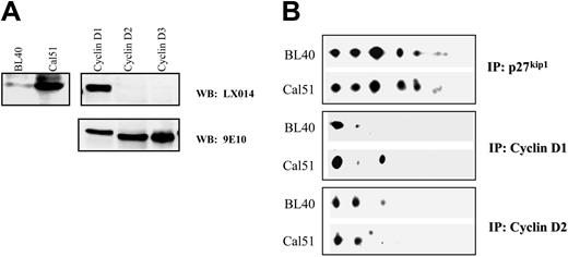
Cyclin D1 and D2 favor unphosphorylated p27kip1 isoforms. Western blot shows the expression levels of cyclin D1 in Cal51 and BL40 cells (A, left panel). The specificity of the anti–cyclin D1 antibody, LX014, is shown in panel A, right 2 panels. Human cyclin D1, D2, and D3 were all tagged with a 9E10 epitope, and the expression levels of these cyclins in SF9 cells were detected by the mouse monoclonal antibody 9E10 (A, bottom right). 2D gel analysis shows that both cyclin D1 and cyclin D2 favor unphosphorylated p27kip1. Although cyclin D1 favors unmodified p27kip1, isoform 1, cyclin D2 binds to both p27kip1 isoforms 1 and 2 (B).
Cyclin D1 and D2 favor unphosphorylated p27kip1 isoforms. Western blot shows the expression levels of cyclin D1 in Cal51 and BL40 cells (A, left panel). The specificity of the anti–cyclin D1 antibody, LX014, is shown in panel A, right 2 panels. Human cyclin D1, D2, and D3 were all tagged with a 9E10 epitope, and the expression levels of these cyclins in SF9 cells were detected by the mouse monoclonal antibody 9E10 (A, bottom right). 2D gel analysis shows that both cyclin D1 and cyclin D2 favor unphosphorylated p27kip1. Although cyclin D1 favors unmodified p27kip1, isoform 1, cyclin D2 binds to both p27kip1 isoforms 1 and 2 (B).
Isolation of human primary B cells
Primary B cells were prepared from buffy coat residues by Ficoll gradient and purified with an anti-CD19 antibody conjugated to Dynabeads as described previously.21 Approval was obtained from the Ludwig Institute for Cancer Research institutional review board and informed consent was provided according to the Declaration of Helsinki. Fluorescein isothiocyanate (FITC)–conjugated mouse monoclonal antibodies to CD20 and CD3 (DAKO, Glostrup, Denmark) were used to measure the purity of each batch of purified primary B cells. In general, 95% of purified cells were B cells, while 4% of the cells were T cells (data not shown). The purified B cells were maintained for 42 hour in RPMI supplemented with 15% fetal calf serum. For B-cell stimulation, 30 ng/mL PMA (Sigma, Saint Louis, MO) was added 72 hours before harvesting.
Phosphatase treatment
The cells were lysed in radioimmunoprecipitation (RIPA) buffer. Samples of cell lysates containing 200 μg total cell protein were incubated at 37°C for 3 hours with 60 units of calf intestinal alkaline phosphatase (CIAP; Boehringer Mannheim, Mannheim, Germany) in a phosphatase reaction buffer in a total reaction volume of 20 μL. The treated lysates were then used in 2-dimensional (2D) gel analysis.
Roscovitine treatment in vivo and in vitro
Cal51 cells were treated with 10 or 30 μM roscovitine (Sigma) or a dimethyl sulfoxide (DMSO) vehicle control for 24 hours as indicated. The treated cells were then lysed and immunoprecipitated with the anti-p27kip1 antibody SX53G8 in the absence of roscovitine. For Figure 3C, Cal51 cells were grown as normal but lysed with lysis buffer containing 60 μM roscovitine or DMSO. The same amount of roscovitine or DMSO was added to all buffers used in immunoprecipitation.
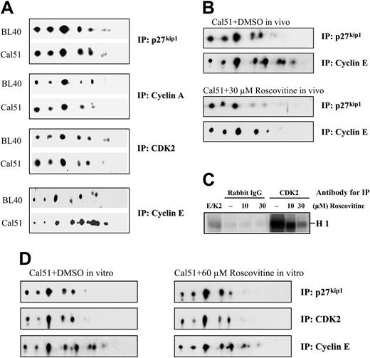
Cyclin E but not cyclin A selectively interacts with phosphorylated p27kip1 isoforms in vivo. 2D gel analysis shows that cyclin E but not cyclin A or cdk2 selectively binds phosphorylated p27kip1 in vivo (A). The isoforms of p27kip1 coimmunoprecipitated with cyclin A, cyclin E, and cdk2 are indicated with arrows. Rabbit polyclonal antibodies specific to cyclin A, cyclin E, cdk2, and p27kip1 were used to carry out immunoprecipitation. Coimmunoprecipitated p27kip1 isoforms were detected with a mouse monoclonal anti-p27kip1 antibody SX53G8. Similar results were also obtained with another anti–p27kip1 mouse monoclonal antibody with a different epitope from SX53G8 (data not shown). The patterns of p27kip1 isoforms associated with cyclin E in Cal51 cells treated with 30 μM roscovitine or DMSO for 24 hours are shown (B). The ability of 10 or 30 μM roscovitine to inhibit the cdk2 kinase activity obtained from the anti-cdk2 immunoprecipitate of Cal51 cells in vitro is shown (C). Panel D shows the patterns of p27kip1 isoforms detected from the anti–cyclin E immunoprecipitates of Cal51 cell lysates incubated with DMSO or 60 μM roscovitine throughout the immunoprecipitation process (indicated as DMSO or roscovitine, respectively).
Cyclin E but not cyclin A selectively interacts with phosphorylated p27kip1 isoforms in vivo. 2D gel analysis shows that cyclin E but not cyclin A or cdk2 selectively binds phosphorylated p27kip1 in vivo (A). The isoforms of p27kip1 coimmunoprecipitated with cyclin A, cyclin E, and cdk2 are indicated with arrows. Rabbit polyclonal antibodies specific to cyclin A, cyclin E, cdk2, and p27kip1 were used to carry out immunoprecipitation. Coimmunoprecipitated p27kip1 isoforms were detected with a mouse monoclonal anti-p27kip1 antibody SX53G8. Similar results were also obtained with another anti–p27kip1 mouse monoclonal antibody with a different epitope from SX53G8 (data not shown). The patterns of p27kip1 isoforms associated with cyclin E in Cal51 cells treated with 30 μM roscovitine or DMSO for 24 hours are shown (B). The ability of 10 or 30 μM roscovitine to inhibit the cdk2 kinase activity obtained from the anti-cdk2 immunoprecipitate of Cal51 cells in vitro is shown (C). Panel D shows the patterns of p27kip1 isoforms detected from the anti–cyclin E immunoprecipitates of Cal51 cell lysates incubated with DMSO or 60 μM roscovitine throughout the immunoprecipitation process (indicated as DMSO or roscovitine, respectively).
2-dimensional gel analysis
Cells were lysed in a solution of 8 M urea, 2 M thiourea, 3% CHAPS (3-[(3-cholamidopropyl)dimethylammonio]-1-propanesulfonate), 1% NP40 (Nonidet P40), 50 mM DTT (dithiothreitol), 24 mM spermine, 50 mM NaF, 1 mM benzamidine, and 120 nM okadaic acid at room temperature or in RIPA-PI (phosphatidylinositol) buffer (supplemented with 50 mM NaF, 1 mM benzamidine, and 120 nM okadaic acid) on ice for 30 minutes. The RIPA cell lysates (200-800 μg) were used for immunoprecipitation or the treatment with calf intestine alkaline phosphatase. For isoelectric focusing, both cell lysates and immunoprecipitates were denatured in the urea solution, loaded onto pH 3 to 10, nonlinear Immobiline DryStrips (Amersham Biosciences, Buckinghamshire, England), and focused for 70 000 V/hour, using the Multiphor II apparatus. The isoelectric-focusing strip was then equilibrated in 50 mM Tris (tris(hydroxymethyl)aminomethane), pH 8.8, 6 M urea, 30% glycerol, and 2% sodium dodecyl sulfate (SDS) and 1% DTT for 20 minutes before subjecting to SDS–polyacrylamide gel electrophoresis. SDS gels were transferred to a nitrocellulose membrane, and p27kip1 isoforms were detected by immunoblotting using p27kip1 antibodies (SX53G8 or C-19). Signals were quantified with the GeneTools program from SynGene (Cambridge, United Kingdom). The isoelectric points were determined using creatine phosphokinase (CPK) in the Carbamylyte Calibration Kit For 2-D Electrophoresis (Amersham Pharmacia Biotech, Buckinghamshire, United Kingdom) run in pH 3 to 10 nonlinear and pH 3 to 10 linear Immobiline DryStrips (Amersham Biosciences).
Kinase assay
Histone H1 or GST-pRb C-terminal fragment (amino acids 763∼928) were used as substrates in kinase assays as described.22 SF9 insect cells were coinfected with baculovirus expressing recombinant cdk2 and 9E10 epitope-tagged human cyclin E. The cyclin E/cdk2 complex was immunoprecipitated with purified 9E10 antibody as described22 and used as cyclin E/cdk2 kinase. Cyclin D1/cdk4, D2/cdk4, D3/cdk4, D1/cdk6, D2/cdk6, D3/cdk6 kinases were prepared in a similar manner with 9E10 epitope-tagged cyclin D. For the kinase inhibition assay, the kinase lysate was incubated with increasing amounts of GST-p27kip1, GST-p21cip1, or GST-p57kip2. For a kinase inhibition and sequestration assay (Figure 6C, lanes 4-9), the amounts of GST-p27kip1, GST-p21cip1, or GST-p57kip2 used were derived from previous titration experiments (data not shown) so as to inhibit the cyclinE/cdk2 kinase activity by about 75% (Figure 6C, lane 3 for both panels). Similarly, the amounts of cyclin D/cdk complexes used were derived from previous titration experiments, and they all have comparable kinase activities to phosphorylated GST-Rb in vitro (Figure 6B). Finally, SF9 cells were coinfected with a fixed amount of cdk4 or cdk6 expressing baculovirus together with a titrated amount of D-type cyclin expressing baculovirus to achieve the same expressing levels of cyclin D1, D2, and D3 in complex with cdk4 or cdk6 (Figure 6B, right panel).
Cyclin D3/cdk4 is the kinase that is most able to sequester the inhibitory activity of p27kip1 but not p21cip1 and p57kip2. Increasing amounts of GST-p27kip1, GST-p21cip1, or GST-p57kip2 were used to inhibit the kinase activity of cyclin E/cdk2 in vitro (A, left panel). The right-hand of panel A shows the expression levels of cyclin D1, D2, D3, cdk4, and cdk6 in individual D-type cyclin/cdk4 or cdk6 complexes (cyclin D1, cyclin D2, and cyclin D3 are labeled as D1, D2, and D3, whereas cdk4 and cdk6 were labeled as K4 and K6, respectively). The expression levels of D-type cyclins and cdk4 or cdk6 were detected by 9E10 antibody and anti-cdk4 or anti-cdk6 antibodies, respectively. The kinase activity of various D-type cyclin/cdk's was measured in vitro using GST-Rb as a substrate (B, left). Cyclin E/cdk2 was used as a positive control. The ability of GST-p27kip1, GST-p21cip1, and GST-p57kip2 but not GST to inhibit the kinase activity of cyclinE/cdk2 is shown in panel C, left (lanes 1-3). The left subpanel of C shows the ability of various D-type cyclin/cdk's to sequester the inhibitory activity of p27kip1, p21cip1, and p57kip2 on cyclin E/cdk2 kinase in vitro. The amounts of different combinations of D-type cyclin/cdk used in this experiment were the same as those shown in panel B, and they were all derived from the same extracts. The amounts of individual D-type cyclin/cdk4 or cdk6 lysates, GST-p27kip1, GST-p21cip1, and GST-p57kip2 used in the kinase assay are also indicated in the right-hand subpanels of B and C. The bar graphs in panel D show quantitatively how efficient each combination of D-type cyclin/cdk is in sequestering the inhibitory activity of p27kip1, p21cip1, and p57kip2 on cyclin E/cdk2 kinase. The cyclin E/cdk2 kinase activity detected in the presence of p27kip1 or p21cip1 or p57kip2 (left graph, lane 3 for all blots in C; right graph, lane 3 for all blots in C) was used to set the baseline and was valued at 1.
Cyclin D3/cdk4 is the kinase that is most able to sequester the inhibitory activity of p27kip1 but not p21cip1 and p57kip2. Increasing amounts of GST-p27kip1, GST-p21cip1, or GST-p57kip2 were used to inhibit the kinase activity of cyclin E/cdk2 in vitro (A, left panel). The right-hand of panel A shows the expression levels of cyclin D1, D2, D3, cdk4, and cdk6 in individual D-type cyclin/cdk4 or cdk6 complexes (cyclin D1, cyclin D2, and cyclin D3 are labeled as D1, D2, and D3, whereas cdk4 and cdk6 were labeled as K4 and K6, respectively). The expression levels of D-type cyclins and cdk4 or cdk6 were detected by 9E10 antibody and anti-cdk4 or anti-cdk6 antibodies, respectively. The kinase activity of various D-type cyclin/cdk's was measured in vitro using GST-Rb as a substrate (B, left). Cyclin E/cdk2 was used as a positive control. The ability of GST-p27kip1, GST-p21cip1, and GST-p57kip2 but not GST to inhibit the kinase activity of cyclinE/cdk2 is shown in panel C, left (lanes 1-3). The left subpanel of C shows the ability of various D-type cyclin/cdk's to sequester the inhibitory activity of p27kip1, p21cip1, and p57kip2 on cyclin E/cdk2 kinase in vitro. The amounts of different combinations of D-type cyclin/cdk used in this experiment were the same as those shown in panel B, and they were all derived from the same extracts. The amounts of individual D-type cyclin/cdk4 or cdk6 lysates, GST-p27kip1, GST-p21cip1, and GST-p57kip2 used in the kinase assay are also indicated in the right-hand subpanels of B and C. The bar graphs in panel D show quantitatively how efficient each combination of D-type cyclin/cdk is in sequestering the inhibitory activity of p27kip1, p21cip1, and p57kip2 on cyclin E/cdk2 kinase. The cyclin E/cdk2 kinase activity detected in the presence of p27kip1 or p21cip1 or p57kip2 (left graph, lane 3 for all blots in C; right graph, lane 3 for all blots in C) was used to set the baseline and was valued at 1.
Flow cytometry
To measure the purity of the isolated cells from buffy coat residues, 1 × 10⁶ cells were incubated for 60 minutes at 4°C with saturating amounts of monoclonal antibodies in 2% bovine serum albumin–phosphate-buffered saline (BSA-PBS). The following antibodies were used: CD3 (CT-CD3), CD20, CD23, and sheep anti–mouse FITC IgG (immunoglobulin G). For cell-cycle analysis, 1 × 10⁶ cells were stained with propidium iodide, and fluorescence-activated cell sorting (FACS) analysis was performed on a Becton Dickinson (Oxford, United Kingdom) flow cytometer as described previously.23 Data were analyzed with CELLQuest (Becton Dickinson).
Results
Cell-cycle entry induces posttranslational modifications of p27kip1 in vivo
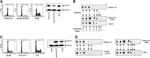
To understand whether posttranslational modifications of p27kip1 regulate its ability to selectively interact with different cyclin/cdk complexes, we initially investigated posttranslational modifications of p27kip1 in primary B cells in vivo in response to the mitogenic stimulus PMA, using 2-dimensional gel electrophoresis (2D-gel). As shown in Figure 1A, about 95% of purified human primary B cells are at G0 stage of the cell cycle. Exposure to PMA for 72 hours reduced the proportion of primary B cells at G0/G1 to around 78%. This was associated with a slight decrease in p27kip1 levels and an increase in the number of cells entering the cell cycle. Using 2D-gel analysis to compare the posttranslational modifications of p27kip1 in primary B cells before or after the treatment of PMA, we observed a dramatic change in the pattern of p27kip1 isoforms (Figure 1B). In primary B cells, there are 5 isoforms, isoforms 1 to 5, with PIs of 6.76, 6.59, 6.37, 6.12, and 6.05, respectively. The signals detected with isoforms 4 and 5 are minimal. Interestingly, the treatment with PMA induced a significant increase in the amount of isoforms 3, 4, and 5 of p27kip1. Additionally, 2 more isoforms with lower PI (5.93 and 5.83 for isoforms 6 and 7, respectively) were detected. The PMA-dependent changes suggested that cell-cycle entry is associated with an increase in posttranslational modification of p27kip1 in vivo in primary human B cells.
Increased posttranslational modification of p27kip1 correlates with cell-cycle entry. FACS analysis shows the percentage of cells arrested in G1/G0 (A, left panel) in primary B cells with or without treatment with PMA for 72 hours (labeled as primary B or primary B + PMA, respectively), Burkitt lymphoma cell line BL40 (labeled as BL40), (C) in H1299 cells with or without the inducible expression of p27kip1 (labeled as H1299, H1299 + p27kip1) and in Cal51 cells. Right-hand panels of A and C are Western blots showing the expression levels of p27kip1 in the corresponding cell populations used to carry out the FACS and 2D gel analysis. The isoforms 1 to 7 are indicated with arrows in panels B and D and their isoelectric points are 6.76, 6.59, 6.37, 6.12, 6.05, 5.93, and 5.83, respectively. Horizontal bars in panels A and C represent the marker used to calculate mean values of G0/G1 peak cell populations. PCNA indicates proliferating cell nuclear antigen.
Increased posttranslational modification of p27kip1 correlates with cell-cycle entry. FACS analysis shows the percentage of cells arrested in G1/G0 (A, left panel) in primary B cells with or without treatment with PMA for 72 hours (labeled as primary B or primary B + PMA, respectively), Burkitt lymphoma cell line BL40 (labeled as BL40), (C) in H1299 cells with or without the inducible expression of p27kip1 (labeled as H1299, H1299 + p27kip1) and in Cal51 cells. Right-hand panels of A and C are Western blots showing the expression levels of p27kip1 in the corresponding cell populations used to carry out the FACS and 2D gel analysis. The isoforms 1 to 7 are indicated with arrows in panels B and D and their isoelectric points are 6.76, 6.59, 6.37, 6.12, 6.05, 5.93, and 5.83, respectively. Horizontal bars in panels A and C represent the marker used to calculate mean values of G0/G1 peak cell populations. PCNA indicates proliferating cell nuclear antigen.
We showed previously that the growth inhibitory activity of p27kip1 is often compromised in Burkitt lymphoma cells which are of B-cell origin.4 In the Burkitt lymphoma cell line BL40, for example, the expression level of p27kip1 is very similar to that detected in untreated primary B cells. However, unlike primary B cells where 95% of cells are in G0/G1, only 52% of BL40 cells are in G1 (Figure 1A). If an increase in posttranslational modifications is responsible for the inactivation of p27kip1, the posttranslational modifications of p27kip1 expressed in BL40 cells would only resemble that seen in PMA-treated primary B cells. Indeed, the number and the intensity of p27kip1 isoforms detected in BL40- and PMA-treated primary B cells are almost identical. The significant differences in the patterns of p27kip1 isoforms between the untreated primary B and BL40 cells is particularly interesting, since these 2 cell populations express very similar amounts of p27kip1 (Figure 1A).
To investigate further whether the phenomenon observed here is restricted to B cells, we also compared the pattern of p27kip1 isoforms in another 2 epithelial cell lines, H1299 cells expressing inducible p27kip1 4 and Cal51 cells.3 Induced expression of p27kip1 arrested H1299 cells in G1, while an amount of p27kip1 similar to that seen in H1299 cells failed to inhibit the cell growth of Cal51 cells (Figure 1C). Consistent with the data from primary B and BL40 cells, the pattern of isoforms of p27kip1 in the inducible H1299 cells was very similar to that seen in the untreated primary B cells. In contrast, the posttranslational modification pattern of p27kip1 in Cal51 cells was almost identical to that of PMA-treated primary B and BL40 cells (Figure 1D). Hence, there is a very good correlation between an increase in posttranslational modifications of p27kip1 and cell-cycle entry.
Cell proliferation is associated with p27kip1 phosphorylation in vivo
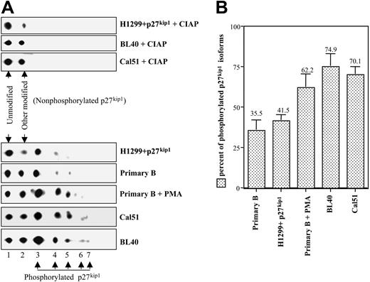
To characterize the posttranslational modifications of p27kip1 that are increased when cells enter the cell cycle, lysates derived from p27kip1-inducible H1299, BL40, and Cal51 cells were treated with calf intestinal alkaline phosphatase. As shown in Figure 2A, alkaline phosphatase treatments reduced the number of isoforms of p27kip1 detected in these cells. Only 2 isoforms with a PI of 6.76 and 6.59 were present, demonstrating that isoforms 3 to 7 are phosphorylated p27kip1. This is in agreement with a previous publication that isoform 1 of p27kip1 is the unmodified protein, while isoform 2 is modified p27kip1 by a currently unknown posttranslational modification.24 The signal intensities of the nonphosphorylated isoforms 1 and 2 were compared with those of phosphorylated p27kip1 (isoforms 3-7 altogether). In primary B cells, 36% of p27kip1 is phosphorylated, but the percentage of phosphorylated p27kip1 increased to 62% after exposure of the cells to PMA for 72 hours. In the 2 fast-growing tumor cell lines BL40 and Cal51, 75% and 70% of p27kip1 was phosphorylated, respectively. However, in the H1299 cells expressing inducible p27kip1 (p27kip1 ind H1299), only 42% of p27kip1 was phosphorylated (Figure 2B). It is interesting to note that regardless of the cell origin, the percentage of phosphorylated p27kip1 was remarkably similar among arrested cells (35% and 41% for primary B cells and H1299 + p27kip1, respectively) versus proliferating cells (62%, 75%, and 70% for PMA-treated primary B, BL40, and Cal51 cells, respectively). An increase in p27kip1 phosphorylation was closely associated with cell proliferation in vivo. Phosphorylation of p27kip1 could be one of the main factors controlling the growth inhibitory activity of p27kip1 in vivo, since p27kip1 expressed in BL40 and Cal51 cells is biologically compromised.
Posttranslational modification of p27kip1 linked to cell-cycle entry is phosphorylation. After treatment with CIAP, only 2 isoforms of p27kip1 were detected on 2D gel analysis (A, upper panels). Using bacterial-produced p27kip1 as a control, it is clear that isoform 1 is unmodified p27kip1, while isoform 2 is modified by a mechanism other than phosphorylation (data not shown). The disappearance of the other isoforms of p27kip1 in response to CIAP treatment demonstrated that isoforms 3 to 7 are phosphorylated p27kip1 isoforms (A, lower panels). The percentage of phosphorylated p27kip1 at a steady-state level in the various indicated cell population was obtained as follows: signals from all isoforms/signals of isoforms 1 and 2. The bar graph shown in panel B was derived from at least 3 independent experiments. Error bars indicate standard deviation; values above error bars indicate the value of the graph bar (not the error bar).
Posttranslational modification of p27kip1 linked to cell-cycle entry is phosphorylation. After treatment with CIAP, only 2 isoforms of p27kip1 were detected on 2D gel analysis (A, upper panels). Using bacterial-produced p27kip1 as a control, it is clear that isoform 1 is unmodified p27kip1, while isoform 2 is modified by a mechanism other than phosphorylation (data not shown). The disappearance of the other isoforms of p27kip1 in response to CIAP treatment demonstrated that isoforms 3 to 7 are phosphorylated p27kip1 isoforms (A, lower panels). The percentage of phosphorylated p27kip1 at a steady-state level in the various indicated cell population was obtained as follows: signals from all isoforms/signals of isoforms 1 and 2. The bar graph shown in panel B was derived from at least 3 independent experiments. Error bars indicate standard deviation; values above error bars indicate the value of the graph bar (not the error bar).
Cyclin E but not cyclin A selectively interacts with phosphorylated p27kip1 isoforms in vivo
It is well established that p27kip1 arrests cells when it binds to cyclinE/cdk2 but not D-type cyclins/cdk's. Perhaps the phosphorylation status of p27kip1 could determine its binding specificity to different cyclin/cdk complexes. Because of the practical difficulties in obtaining sufficient numbers of primary B cells and the low-level expression of cyclins, this issue was only addressed in 2 fast-growing tumor cell lines, BL40 and Cal51. 2D-gel was used to identify p27kip1 isoforms that specifically associate with different cyclins and cdk's. Lysates derived from BL40 and Cal51 cells were immunoprecipitated with antibodies specific for p27kip1, cdk2, cyclin A, or cyclin E. The immunoprecipitates were then analyzed in 2D-gels. The p27kip1 isoforms that coimmunoprecipitated with cdk2, cyclin A, or cyclin E were detected using an anti-p27kip1 antibody from a different species. As shown in Figure 3A, the pattern of p27kip1 isoforms coimmunoprecipitated with the anti-cdk2 antibody was very similar to that immunoprecipitated with the anti-p27kip1 antibody. The pattern of p27kip1 isoforms coimmunoprecipitated with cyclin A was also very similar to that seen with cdk2. These results suggested that cdk2 and cyclin A do not interact selectively with p27kip1 isoforms. In contrast the anti–cyclin E antibody preferentially immunoprecipitated phosphorylated p27kip1 isoforms. The p27kip1 isoforms with the lowest PI were significantly enriched in the precipitates immunoprecipitated with anti–cyclin E antibody in both BL40 and Cal51 cell lines (Figure 3A, bottom panel). The striking difference between the p27kip1 isoforms associated with cyclin A, cdk2 on one hand, and cyclin E on the other hand indicates for the first time that distinct p27kip1 isoforms are involved in binding to cyclin A and cyclin E. Moreover, it suggested that in BL40 and Cal51 cells, there is a substantial fraction of p27kip1 that interacts with cyclin E independently of cdk2 in vivo.
The enrichment of phosphorylated p27kip1 isoforms detected in the cyclin E immunoprecipitates could be caused by the ability of cyclin E to selectively interact with phosphorylated p27kip1 isoforms. Alternatively, the phosphorylated p27kip1 isoforms are the result of cyclin E/cdk2 phosphorylation when they are in the same complex together. To address these issues, Cal51 cells were treated with roscovitine, an inhibitor of cdk2 kinase.25 As shown in Figure 3B, roscovitine was able to inhibit p27kip1 phosphorylation in vivo to a large extent since there was a clear reduction in the amount of phosphorylated p27kip1 isoforms being detected. Under this condition, cyclin E was once again able to selectively bind to phosphorylated p27kip1 isoforms (Figure 3B, lower panels). In vitro, 30 μM roscovitine inhibited the immunoprecipitated cdk2 kinase activity from Cal51 cells by 87%, using histone H1 as substrate (Figure 3C). To further demonstrate that the phosphorylated p27kip1 isoforms detected in cyclin E immunoprecipitate was not caused by the kinase activity of cyclin E/cdk2 in vitro, 60 μM roscovitine was included in all buffers used to immunoprecipitate cyclin E from Cal51 cell lysates to eliminate the cyclinE/cdk2 kinase activity. Interestingly, the presence of roscovitine did not alter the pattern of p27kip1 isoforms associated with cyclin E (Figure 3D, comparing left with right panels). These results demonstrated clearly that cyclin E has higher binding affinity to phosphorylated p27kip1 isoforms.
Cyclin D1 and D2 favor unphosphorylated p27kip1 isoforms
The ability of different D-type cyclins and cdk's to interact with p27kip1 isoforms in vivo was also investigated, using 2D gel analysis in BL40 and Cal51 cells. Although lymphoid cells, including BL40 cells, express low levels of cyclin D1, however, our newly generated monoclonal anti–cyclin D1 antibody LX014 enabled us to detect the expression of cyclin D1 in BL40 cells (Figure 4A). The specificity of this anti–cyclin D1 antibody is also illustrated. Interestingly, the anti–cyclin D1 antibody LX014 selectively enriched the unmodified p27kip1 isoform, isoform 1. The propensity of cyclin D1 to interact with unmodified p27kip1 isoform 1 was so high that in BL40 cells almost only isoform 1 was coimmunoprecipitated with cyclin D1. In Cal51 cells, around 70% of p27kip1 associated with cyclin D1 was isoform 1 (Figure 4B).
Cyclin D2 also selectively interacts with unphosphorylated p27kip1 in the 2 cell lines tested. However, the pattern of p27kip1 isoforms interacting with cyclin D2 was different from that seen with cyclin D1. Unlike that seen with cyclin D1, less than 50% of the cyclin D2–bound p27kip1 was unmodified p27kip1 (isoform 1). The other major form of p27kip1 coimmunoprecipitated with cyclin D2 was isoform 2 (Figure 4B). These results indicated that unlike cyclin A, cyclin E and cdk2, both cyclin D1 and cyclin D2 selectively bind to unphosphorylated p27kip1 isoforms.
Cyclin D3 and cdk4 selectively interact with phosphorylated p27kip1 in vivo
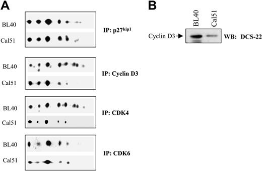
We also investigated the ability of cyclin D3, cdk4, and cdk6 to interact with different p27kip1 isoforms in vivo. Using the same set of cell lysates, we observed that cdk6 does not bind p27kip1 in a highly selective manner. In contrast, cdk4 selectively interacts with phosphorylated p27kip1 isoforms in BL40 but not Cal51 cells. Moreover, the pattern of p27kip1 isoforms coimmunoprecipitated with cyclin D3 was almost identical to that seen with cdk4 in BL40 and Cal51 cells, respectively (Figure 5A). The failure to see the selective interaction of phosphorylated p27kip1 isoforms with cyclin D3 and cdk4 in Cal51 cells may be due to the low-expression levels of D3 (Figure 5B). Nevertheless, the binding spectrum of cyclin D3 and cdk4 to p27kip1 isoforms was similar in vivo. This is in contrast to that seen with cyclin D1 and cyclin D2 in which their binding patterns are completely independent of their partners cdk4 and cdk6. Hence, cyclin D3 and cdk4 may bind p27kip1 as a complex in vivo, while cyclin D1 and cyclin D2 can bind p27kip1 independently from their cdk partners.
Cyclin D3 and cdk4 selectively interact with phosphorylated p27kip1 in vivo. 2D gel analysis shows that cyclin D3 and cdk4 (A) but not cdk6 (B) selectively interact with p27kip1 isoforms 5 to 8 in BL40 cells (A). Western blot shows the expression level of cyclin D3 in Cal51 and BL40 cells detected by the anti–cyclin D3 antibody DCS-22 (B).
Cyclin D3 and cdk4 selectively interact with phosphorylated p27kip1 in vivo. 2D gel analysis shows that cyclin D3 and cdk4 (A) but not cdk6 (B) selectively interact with p27kip1 isoforms 5 to 8 in BL40 cells (A). Western blot shows the expression level of cyclin D3 in Cal51 and BL40 cells detected by the anti–cyclin D3 antibody DCS-22 (B).
Cyclin D3/cdk4 is the most active kinase complex to sequester p27kip1 in vitro
To provide some biochemical evidence that the different binding spectrums of D-type cyclins and cdk4 and cdk6 may have different influences on the growth inhibitory activity of p27kip1, we turned to an in vitro cyclin E/cdk2 kinase assay. Our in vivo observations suggested that phosphorylation plays an important role in determining the binding specificity of p27kip1 isoforms to different cyclins and cdk's. As it is the major kinase of p27kip1 in vivo, cyclin E/cdk2 phosphorylates p27kip1 very effectively in vitro. Therefore, under the experimental conditions described here, it is possible that cyclin E/cdk2 could phosphorylate the recombinant p27kip1 used in the in vitro kinase assay. Such phosphorylation may even enhance the inhibitory activity of p27kip1 and would agree with our in vivo finding that cyclin E selectively interacts with phosphorylated p27kip1. If so, one would predict that in vitro cyclin D3 would be more effective than either cyclin D1 or cyclin D2 at sequestering the inhibitory effect of p27kip1 on the cyclin E/cdk2 kinase because cyclin D3 selectively interacts with phosphorylated p27kip1 isoforms in vivo. Cyclin D1 and D2, however, selectively interact with unphosphorylated p27kip1 in vivo. Similarly, the ability of cdk4 to sequester the inhibitory activity of p27kip1 may be greater than that of cdk6. The D-type cyclin/cdk4 or cdk6 complexes are kinases; it is also possible that the kinase activity of different D-type cyclin/cdk4 or cdk6 complexes would determine their ability to sequester p27kip1. To test this, an in vitro cyclin E/cdk2 kinase assay was used to measure the inhibitory activity of p27kip1. We also investigated the ability of various D-type cyclins/cdk's to sequester the inhibitory activity of p27kip1 in vitro. The potential effects of D-type cyclins/cdk's on the inhibitory activity of p21cip/kip and p57kip2 were also investigated. Increasing amounts of GST-p27kip1, GST-p21cip1, and GST-p57kip2 were incubated with a fixed amount of cyclin E/cdk2 and then tested in a kinase assay using histone H1 as a substrate. An inverse correlation between the amounts of p27kip1, p21cip1, p57kip2, and the kinase activity of cyclin E/cdk2 was observed. Under the same conditions, GST did not inhibit cyclin E/cdk2 kinase activity (Figure 6C, lane 2; also data not shown). After a careful titration, the amounts of recombinant p27kip1, p21cip1, and p57kip2 that inhibited cyclin E/cdk2 kinase activity to a similar extent were obtained (Figure 6A, left panel).
To investigate the ability of different D-type cyclin/cdk complexes to sequester the inhibitory activity of p27kip1, p21cip1, and p57kip2, various combinations of D-type cyclin/cdk's were generated by coinfecting SF9 cells with baculoviruses expressing different D-type cyclins tagged with a 9E10 epitope alongside cdk's (untagged).25 To study the requirement of the kinase activity of D-type cyclin/cdk4 or cdk6 complexes, titrations of the infected SF9 cell lysates expressing each D-type cyclin/cdk were also used in a kinase assay using Rb as a substrate to measure their kinase activity. Under the same conditions, lysates derived from SF9 cells infected only with any of the D-type cyclins or cdk's alone failed to phosphorylate Rb (data not shown). They also failed to sequester the inhibitory activity of p27kip1 in vitro (data not shown), suggesting that a protein complex is required. Because of the practical difficulty in obtaining the protein complexes expressing the same amount of the different D-type cyclin/cdk4 or cdk6 complexes, cell lysates expressing D-type cyclin/cdk with comparable kinase activity on Rb were then chosen to carry out the sequestration experiment (Figure 6B, left panel).
In the same assay, around 75% of cyclin E/cdk2 kinase activity was inhibited by GST-p27kip1, GST-p21cip1, or GST-p57kip2 but not by GST alone (Figure 6C, left panel, lanes 1-3). The cyclin E/cdk2 kinase activity detected in the presence of GST-p27kip1, GST-p21cip1, or GST-p57kip2 (Figure 6C, left panel, lane 3 for all panels) was used as a baseline, set to a value of 1. The ability of different D-type cyclin/cdk complexes to sequester the inhibitory activities of GST-p27kip1, GST-p21cip1, or GST-p57kip2 was measured by their ability to increase the cyclinE/cdk2 kinase activity in the presence of GST-p27kip1, GST-p21cip1, or GST-p57kip2. These data shown in Figure 6B, left panel (lanes 4-9) suggested that D-type cyclins/cdk's are more able to sequester the inhibitory activity of p27kip1 than that of p21cip1 or p57kip2. There is a large variation among all the D-type cyclin/cdk's in their ability to sequester the inhibitory activity of p27kip1 (Figure 6C, left panel, lanes 4-9). Interestingly, the cyclinE/cdk2 kinase activity detected in the presence of p27kip1 and cyclin D3/cdk4 was the highest. Although the cyclin E/cdk2 kinase activity detected in the presence of GST-p27kip1 + cyclin D3/cdk4 was similar to that of GST-p27kip1 + cyclin D1/cdk6, the kinase activity of cyclin D3/cdk4 was less than that of cyclin D1/cdk6 (Figure 6B, left panel). When the kinase activity of individual D-type cyclin/cdk's was taken into consideration, it became clear that cyclin E/cdk2 kinase activities in the presence of cyclin D3/cdk4 or cyclin D1/cdk6 were about 7- and 4-fold higher than that detected in the absence of cyclin D3/cdk4 or cyclin D1/cdk6 kinases, respectively (Figure 6B-D, left panels, comparing lanes 6 and 7 with lane 3). The cyclin E/cdk2 kinase activity detected in the presence of the remaining D-type cyclins/cdk's were 1- to 4-fold higher than that with p27kip1 alone. Additionally, the effect of cyclin D3/cdk4 on p27kip1 was very specific, since its effect on p21cip1 and p57kip2 was minimal under the same conditions (Figure 6C, left panel, lower 2 panels). These results provide another line of evidence that the kinase activity detected in Figure 6C was due to the ability of cyclin D3/cdk4 to sequester the inhibitory activity of p27kip1 rather than cyclin D3/cdk4 itself phosphorylating histone H1 directly (Figure 6C-D, left panels).
Finally, similar amounts of different D-type cyclins/cdk4 or cdk6 complexes were also used in the kinase assay (Figure 6A, right panel). The result shown in the right panel of Figure 6B illustrates that, under the same molar concentration, cyclin D3/cdk4 is the most active kinase to phosphorylate GST-Rb compared with other D-type cyclin/cdk complexes. Importantly, under the same conditions cyclin D3/cdk4 is also the most effective kinase at sequestering the inhibitory activity of p27kip1 but has little effect on either p21cip1 or p57kip2 (Figure 6C-D, right panels). Taken together, the results shown here demonstrate for the first time that, among all D-type cyclins/cdk's, cyclin D3/cdk4 is the most active complex able to sequester the inhibitory activity in vitro of p27kip1 but not p21cip1 or p57kip2.
Discussion
Through a detailed analysis of p27kip1 modification in vivo, we observed that there is a tight correlation between cell-cycle entry and an increase in p27kip1 phosphorylation. Using 2D gel analysis, we show here for the first time that different cyclins and cdk's interact with distinct isoforms of p27kip1 in vivo. Five different cyclins bind to 4 different patterns of p27kip1 isoforms in vivo in 2 fast-growing tumor cell lines. Cyclin E and cyclin D3 favor phosphorylated p27kip1 isoforms, but cyclin D1 and cyclin D2 preferentially bind unphosphorylated p27kip1. Moreover, while cyclin D1 favors the unmodified isoform of p27kip1 (isoform 1), cyclin D2 preferentially binds unphosphorylated isoforms of p27kip1 (isoforms 1 and 2). Only cyclin A did not show any selectivity toward specific p27kip1 isoforms. Interestingly, cyclin A and cyclin D3 share similar binding patterns to their respective cdk partners, cdk2 and cdk4. The binding patterns of cyclin E, cyclin D1, and cyclin D2 are unique and have no resemblance to those of their respective partners cdk2 (for E), cdk4, and cdk6. Thus, the binding specificity of various cyclins to p27kip1 is determined by the posttranslational modification. The findings also agree with an earlier in vitro observation that the cip/kip family of proteins bind to cdk's via cyclins.26 The binding patterns of cyclins and cdk's to different p27kip1 isoforms were similar in 2 cell lines with completely different cell origins, suggesting that the observations shown here reflect the general binding spectrum of p27kip1 to different cyclins and cdk's in vivo. The unique binding patterns of p27kip1 isoforms to different cyclins and cdk's were also not specific to the antibodies used as similar results were also obtained with other anti–cyclin and cdk antibodies (data not shown). The p27kip1 expressed in these 2 cell lines is inactive and heavily phosphorylated in comparison with its normal counterpart, for example, that was seen in primary B cells. It is tempting to speculate that one of the mechanisms by which B-cell lymphomas inactivate the growth inhibitory properties of p27kip1 in vivo is to prevent p27kip1 from binding to cyclin E/cdk2 complexes, just as a substantial amount of cyclin E binds to p27kip1 independently of its partner cdk2 in BL40 and Cal51 cells. This ability of cyclin E to bind phosphorylated p27kip1 alone may be an underlying explanation of why p27kip1 is expressed at a high level in these 2 cell lines, since efficient ubiquitination of p27kip1 requires its stable association with cyclin E/cdk2 or cyclin A/cdk2 complexes.11 Overexpression of cyclin E is often inversely associated with high expression levels of p27kip1. Nevertheless, the expression levels of cyclin E were not significantly lower in BL40 and Cal51 cells in comparison to other Burkitt lymphoma or breast carcinoma cell lines with lower p27kip1 levels.3-5 Therefore, it is possible that phosphorylation of p27kip1 at sites other than Thr187 may result in the enhanced stability of p27kip1 through its specific interaction with cyclin E independently of cdk2. Whether the specific interaction with cyclin D1 and D2 independently of cdk4 and cdk6 could also enhance the stability of p27kip1 remains unknown.
The identification of cyclin D3/cdk4 as the complex that is most able to overcome the inhibitory activity of p27kip1 on cyclin E/cdk2 kinase in vitro is particularly interesting, since our data shown here suggested that cyclin D3 but not cyclin D1 or D2 binds p27kip1 in vivo as a protein complex with cdk4. Although cyclin D3 and cdk4 share a similar binding specificity to p27kip1, cyclin D3/cdk6, cyclin D1/cdk4, and cyclin D2/cdk4 were unable to sequester the inhibitory activity of p27kip1 on cyclin E/cdk2 effectively. Hence, neither cyclin D3 nor cdk4 alone was sufficient to sequester the inhibitory activity of p27kip1. Despite the fact that the kinase activities or the molar concentrations of different D-type cyclins/cdk4 and cdk6 complexes used in the kinase assays shown in Figure 6 were similar, their ability to sequester the inhibitory activity of p27kip1 differed dramatically. This finding suggested that in addition to the kinase activity, other properties of the D-type cyclin/cdk's could influence their ability to sequester the inhibitory activity of p27kip1. Cyclin D3/cdk4 may acquire the binding specificity for p27kip1 as a complex but not as an individual protein. Nevertheless, the observation that cyclin D3/cdk4 is the complex that is most able to prevent the inhibitory activity of p27kip1 on cyclin E/cdk2 provided a molecular explanation as to why, among all cdk's, the amplification of cdk4 is very frequent in human tumors. It also explains why increased expression of cyclin D3 is often observed in B-cell lymphomas such as diffuse large B-cell lymphomas and Burkitt lymphomas. More importantly, high-expression levels of p27kip1 and cyclin D3 are associated with adverse clinical outcome. Thus, the finding reported here provides an additional molecular insight into how the growth inhibitory activity of p27kip1 is deregulated in human tumors, B-cell lymphomas in particular. Although the other D-type cyclin/cdk complexes are not as active as cyclin D3/cdk4 in overcoming the inhibitory activity of p27kip1, overexpression of cyclin D1 occurs frequently in carcinomas of the breast, head, and neck. At a higher concentration, cyclin D1/cdk4 can sequester the inhibitory activity of p27kip1. Perhaps in these tumors, increased expression of cyclin D1 enables the efficient sequestration of the inhibitory activity of p27kip1. Future understanding of the regulation of p27kip1 phosphorylation in vivo could provide new strategies to inhibit tumor growth by reactivating the growth inhibitory activity of p27kip1.
Prepublished online as Blood First Edition Paper, January 21, 2005; DOI 10.1182/blood-2003-07-2558.
Supported by the Leukaemia Research Fund and the Ludwig Institute for Cancer Research. W.Z. was a fellow of the Chinese Scholars Council.
The publication costs of this article were defrayed in part by page charge payment. Therefore, and solely to indicate this fact, this article is hereby marked “advertisement” in accordance with 18 U.S.C. section 1734.
We thank Dr Mittnacht for reagents and Drs Farrell and Watson for the critical reading of this manuscript.


This feature is available to Subscribers Only
Sign In or Create an Account Close Modal